-
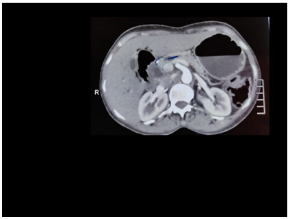
Figure 1.
Pancreatic protocol CT scan showing pancreatic head lesion (marked in semicircular blue) causing dilatation of CBD. Lesion is placed between MPD (marked in linear blue) and CBD.
-
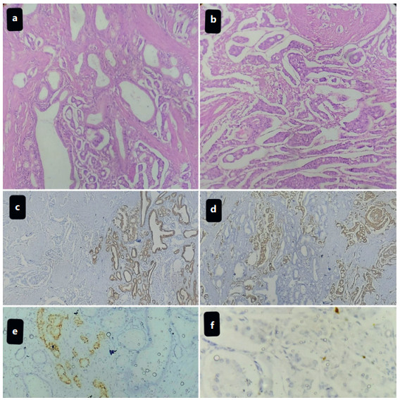
Figure 2.
Histopathology and immunohistochemistry of MINEN. a High power (×40) (H&E); tumor cells arranged in glandular pattern suggestive of adenocarcinoma component. b High power (×40) (H&E); tumor cells arranged in nesting pattern suggestive of NEN component. c Low power (×10); adenocarcinoma component of tumor strongly highlighted by PanCK immunohistochemistry marker. d Low power (×10); NEN component of tumor strongly highlighted by synaptophysin immunohistochemisty marker. e High power (×40); NEN component of tumor highlighted by chromogranin. f High power (×40); Ki 67 index <1%.
-
Genes common to NEN Genes common to nNEN Genes common to both ATRX ARID1A TP53 RB1 PIK3CA Loss of heterozygosity CTNNB1 KRAS MYC BRAF APC PI3KCA MUTYH CHECK2 BRCA2
Figures
(2)
Tables
(1)