-
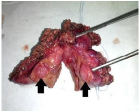
Figure 1.
Case 1. Postoperative cut-opened specimen of the liver from extended cholecystectomy showing nodular lesions (black arrows) showing liver metastases from the primary small cell carcinoma neuroendocrine carcinoma of the gallbladder.
-
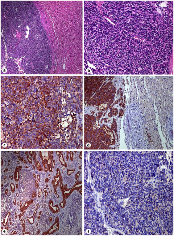
Figure 2.
Case 1. Small cell neuroendocrine tumor. a The tumor is composed of poorly differentiated oval to polygonal cells with hyperchromatic nuclei (H&E stain. ×100). b Tumor cells show pleomorphic nuclei with mitotic activity (H&E. ×400). c CK7 positivity in the tumor cells. d Synaptophysin positivity in the tumor cells. e CK7 positivity in the metastatic carcinoma in the lymph node. f MIB-1 proliferation in the tumor cells.
-
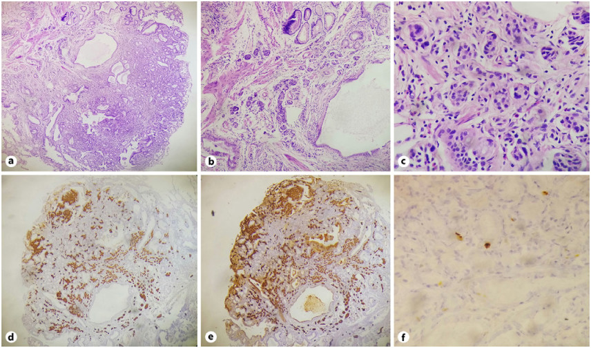
Figure 3.
Case 2. a Gallbladder NET polyp. b Tumor infiltrating the muscularis mucosa. c Tumor cells in cords and nests in high power. d Positive immunohistochemistry for synaptophysin in low power. e Positive immunohistochemistry for chromogranin in low power. f MIB-1 labeling index 1–2% in high power.
Figures
(3)
Tables
(0)