-
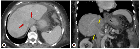
Figure 1.
a Axial contrast-enhanced CT image demonstrating diffuse hypoattenuating liver metastases (red arrows). b Coronal contrast-enhanced CT image demonstrating 2 metastases, seen to exert marked mass effect on the IVC (yellow arrows). IVC, inferior vena cava; CT, computed tomography.
-
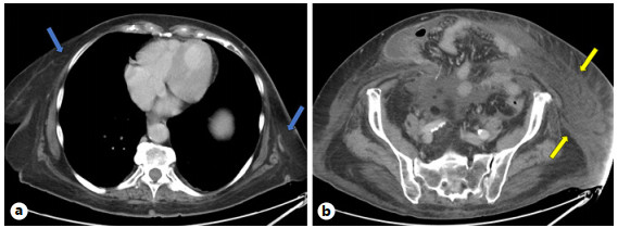
Figure 2.
a Axial contrast-enhanced CT image at the level of the heart. Subcutaneous soft tissue above the diaphragm is unremarkable (red arrows). b Axial contrast-enhanced CT image at the level of the sacroiliac joints demonstrating diffuse soft tissue infiltration below the diaphragm (yellow arrows). CT, computed tomography.
-
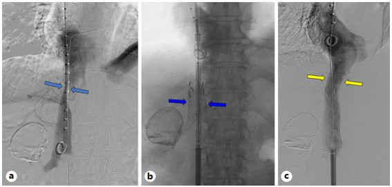
Figure 3.
a–c IVC venograms which demonstrate (a) severe external compression (red arrows), (b) stent deployment (blue arrows), and (c) post-stenting and angioplasty with improved patency of the IVC (yellow arrows). IVC, inferior vena cava.
-
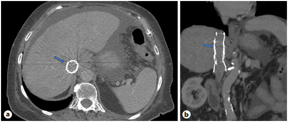
Figure 4.
a, b Axial and coronal contrast-enhanced CT images demonstrating patent stents within a widely patent IVC (red arrows). IVC, inferior vena cava; CT, computed tomography.
-
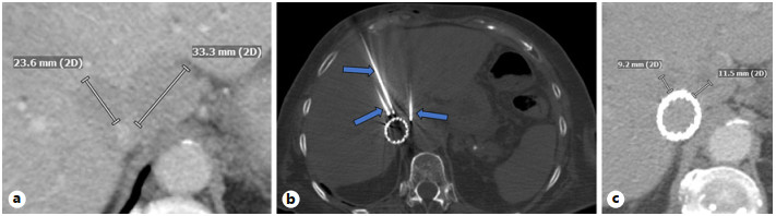
Figure 5.
a Axial contrast-enhanced CT images before cryoablation. b Axial intraoperative CT image demonstrating cryoablation probes within the previously noted segment I metastatic lesions (red arrows). c Postcryoablation 2- month follow-up demonstrating reduction in the size of segment I metastatic lesions and patent stent within the IVC. IVC, inferior vena cava; CT, computed tomography.
-
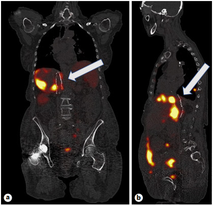
Figure 6.
a Coronal Ga68 Dotatate PET-CT scan showing maintenance of IVC stent patency at 6-month follow-up (white arrow). b Sagittal Ga68 Dotatate PET-CT scan showing maintenance of IVC stent patency at 6-month follow-up (white arrow). IVC, inferior vena cava; CT, computed tomography.
-
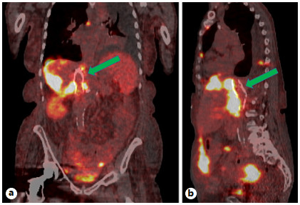
Figure 7.
a Coronal Ga68 Dotatate PET-CT scan showing maintenance of IVC stent patency at 9-month follow-up (green arrow). b Sagittal Ga68 Dotatate PET-CT scan showing maintenance of IVC stent patency at 9-month follow-up (green arrow). IVC, inferior vena cava; CT, computed tomography.
Figures
(7)
Tables
(0)