-
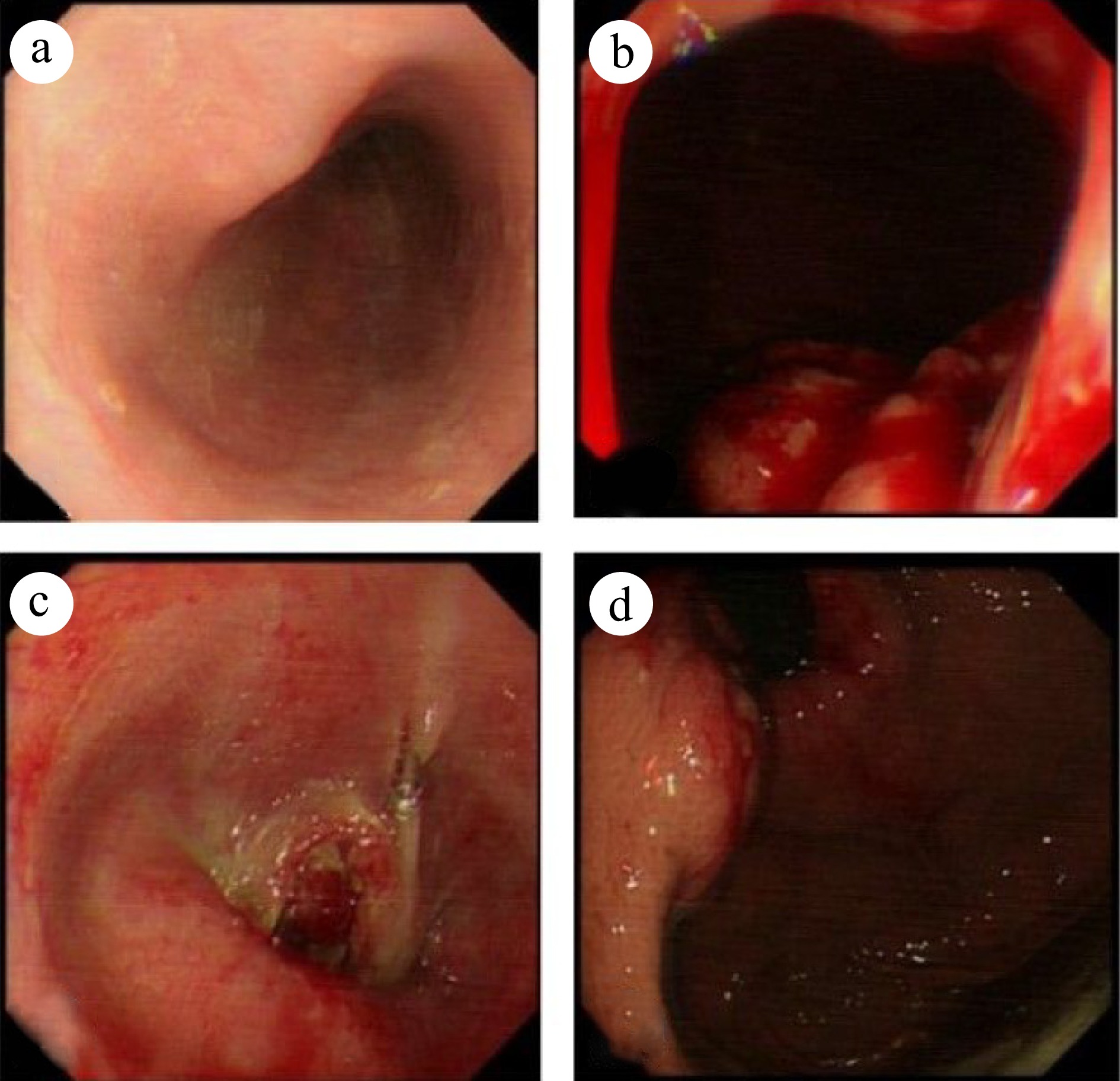
Figure 1.
Gastroscopy reveals an unobstructed esophagus with smooth mucosa. The dentate line is indistinct, and infiltrative lesions are evident in the cardia gastric body. The gastric fundus mucosa appears smooth and displays a normal coloration. (a) Esophagus. (b) Inferior cardia. (c) Esophagogastric junction. (d) Fundus of the stomach.
-
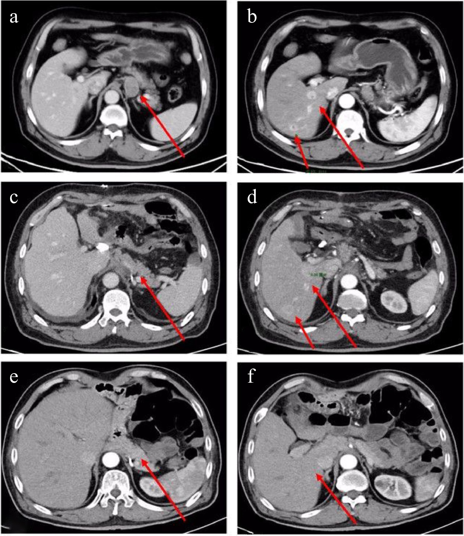
Figure 2.
Enhanced CT scan of the chest and abdomen: Preoperative: (a) Enlarged lymph nodes below the cardia (maximum size 3.3 cm × 2.3 cm) (indicated by an arrow); (b) Multiple abnormally enhanced nodules (maximum size 1.6 cm × 2.1 cm) in the right lobe of the liver (indicated by an arrow). Postoperative: (c) No enlarged lymph nodes are observed at the approximate position of the original cardia (indicated by an arrow); (d) Two abnormally enhanced nodules in the right lobe of the liver, larger than before (3.1 cm × 2.8 cm) (indicated by an arrow). The latest images: (e) No enlarged lymph nodes detected at the approximate position of the original cardia; (f) No abnormal enhancement nodules in the right lobe of the liver.
-
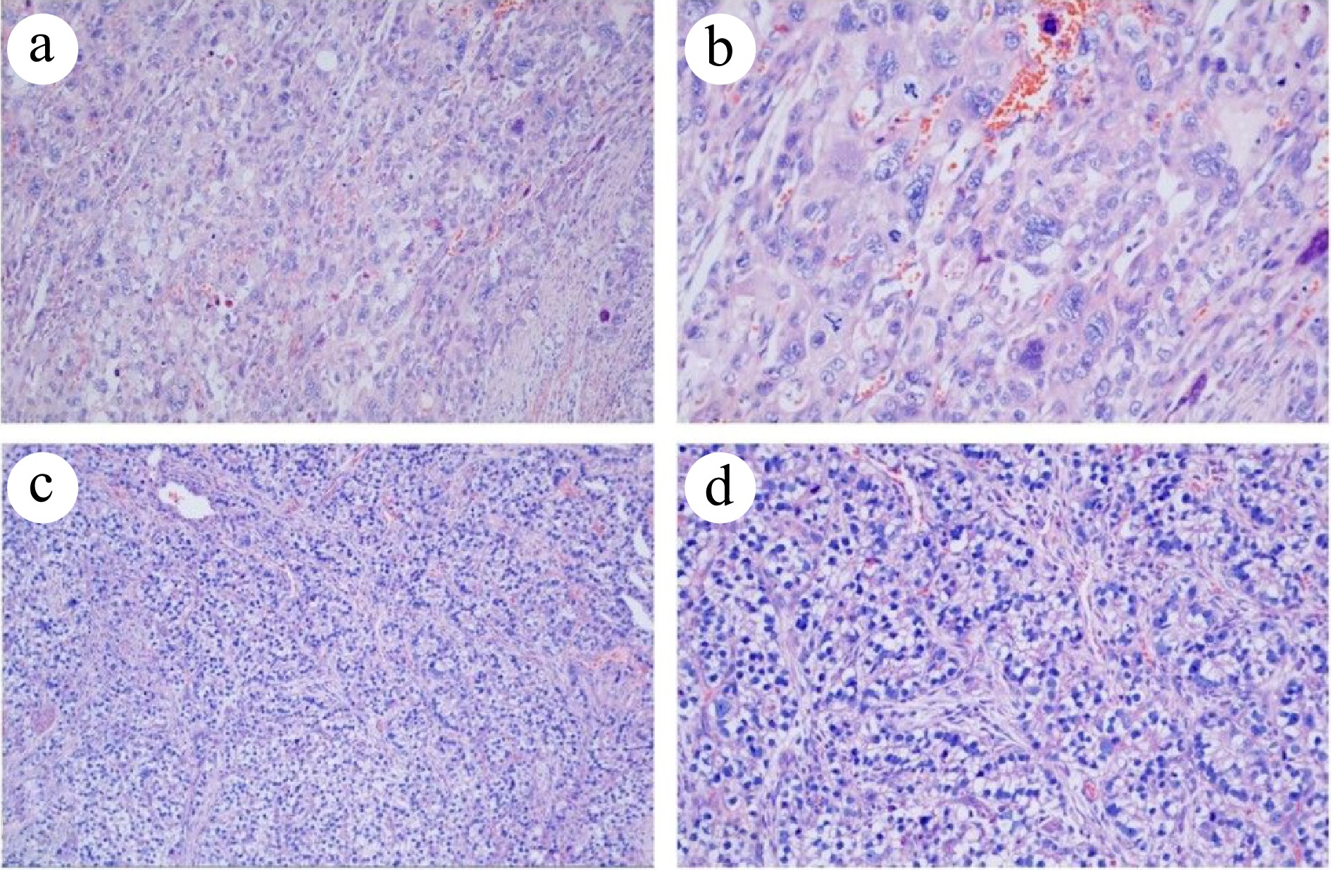
Figure 3.
Histopathological analysis of the resected specimen (HE staining). (a) PGC at 100× magnification. (b) PGC at 200× magnification. (c) Intestinal fibroblast-differentiated AFPGC at 100× magnification. (d) Intestinal fibroblast- differentiated AFPGC at 200× magnification.
Figures
(3)
Tables
(0)