-
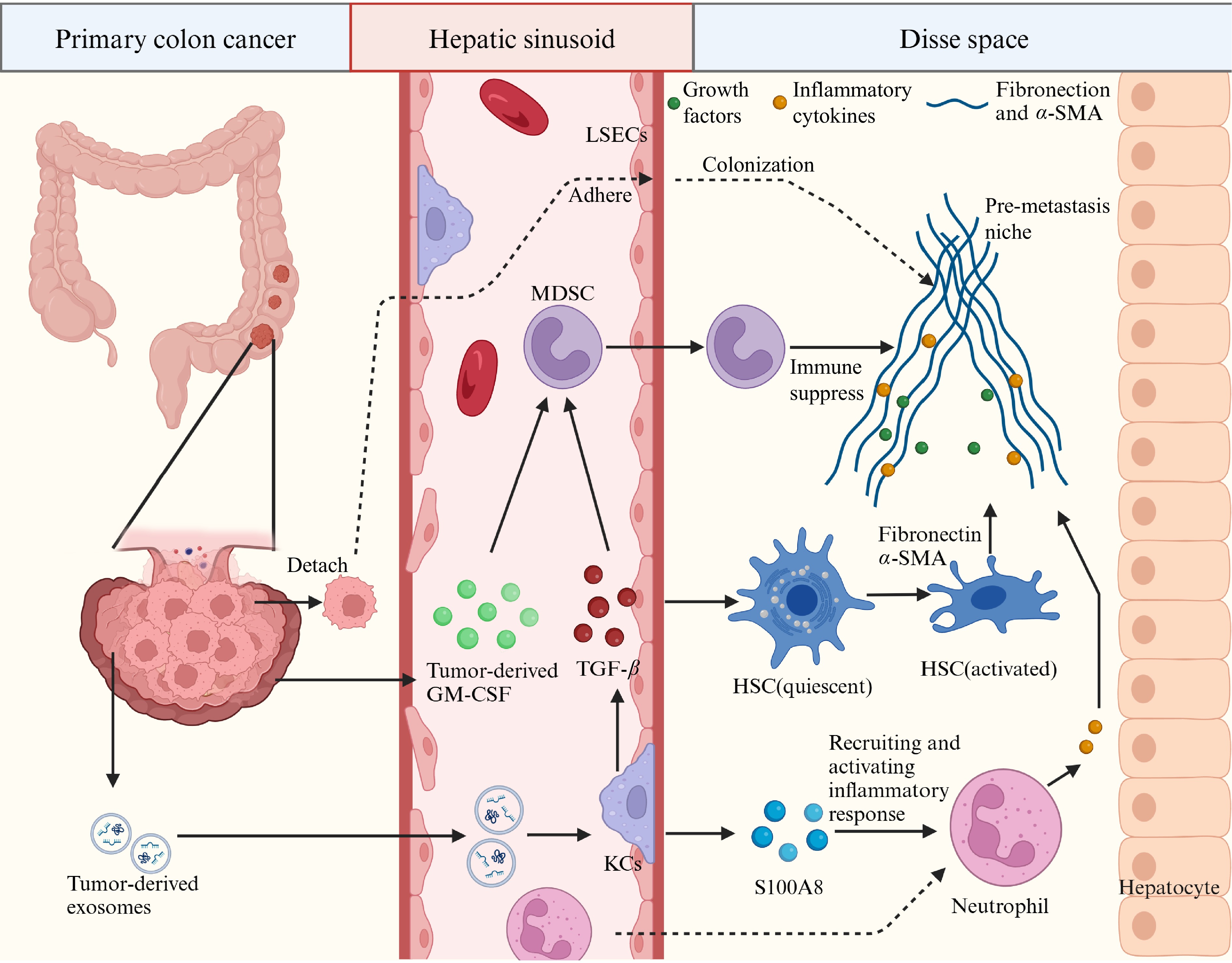
Figure 1.
The pre-metastatic niche. Shown is a diagrammatic representation of the cells and molecules involved in the formation of premetastatic niches in the liver. Disse space, also known as perisinusoidal space, is the liver blood sinusoidal endothelial cells and hepatocytes between a narrow gap.
-
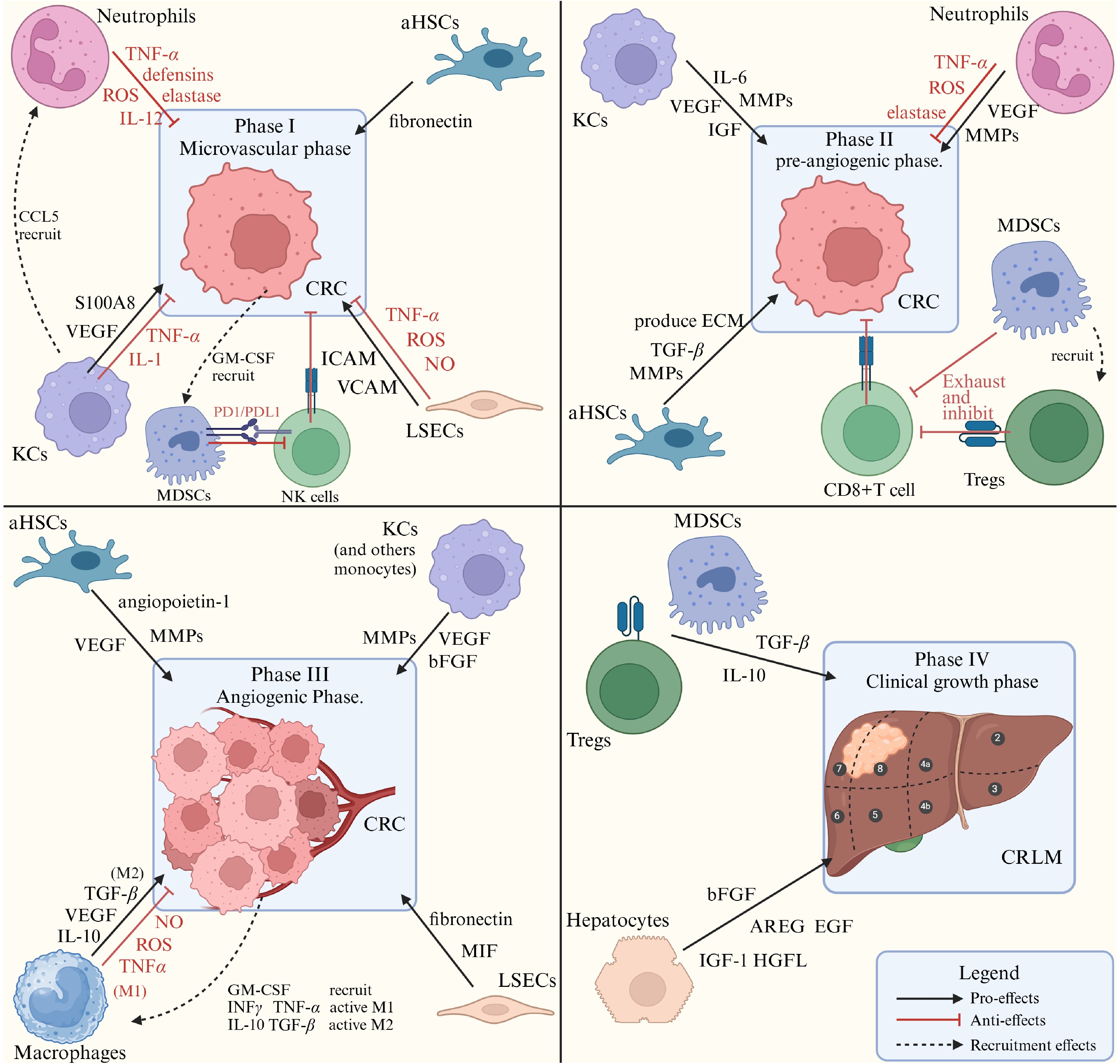
Figure 2.
Different kinds of cells in the liver immune microenvironment and their roles in CRLM phases. The secreting factors that promote and inhibit tumor colonization and growth, from varieties of liver cells are listed. MMPs: matrix metalloproteinases; VEGF: vascular endothelial growth factor; MIF: macrophage migration inhibitory factor; AREG: amphiregulin, an EGF-like protein; bFGF: basic fibroblast growth factor; HGFL: hepatocyte growth factor-like protein.
-
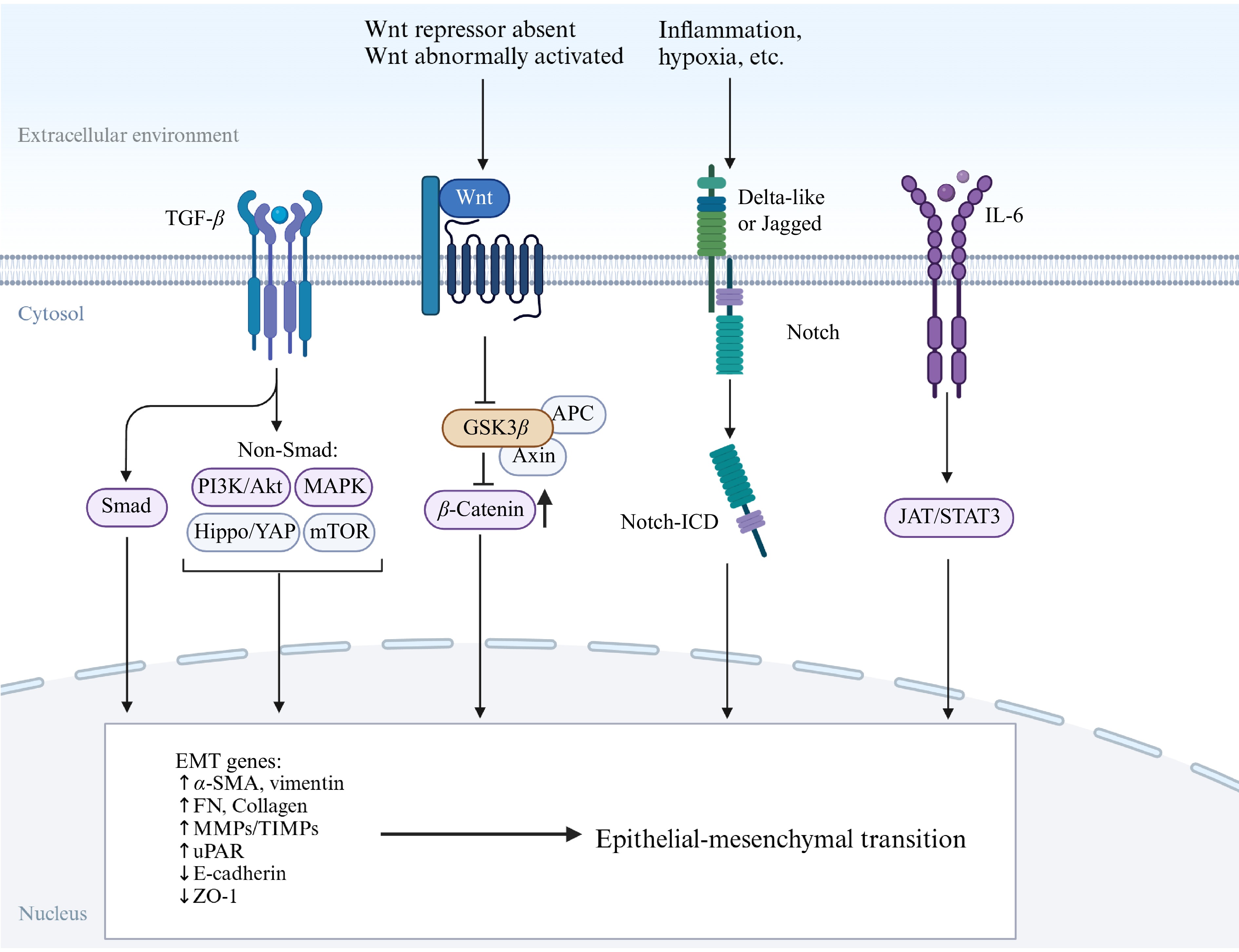
Figure 3.
Several main signaling pathways of EMT. The Wnt pathway is active because the Wnt/β-catenin pathway inhibitor is absent and β-catenin accumulates in the cytoplasm. The Notch pathway is activated by contact with neighboring cells, which comes from inflammation, ER stress, hypoxia, and others.
-
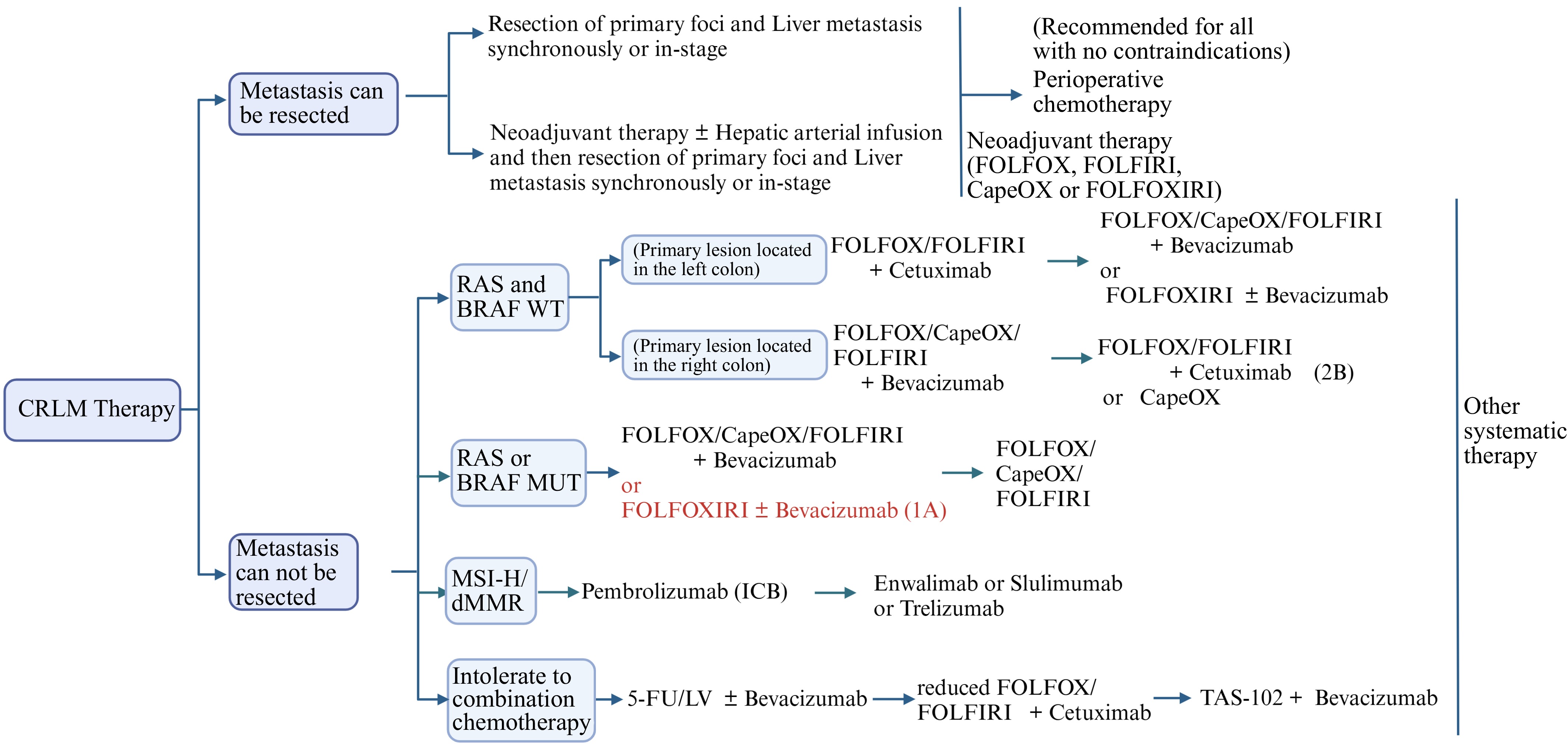
Figure 4.
CRLM's recommended treatment flowchart. FOLFOX: Calcium folinate + Fluorouracil (5-FU) + Oxaliplatin; CapeOX: Oxaliplatin + Capecitabine; LV: Leucovorin (folinate); FOLFIRI: Calcium folinate + Fluorouracil + Irinotecan; FOLFOXIRI: Calcium folinate + Fluorouracil + Oxaliplatin + Irinotecan.
Figures
(4)
Tables
(0)