-
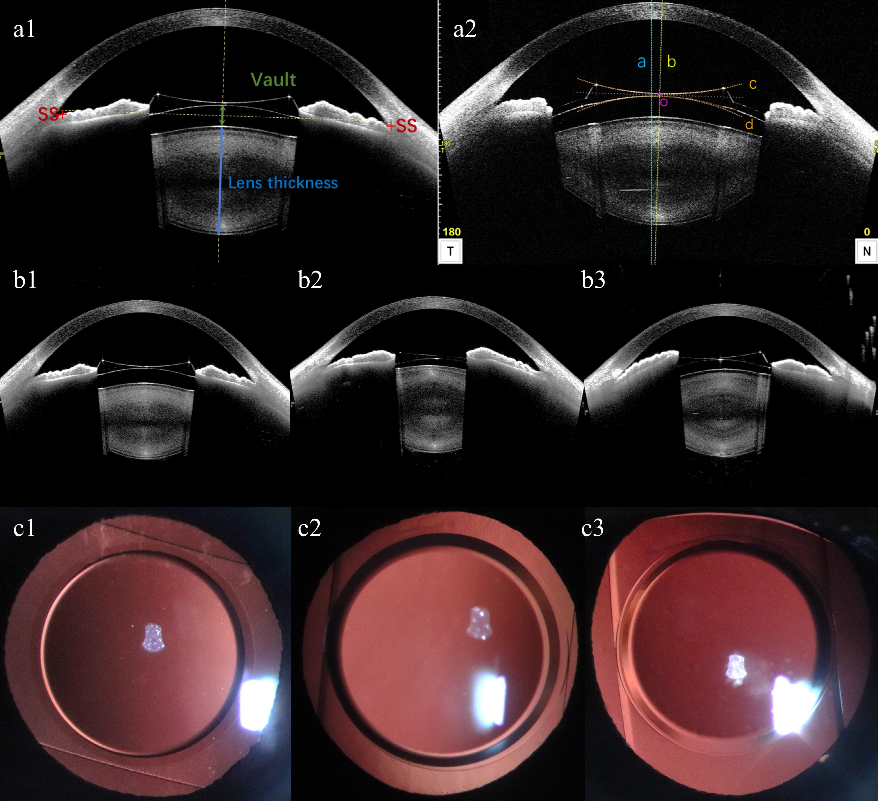
Figure 1.
(a1), (a2) Schematic diagram of anterior segment parameters from CASIA2. (a1) Vault, postoperative ACD, ACW, and lens thickness measured by CASIA2. SS: scleral spur. ACW: anterior chamber width, the distance between the bilateral scleral spurs. Postoperative ACD: postoperative anterior chamber depth, distance from corneal endothelium to anterior surface of EPRL. Vault: green line with double arrowheads, distance from the back surface of EPRL to the front surface of natural crystalline lens. Lens thickness: blue line with double arrowheads, natural crystalline lens thickness. (a2) A representative image showing the location of EPRL in the eye from the manually corrected Lens Analysis mode of CASIA2. The horizontal and vertical decentration and tilt were the values on the 0- and the 90- degree-images. The blue dashed line (line a) represents the visual axis passing through the corneal vertex. The yellow dashed line (line b) represents the central axis of EPRL, which passing through the centers of the two circles formed by the manually corrected front surface line (line c) and back surface line (line d) of EPRL. The midpoint of the intersections between the central axis and the front and back surfaces of EPRL is the center point of EPRL (point o). The decentration of the EPRL is quantified by calculating the vertical distance between point o and the visual axis. The tilt of the EPRL were quantified by calculating the rotation degree between line a and b. (b1)−(b3) EPRL and iris morphology observed by CASIA2. (b1) EPRL is relatively centered, with bilateral iris symmetry. (b2) EPRL decenters to the right, with a higher position of the iris on the right side compared to the left. (b3) EPRL decenters to the left, with the iris on the left side positioned higher than the right. (c1)−(c3) Anterior segment photography with the slit lamp backlighting method was used to capture the position of EPRL rotation.
-
Characteristics Value Case (eyes) 51 Age (yrs) 30.27 ± 7.76 (range, 20−53) Gender Male 5 (10 eyes) Female 21 (41 eyes) Follow-up period (mos) 9.75 ± 6.88 (range, 0.25−20.00) EPRL diopter (D) −19.33 ± 5.08 (range, −28.00 to −10.00) EPRL model BK108 9 (18 eyes) BK113 17 (33 eyes) EPRL = Ejinn phakic refractive lens. Table 1.
Demographics and clinical characteristics of 26 patients with EPRL.
-
Preoperative variables (range) Postoperative variables (range) p-value SE (D) −19.09 ± 6.17
(−29.00 to −7.87)−1.00 ± 1.19
(−4.50 to 1.00)< 0.001* UDVA (logMAR) 0.04 ± 0.03; (0.01−0.12) 0.50 ± 0.23; (0.10−1.00) < 0.001* CDVA (logMAR) 0.55 ± 0.29; (0.15−1.00) 0.70 ± 0.26; (0.30−1.00) 0.006* IOP (mmHg) 15.28 ± 2.70; (9.7−20.1) 15.30 ± 2.59; (10.2−20.8) 0.964 ACD (mm) 2.81 ± 0.27; (2.45−3.40) 2.73 ± 0.26; (2.36−3.27) 0.096 ECD (cells/mm²) 2,976.82 ± 306.38; (2,301−3,642) 2,854.43 ± 335.693; (2,069−3,458) 0.057 AL (mm) 31.10 ± 3.09; (25.34−36.05) 31.20 ± 3.03; (25.94−36.19) 0.869 SE = spherical equivalent; UDVA = uncorrected distance visual acuity; CDVA = corrected distance visual acuity; IOP = intraocular pressure; ACD = anterior chamber depth; ECD = endothelial cell density; AL = axial length. Table 2.
Changes of preoperative and postoperative variables.
-
Partial regression coefficient (B) Partial regression coefficient (β) (95% CI) p-value† Vault (μm) (R2 = 0.791, Adjust R2 = 0.739) Gender, Male 203.45 (64.76, 342.15) 0.51 (0.16, 0.84) 0.014* EPRL, BK113 193.02 (66.69, 342.15) 0.56 (0.18, 0.87) 0.014* EPRL diopter (D) 12.42 (1.18, 23.66) 0.39 (0.04, 0.71) 0.032* Lens thickness (mm) −220.34 (−359.14, −41.54) −0.40 (−0.72, −0.08) 0.023* Constant 779.460 EPRL = Ejinn phakic refractive lens; R2 = coefficient of determination; † False discovery rate adjusted p value. Table 3.
Multiple regression analysis of postoperative vault.
-
Variables Horizontal Vertical Decentration (mm) Tilt (°) Decentration (mm) Tilt (°) β (95% CI) p-value† β (95% CI) p-value† β (95% CI) p-value β (95% CI) p-value† Age (year) 0.02 (−0.00, 0.03) 0.101 −0.07 (−0.25, 0.11) 0.560 0.01 (−0.01, 0.02) 0.563 0.04 (−0.08, 0.16) 0.660 Gender, male 0.42 (0.12, 0.72) 0.036* 0.01 (−3.34, 3.36) 0.996 −0.21 (−0.44, 0.02) 0.297 −0.47 (−2.74, 1.80) 0.690 EPRL, BK108 −0.35 (−0.61, −0.09) 0.036* −2.07 (−4.93, 0.80) 0.560 −0.04 (−0.23, 0.16) 0.705 1.08 (−0.86, 3.02) 0.438 Postoperative ACD (mm) −0.79 (−1.41, −0.17) 0.039* 2.87 (−4.05, 9.79) 0.560 −0.11 (−0.58, 0.37) 0.705 −3.28 (−7.96, 1.41) 0.438 Lens thickness (mm) −0.36 (−0.81, 0.09) 0.138 2.00 (−3.02, 7.02) 0.560 −0.16 (−0.50, 0.18) 0.563 −2.99 (−6.39, 0.41) 0.438 ACW (mm) 0.52 (0.16, 0.89) 0.036* −1.56 (−5.63, 2.51) 0.560 0.32 (0.04, 0.59) 0.258 1.58 (−1.18, 4.33) 0.438 EPRL = Ejinn phakic refractive lens; ACD = anterior chamber depth; ACW = anterior chamber width; † False discovery rate adjusted p value. Table 4.
Risk factors for EPRL tilt and decentration postoperatively.
Figures
(1)
Tables
(4)