-
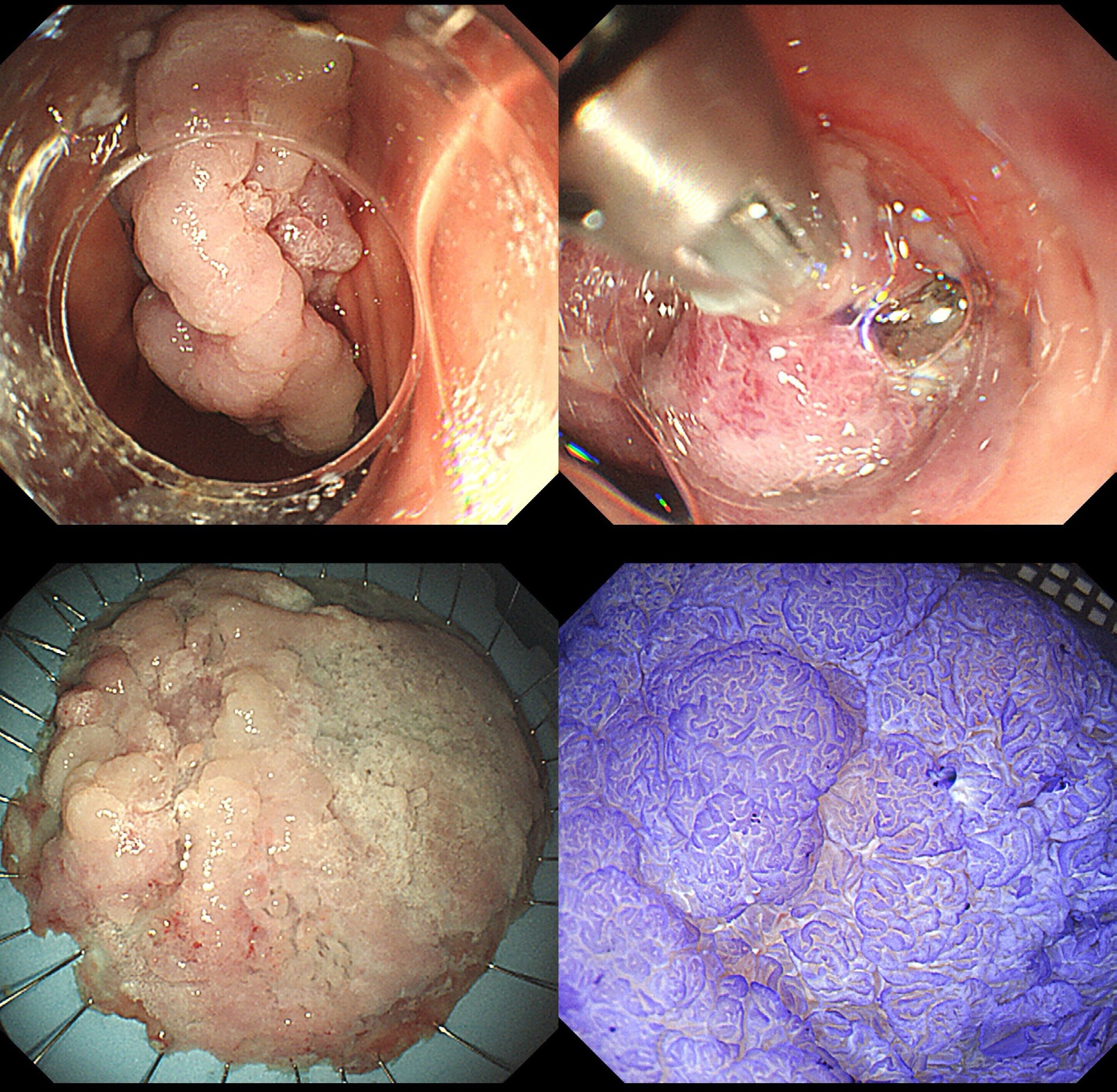
Figure 1.
The procedure of endoscopic papillectomy. Patient No. 9 underwent endoscopic submucosal dissection (ESD). The lesion's morphology was a laterally spreading tumor of the nongranular type with a flat elevation (LST-NG-F). The size of the lesion was 85 mm × 95 mm. Postoperative pathology showed moderately differentiated adenocarcinoma. This case was operated by Qide Zhang.
-
Number Gender Age Diagnosis Surgical method Past medical history 1 Male 48 DPA EMR + ERC + ERBD None 2 Male 53 Duodenal papillary lesion EMR + ERC + ERPD + ERBD Hypertension 3 Male 47 Duodenal papillary lesion EMR + ERC + ERBD Hypertension 4 Female 73 Duodenal papillary tumor ESD + ERC + ERPD Hypertension 5 Female 74 DPA ESD + ERC + ERPD + ERBD None 6 Male 58 Duodenal papillary lesion ESD + ERC + ERPD Hypertension 7 Female 36 DPA EMR + ERC + ERPD Anemia 8 Male 49 DPA ESD + ERC + ERPD Diabetes 9 Male 71 DPA ESD + ERC + ERPD Hypertension 10 Male 69 DPA EMR + ERC + ERPD HTN, DM, HLD, esophageal malignancy 11 Male 59 DPA EMR + ERC + ERPD Diabetes 12 Female 48 DPA + mucosal lesion ESD + UEMR + ERPD None 13 Male 61 DPA ESD + ERC + ERPD Hypertension 14 Female 34 DPA EMR + ERC + ERPD None 15 Male 65 DPA EMR + ERC + ERPD + ERBD None 16 Female 59 DPA EMR + ERCP + ERBD + ERPD Diabetes 17 Female 75 DPA EMR + ERC + ERPD Hyperlipidemia 18 Male 59 DPA EMR + ERCP + ERBD + ERPD None DPA, duodenal papillary adenoma; EMR, endoscopic mucosal resection; ERC, endoscopic retrograde cholangiopancreatography; ERPD, endoscopic retrograde pancreatic drainage; ERBD, endoscopic retrograde biliary drainage; ESD, endoscopic submucosal dissection; HTN, hypertension; DM, diabetes mellitus; HLD, hyperlipidemia; UEMR, underwater endoscopic mucosal resection. Table 1.
General condition.
-
Number Morphology Size (mm) EUS Pathological diagnosis Microscopic structure Margins 1 0-Is 8 × 6 + Polypoid hyperplasia μs Mild disarray − 2 0-Is 20 × 38 + Tubular adenoma with LGIN None − 3 0-Is 6 × 12 + Tubular adenoma with LGIN + Local HGIN None − 4 0-Is 25 × 45 + TSA WOS+ disarray − 5 0-Isp 18 × 20 + Villous tubular adenoma with LGIN μs Regular − 6 0-Is + IIa 15 × 30 − Tubular Adenoma with LGIN + Focal HGIN μv Irregular − 7 0-Is 18 × 40 − Duodenal Adenomatous Hamartoma μs Disarray Vert. + 8 LST-NG-F 30 × 60 − Tubular adenoma with LGIN + Local HGIN No WOS and VCL − 9 LST-NG-F 85 × 95 + Adenocarcinoma WOS+ DL+ μs+ μv+ − 10 0-Is 8 × 10 − Villous tubular adenoma with LGIN μs Irregular − 11 0-Is 12 × 12 − Tubular adenoma with LGIN Adenomatous − 12 0-IIb + IIc 20 × 25 − Tubular adenoma with LGIN WOS+ villous − 13 SSL 12 × 20 − TSA Coarse granular − 14 0-Is + IIa − − Tubular adenoma with LGIN Coarse granular − 15 0-Is − + Hamartoma μs Irregular − 16 0-Is − + Tubular adenoma with LGIN μs Irregular − 17 0-Is 8 × 12 + Villous tubular adenoma with LGIN μs Irregular − 18 0-Is 10 × 12 + Tubular adenoma with LGIN μs Irregular − EUS, endoscopic ultrasound; μs, microscopic structure; LGIN, low-grade intraepithelial neoplasia; HGIN, high-grade intraepithelial neoplasia; TSA, traditional serrated adenoma; WOS, white opaque substance; Vert., vertical margin; LST-NG-F, laterally spreading tumor of the nongranular type with a flat elevation; VCL, villous crypt length; SSL, sessile serrated lesion. Table 2.
Endoscopic morphology and pathology.
-
Number Surgical method Catheterization Bleeding Pancreatitis Other 1 EMR + ERC + ERBD Biliary stent − − None 2 EMR + ERC + ERPD + ERBD Both −+ − None 3 EMR + ERC + ERBD Biliary stent − − None 4 ESD + ERC + ERPD Pancreatic stent − − Hypertension, allergic rash 5 ESD + ERC + ERPD + ERBD Both + − Bleeding from the posterior pharyngeal wall 6 ESD + ERC + ERPD Pancreatic stent − − Mild infection 7 EMR + ERC + ERPD Pancreatic stent −+ − None 8 ESD + ERC + ERPD + PC Pancreatic stent ++ + None 9 ESD + ERC + ERPD Pancreatic stent + − Infection 10 EMR + ERC + ERPD Pancreatic stent − − None 11 EMR + ERC + ERPD Pancreatic stent − − None 12 ESD + UEMR + ERPD Pancreatic stent − − None 13 ESD + ERC + ERPD Pancreatic stent + + None 14 EMR + ERC + ERPD Pancreatic stent ++ + None 15 EMR + ERC + ERPD + ERBD Both ++ − None 16 EMR + ERCP + ERBD + ERPD Both − + None 17 EMR + ERC + ERPD Pancreatic stent − − Mild infection 18 EMR + ERCP + ERBD + ERPD Both −+ − Infection Table 3.
Postoperative complications.
-
Number INR NSAID PPI AMY (3 h postprocedure) SST AB Bleeding pancreatitis Endoscopic management Other management 1 0.98 − + 60 + + − − − − 2 0.92 − + 55 + + −+ − − − 3 0.92 − + 58 + + − − − − 4 1.03 − + 164 + + − − − − 5 1.34 − + 169 + + ++ − + − 6 1.02 − + 111 + + − − − − 7 1.03 − + 116 + + −+ − − − 8 1.15 − + 409 + + ++ + − − 9 1.02 − + 117 + + + − − Gastric artery embolization 10 1.03 − + 100 + + − − − − 11 0.94 − + 30 + + − − − − 12 0.92 − + 82 − − − − − − 13 1.13 − + 618 + + + + + − 14 1.01 − + 485 + + + + + − 15 1.08 − + 74 + + ++ − + 16 0.97 − + 518 + + − ++ − 17 1.01 − + 92 + + − − − 18 1.11 − + 124 + + −+ − − 1 Isolated hyperamylasemia (n = 2) not classified as pancreatitis per diagnostic protocol. INR, international normalized ratio; NSAID, nonsteroidal anti-inflammatory drug; PPI, proton pump inhibitor; AMY, amylase; SST, somatostatin; AB, antibiotics. Table 4.
Auxiliary examinations and management 1.
Figures
(1)
Tables
(4)