-
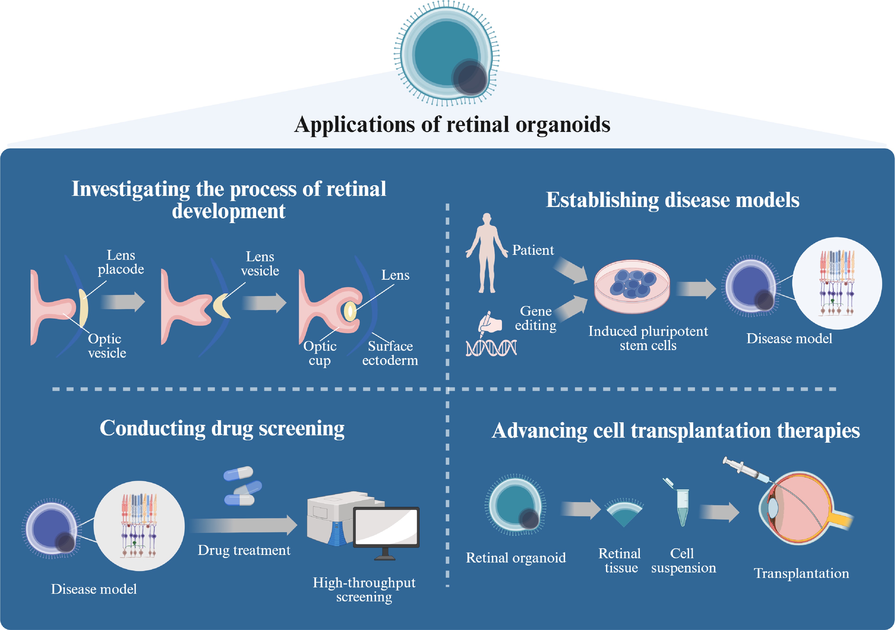
Figure 1.
The application of ROs (created with BioRender,
www.biorender.com ). Retinal organoids demonstrate significant potential across multiple research domains, including developmental biology, modeling disease pathophysiology, therapeutic discovery, and regenerative medicine applications. (1) These 3D in vitro models recapitulate the spatiotemporal progression of retinogenesis from embryonic progenitor commitment to stratified tissue maturation, enabling systematic investigation of molecular regulators and cellular dynamics during neuroretinal development. (2) Disease-specific models can be established through either derivation from patient-specific hiPSCs or CRISPR/Cas9-mediated genome editing, allowing precise replication of inherited retinal disorder phenotypes. (3) Their structural complexity and scalability serve as a high-throughput screening platform for pharmacological compounds, facilitating quantitative assessment of therapeutic efficacy and developmental toxicity. (4) From a translational perspective, retinal organoids represent a promising autologous cell source for PR replacement therapies targeting degenerative retinopathies. -
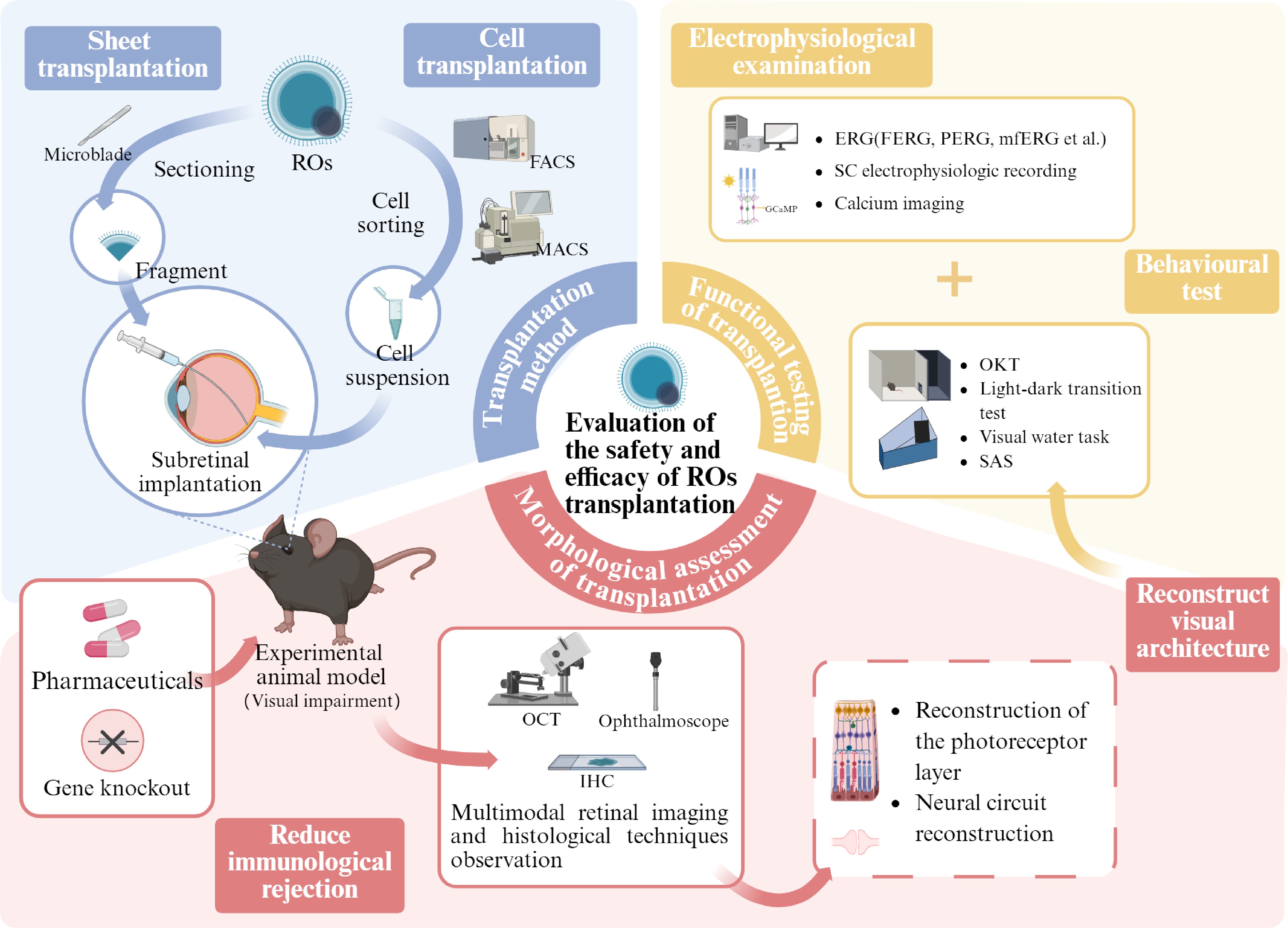
Figure 2.
Retinal organoids: evaluating the safety and efficacy of transplantation (created with BioRender,
www.biorender.com ). In order to advance retinal organoid transplantation therapies toward clinical application, their safety and efficacy must be thoroughly evaluated. Currently, there are two main transplantation methods: lamellar transplantation, in which the organoid is trimmed to a suitable size, and cell suspension transplantation, in which the target cells are isolated from the organoid. Researchers deliver these grafts to the subretinal cavity of the disease model animals using specific tools. In order to reduce immune rejection and increase the survival rate of transplanted cells, researchers have established immunodeficient animal models or used pharmacological treatment protocols. At the same time, multimodal retinal imaging and histologic techniques are used to monitor the contact between the transplanted cells and the host's retina. These assays are designed to assess whether transplantation treatments are successful in reestablishing visual structures, focusing on two key aspects: reconstruction of the PR layer and reconstruction of the neural circuit, i.e., the formation of synaptic connections. In addition, the combined assessment of behavioral tests and electrophysiology can further validate the effective reconstruction of visual structures and provide evidence of improved visual function. OCT, optical coherence tomography; IHC, immunohistochemistry; OKT, optokinetic tracking; SAS, shuttle avoidance behavioral experiment; FERG, flash electroretinogram; PERG, patern electroretinogram. -
Therapeutic agents Efficacy Ref. Dexamethasone, rapamycin Survival and differentiation of organoids were only successful under dexamethasone treatment. Rapamycin treatment showed no efficacy in preventing immune rejection. [86] Prednisolone, cyclosporine Combination therapy resulted in higher donor cell survival rates and reduced immune responses. [76] Prednisolone, cyclosporin A, and mycophenolate mofetil Combined drugs suppressed rejection, supported long-term donor cell survival, and prevented rapid donor cell loss in untreated hosts post-transplantation. [54] MPA, TAC Immunosuppressants enhanced PRs' survival and functional performance. [83] Table 1.
Exploration of dosing regimens to reduce immune rejection in retina-like organ transplantation.
-
Technique Application Can it be used
in the clinic?Fundus camera Enables in vivo real-time monitoring of graft viability, spatial distribution, and host retinal structural changes. Yes OCT Provides high-resolution 3D tomographic retinal imaging for graft tracking, retinal thickness quantification, and detection of immune-mediated complications (e.g., macular edema). Yes CSLO Generates 2D en face retinal images for dynamic morphological assessment of transplant regions, resolving fine structures (e.g., PRs' outer segments). Yes Adaptive optic SLO (AOSLO) Achieves cellular-level resolution for in vivo visualization of RPE/PR morphology, structural alignment, and spatial orientation. Yes FAOSLO Combines AOSLO's resolution with molecular specificity, enabling noninvasive longitudinal tracking of fluorescently labeled donor cells without terminal histology. Yes TEM Validates ultrastructural reconstitution ex vivo, cone inner/outer segment formation, and synaptic ribbon connectivity with the host's bipolar cells. Yes IHC Postmortem analysis of cellular stratification, differentiation status (recoverin+/rhodopsin+), and synaptic connectivity (ribbon synapse quantification). No FISH Post-transplantation mapping of donor cells' distribution and survival kinetics in fixed retinal sections. No TEM, transmission electron microscopy; FISH, fluorescence in situ hybridization. Table 2.
Common methods and uses for detecting graft status in RO transplantation studies.
-
Name Content Advantage Disadvantage Behavioral tests OKT The ability of an animal to produce a normal optokinetic nystagmus (OKN) in response to a rotating streak or moving light spot stimulation indicates basic visual perception. Reflects spatial visualization abilities without training Behavioral variables interfere, limited resolution, inability to distinguish between cell types SAS The animals were induced to actively avoid the light area by light stimulation and electrical or noise stimulation to determine whether their visual function was restored. Rapid, noninvasive, applicable to a wide range of animal models Dependent on the animal's learning ability, interfered with by other senses, unable to differentiate between cell types VGS Training animals to respond oculomotorly to specific visual stimuli to assess their advanced visual processing ability and the integrity of the retina–optic nerve–brain pathway. Evaluate advanced visual processing, quantifiable, clinically relevant Relies on complex training and high-precision equipment Visual acuity testing By training an animal to actively avoid a certain unfavorable environment when a specific visual stimulus is presented, it indirectly reflects its visual perception and behavioral integration abilities. Rapid, noninvasive, applicable to a wide range of animal models Dependent on the animal's learning ability, interfered with by other senses, unable to differentiate between cell types Electrophysiological tests ERG Recording the overall electrical response of the retina to light stimulation by means of electrodes. Noninvasive, easy to operate, reflective of overall functionality Unable to provide localized or single-cell information, limited sensitivity multifocal ERG (mERG) Similar to ERG, it has the ability to differentiate the functional status of different areas of the retina through multipoint stimulation and mathematical modeling. Noninvasive, easy to follow, reflects overall function Complex equipment, high signal interference, high training requirements SC electrophysiology By implanting electrodes in the region of the SC in an animal model and recording the electrical activity of neurons, it can be determined whether retinal signals could be perceived by the superior colliculus after transplantation. High spatial and temporal resolution, direct reflection of visual signaling Highly invasive, complex operation, difficult signal resolution MEA Retinal tissue was placed on the MEA chip, and its electrical activity in response to light stimulation was recorded. High spatial and temporal resolution, can record the synchronized activity of multiple cells Applicable to ex vivo models only Calcium imaging Indirect reflection of neuronal activity by detecting changes inthe intracellular calcium ion concentration in isolated retinal slices. High spatial resolution, simultaneous recording of multiple neurons, ability to distinguish cell types Signal delay, data processing complexity, only for ex vivo Table 3.
Functional tests used in RO transplantation studies.
Figures
(2)
Tables
(3)