-
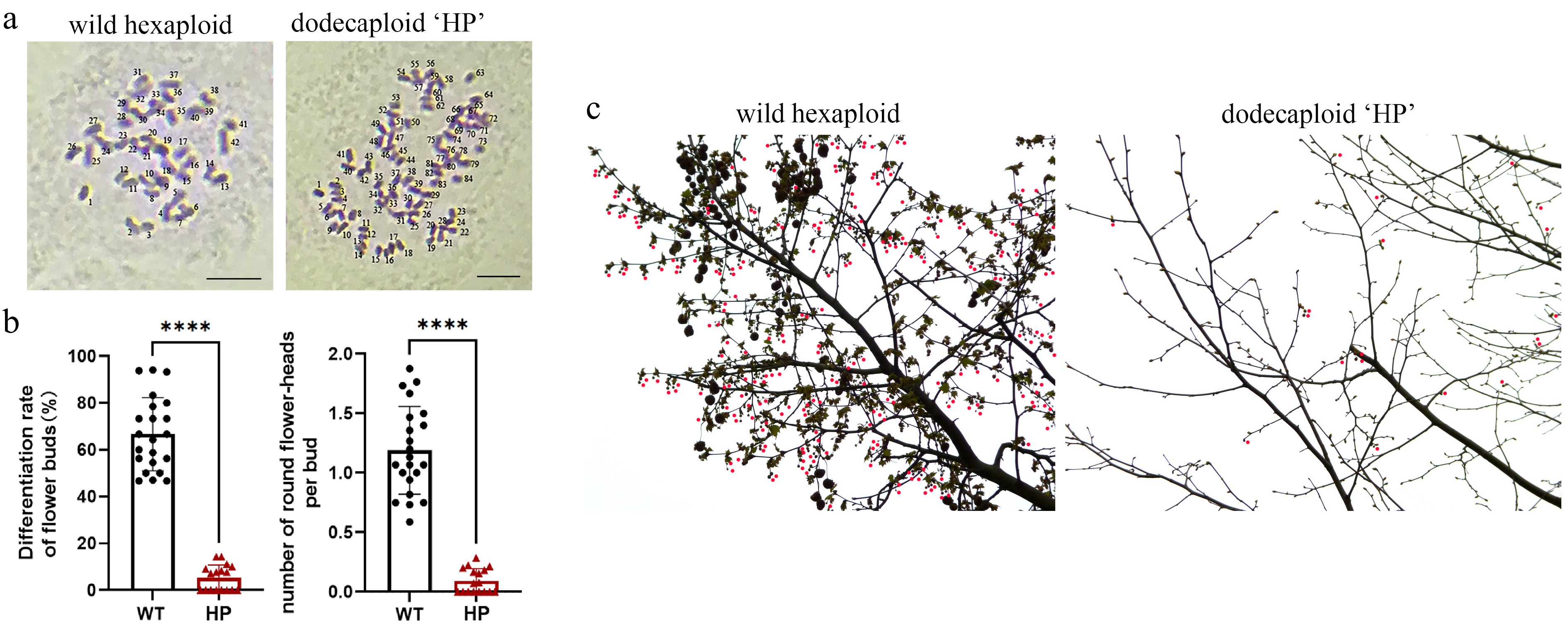
Figure 1.
Comparative reproductive phenotyping between polyploid 'HP' and wild-type P. × acerifolia. (a) Ploidy verification. Chromosome number of bud primordia cells were observed in wild-type P. × acerifolia (left) and 'HP' line (right). Scale bars = 5 μm. (b) Quantitative assessment of reproductive efficiency. Flower bud differentiation frequency (% of total buds) and mean inflorescence count per bud were analyzed in 'WT' and 'HP' lines. Student's t-test of 20 biological replicates; Error bars indicate ± SD; **** p < 0.0001. (c) Inflorescence distribution on mid-crown branches of 'WT' and 'HP' lines. Wild-type displayed characteristic high-density floral clusters, while the 'HP' line exhibited significantly reduced flowering sites. Red dots indicate globose inflorescence.
-
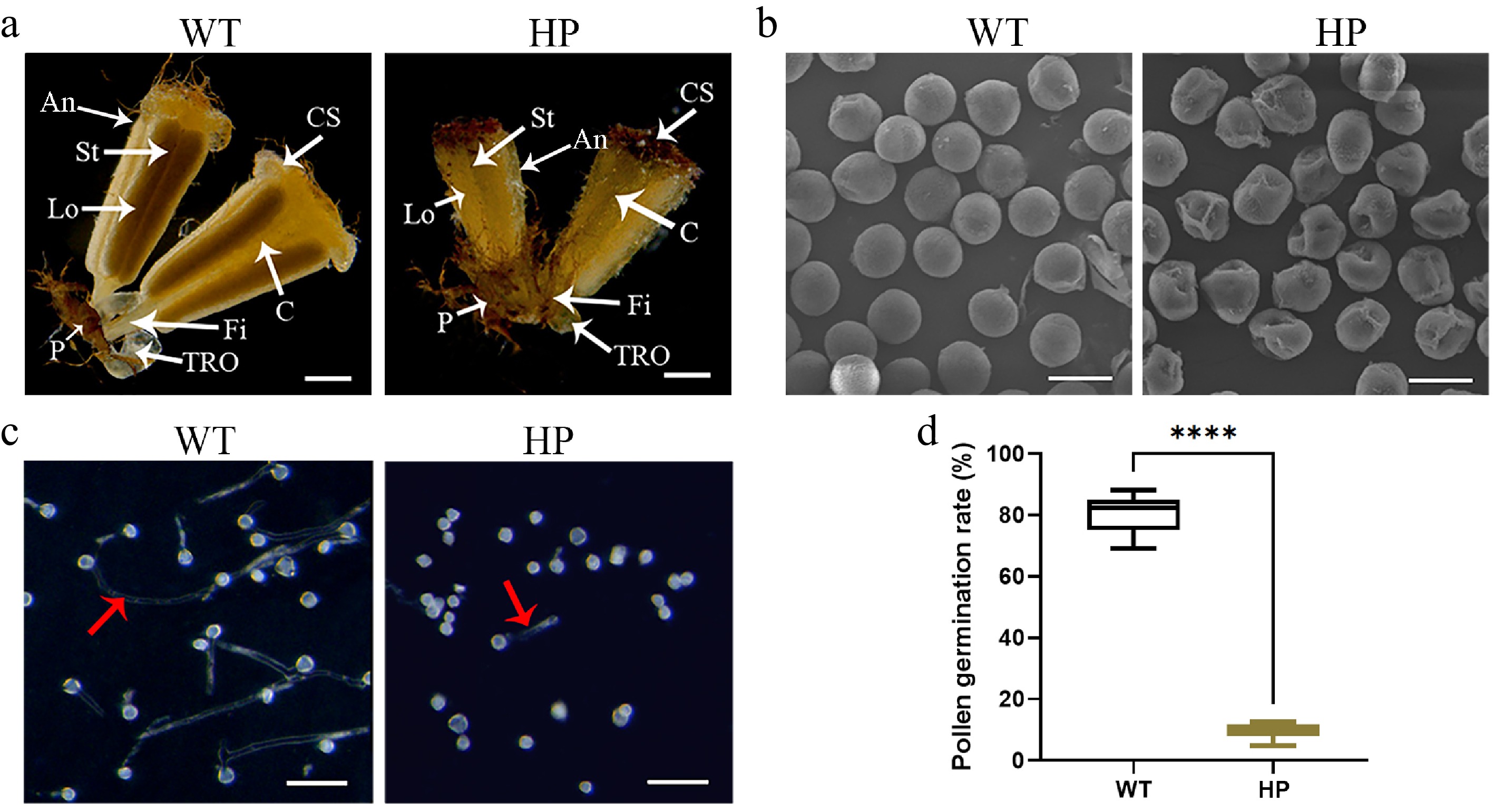
Figure 2.
Pollen fertility analysis in wild-type ('WT') and polyploid ('HP') London plane tree. (a) Morphology of male flowers in 'WT' and 'HP'. Key floral structures: An, anther; C, connective; CS, cap structure; Fi, filament; Lo: locule; P, perianth; St, stomium; TRO, three-ridged organ. Scale bar = 1 mm. (b) Scanning electron micrographs of pollen grains. Scale bar = 20 μm. (c) In vitro pollen germination assays. Red arrow indicates pollen tubes. Scale bar = 100 μm. (d) Comparative analysis of pollen germination rate in 'WT' and 'HP' (mean ± SD; n = 4 flowers, more than a thousand pollens were analyzed).
-
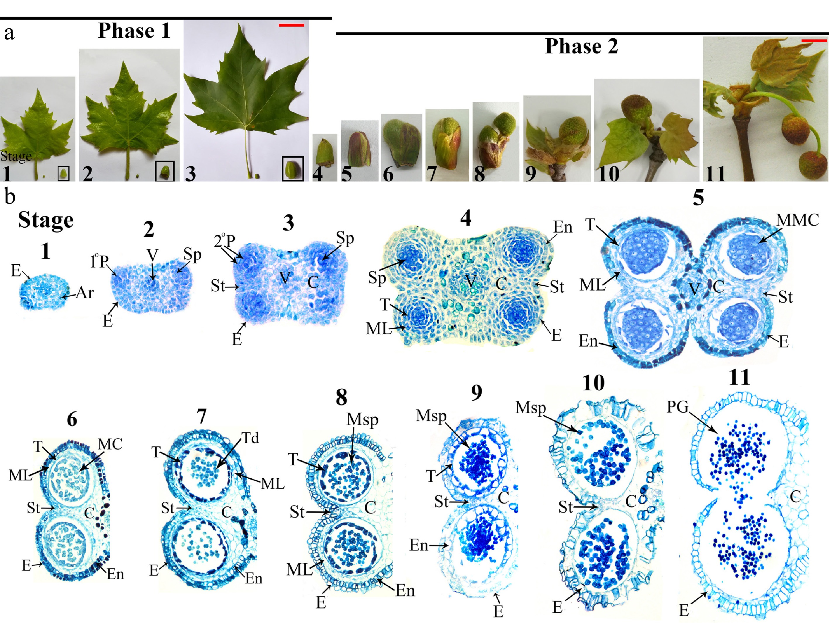
Figure 3.
Male flower and anther development in wild-type London Plane tree. (a) Male flower development progression from male flower bud formation to opening (shown with concomitant leaf morphology). Scale bars: 7 cm (Stages 1−3), 1 cm (Stages 4−11). (b) Bright-field micrographs of anther transverse sections throughout developmental Stages 1−11. Ar, archesporial cells; C, connective; E, epidermis; En, endothecium; MC, meiotic cell; ML, middle layer; MMC, microspore mother cells; Msp, microspores; 1°P, primary parietal layer; 2°P, secondary parietal cell layers; PG, pollen grain; Sp, sporogenous cells; St, stomium; T, tapetum; Td, tetrads; V, vascular bundle. Microscopy magnification factors for Stages 1−4, 5−7, 8−10, and 11 were × 400, × 200, × 100, and × 70, respectively.
-
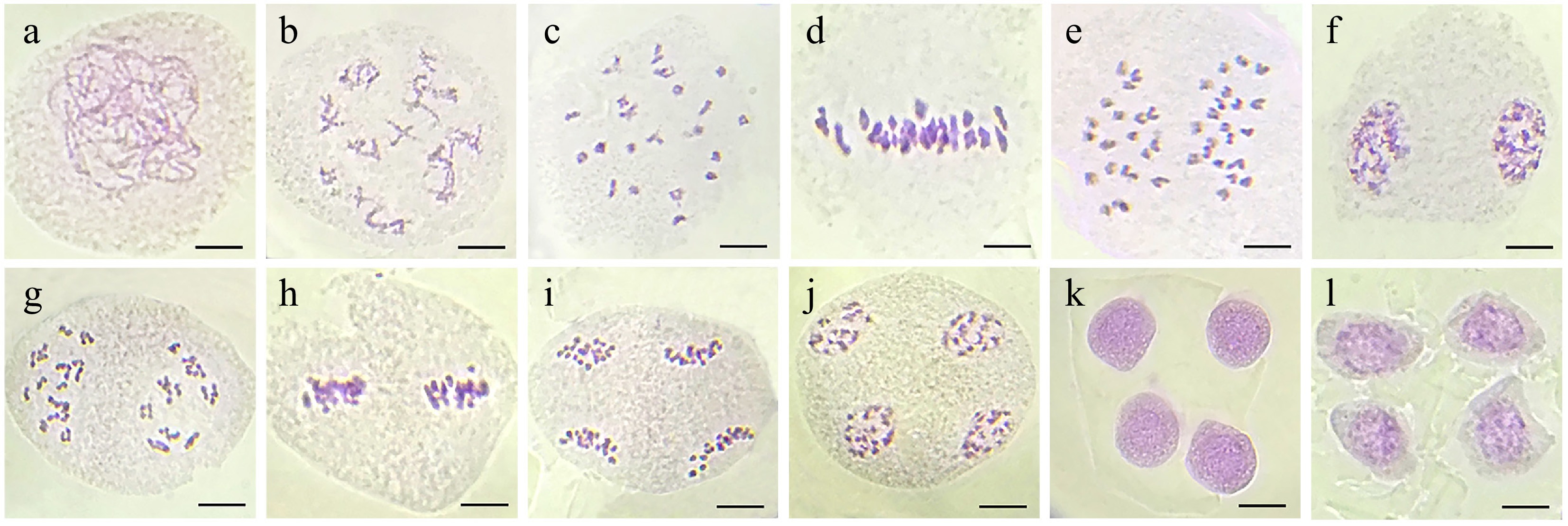
Figure 4.
Normal meiotic divisions in microspore mother cells of wild-type London plant tree. (a) Leptotene. (b) Diplotene with sister chromatid exchanges. (c) Diakinesis with 21 bivalents. (d) Metaphase I. (e) Anaphase I. (f) Telophase I. (g) Prophase II. (h) Metaphase II. (i) Anaphase II. (j) Telophase II. (k) Tetrad. (l) Uninucleate microspores. Bar = 10 μm.
-
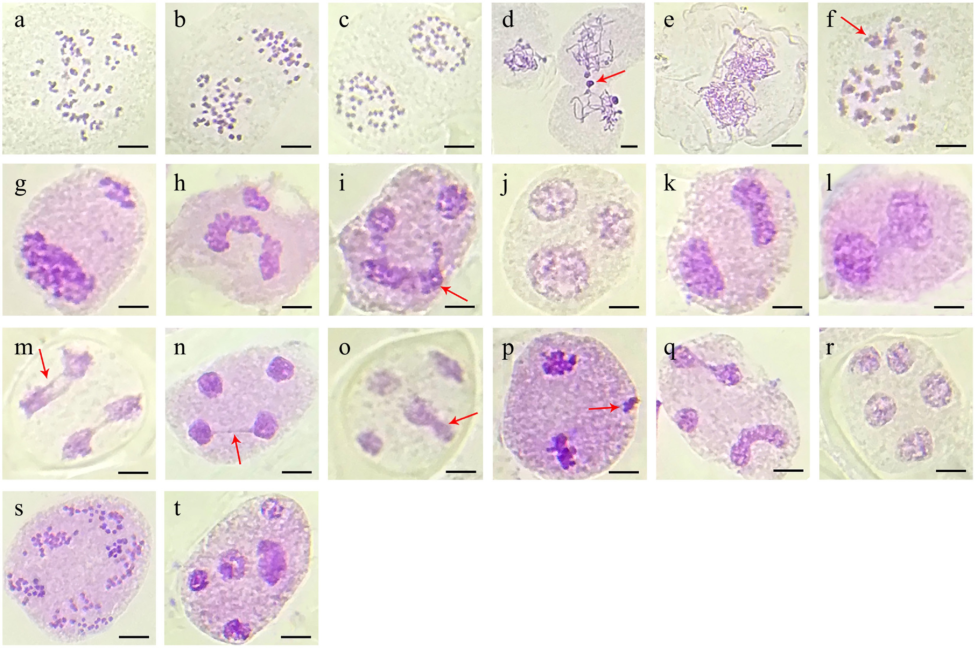
Figure 5.
Meiotic behavior observed in the polyploid 'HP' line of London plane tree. (a) Diakinesis with 42 bivalents. (b) Anaphase I with 42:42 chromosomes. (c) Telophase I with two daughter cells. (d) Early prophase with chromatin transfer (arrow). (e) Proximate pollen mother cell with double chromosome complement. (f) Anaphase I with interbivalent stickiness (arrow). (g), (h) Unbalanced chromosome segregation leading to additional nuclear clusters in telophase I. (i), (j) Anaphase II with severe chromosome stickiness in one daughter cell (arrow) impairing chromosome segregation leading to triad formation. (k), (l) Both daughter cells showing chromosome stickiness and forming dyads. (m), (n) Anaphase II and telophase II with chromosome stickiness in micronuclei through chromosome bridges (arrow). (o) Telophase II with irregular microspore (arrow). (p) Telophase I with one extra nuclear chromosome cluster (arrow). (q), (r) One extra nucleus and abnormal chromosome segregation formed into polyad. (s), (t) Chromosome segregation with abnormal spindle orientation leading to polyad formation with unequal microspores.
-
Anther stage Time Characteristic of male flower bud Capitulum diameter (cm) Major events and morphological markers of anthers 1 Early July Capitulum primordia formed. 0.12 Oval stamen primordium, archesporial cells arise in four corners. 2 Middle of June Young leaves grown rapidly 0.15 Four regions of mitotic activity; 1° parietal, sporogenous cell and vascular region initiated. 3 Early July Adult leaves formed 0.2 Vigorous mitotic activity in four corners; two bilaterally symmetrical pollen sacs began to establishing. 4 December Leaves start shedding, flower bud enter dormancy. 0.35 Four clearly locules established; all anther cell types present. 5 End of February Outer bud-bract cracking. 0.5 Pollen sacs distinct; microspore mother cells with prominent, centrally located nuclei. 6 Beginning of March Inner bud-bract stared cracking. 0.65−0.7 Meiosis begins; middle layer be crushed and degenerate. 7 Early March Inner bud-bract cracked. 0.65−0.7 Meiosis complete; microspores in tetrad; tapetum become large and multinucleate. 8 Middle of March Capitate head out form subpetiolar bud. 0.75 Microspores released; remnants of middle layer present; secondary thickening in outer wall layers. 9 Middle to end of March Young leaves head out, outer bud-bract detachment. 0.9 Tapetum degeneration initiated; expansion of endothecial layer; microspore become vacuolated with an increase size. 10 End of March Young leaves head out. 1.05−1.1 Septum cell degeneration intiated; stomium differentiation begins; endothecium layer shrink, pollen binucleate. 11 Early April Young leaves fully expanded, capitate turn red. 1.3−1.4 Anthers dehisced along stomium; mature pollen grains released. Table 1.
Major events during anther development of London plane tree.
-
Phases Total number of cells Abnormalities Number of cells Diakinesis 207 65 (31.40%) Chromosome transfer 19 Unorganized and pycnotic chromatin 35 Interbivalent connections 11 Metaphase I 51 4 (7.84%) Laggard chromosomes 4 Anaphase I 105 24 (22.85%) Multivalent 9 Asymmetric chromosome separation 7 Telophase I 156 19 (12.18%) Extra-nuclei 7 Asymmetric cell division 12 Prophase II 63 5 (7.94%) Asynchronous nucleus 5 Metaphase II 89 17 (19.10%) Asynchronous nucleus 4 Extra-nuclei 13 Anaphase II 191 122 (63.87%) Extra-nuclei 16 Laggards 20 Stickiness 35 Irregular spindle activity 51 Telophase II 238 172 (72.27%) Abnormal tetrad 64 Triad 61 Dyad 16 Bridges 20 Polyad 11 Table 2.
Meiotic abnormalities in tetraploid 'HP' of London plane tree.
Figures
(5)
Tables
(2)