-
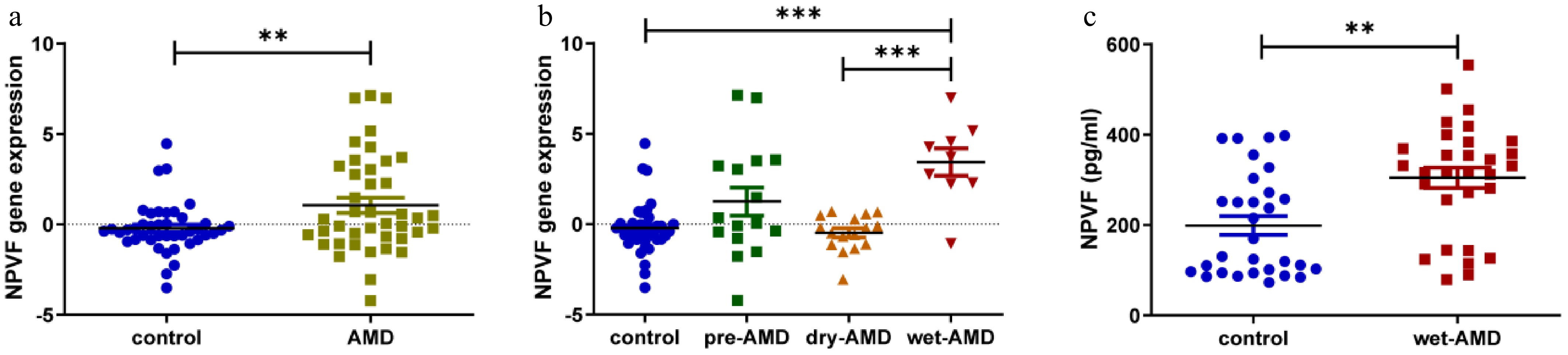
Figure 1.
Neuropeptide VF (NPVF) expression in age-related macular degeneration (AMD) tissue and serum and its effect on hypoxia-inducible factor-1α (HIF-1α). (a) Box plot comparing NPVF gene expression between AMD and control samples from a publicly available database. The y-axis represents gene expression values. (b) Box plot comparing the expression level of NPVF in different groups (control, pre-AMD, dry AMD, and wet AMD). (c) Bar graph showing NPVF protein abundance quantified by enzyme-linked immunosorbent assay (ELISA) in serum samples from 30 healthy and 30 wet AMD donors. NPVF was successfully overexpressed in (d) mRNA, and (e) protein levels. (f) Bar graphs showing the ELISA results of HIF-1α expression after overexpressing NPVF. Two-sided unpaired t-test, ** p < 0.01, *** p < 0.001.
-
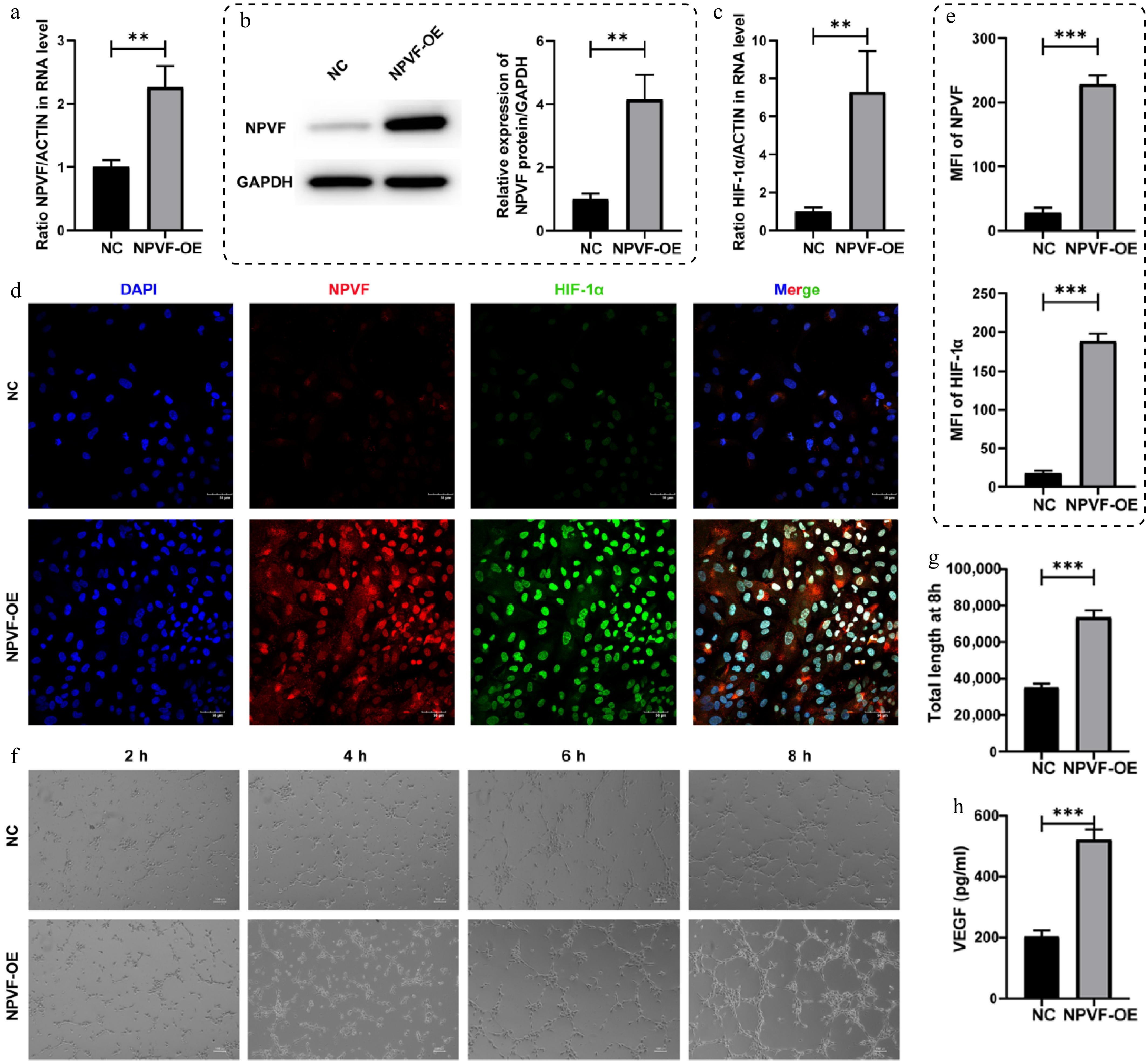
Figure 2.
Effects of neuropeptide VF (NPVF) overexpression on retinal pigment epithelium (RPE) cells. NPVF was successfully overexpressed in (a) mRNA, and (b) protein levels. (c) Bar graphs showing the upregulation of hypoxia-inducible factor-1α (HIF-1α) in mRNA level after overexpressing NPVF. (d) Immunofluorescence staining of NPVF (red) and HIF-1α (green) in NPVF-overexpressing (NPVF-OE) and control (NPVF-NC) ARPE-19 cells. Nuclei were counterstained with DAPI (blue). Scale bar: 50 μm. (e) Quantification of NPVF and HIF-1α immunofluorescence in ARPE-19 cells from (d), showed mean fluorescence intensity (MFI) (n = 3 replicates). (f) Tube formation of human umbilical vein endothelial cells (HUVECs) cultured with conditioned media derived from NPVF-OE or control ARPE-19 cells was tested. (g) Quantification of tube formation at 8h were performed (n = 3 replicates). (h) The concentration of vascular endothelial growth factor (VEGF) in the supernatant of NPVF-overexpressing (NPVF-OE) or control ARPE-19 cells was measured using ELISA. Two-sided unpaired t-test, ** p < 0.01, *** p < 0.001.
-
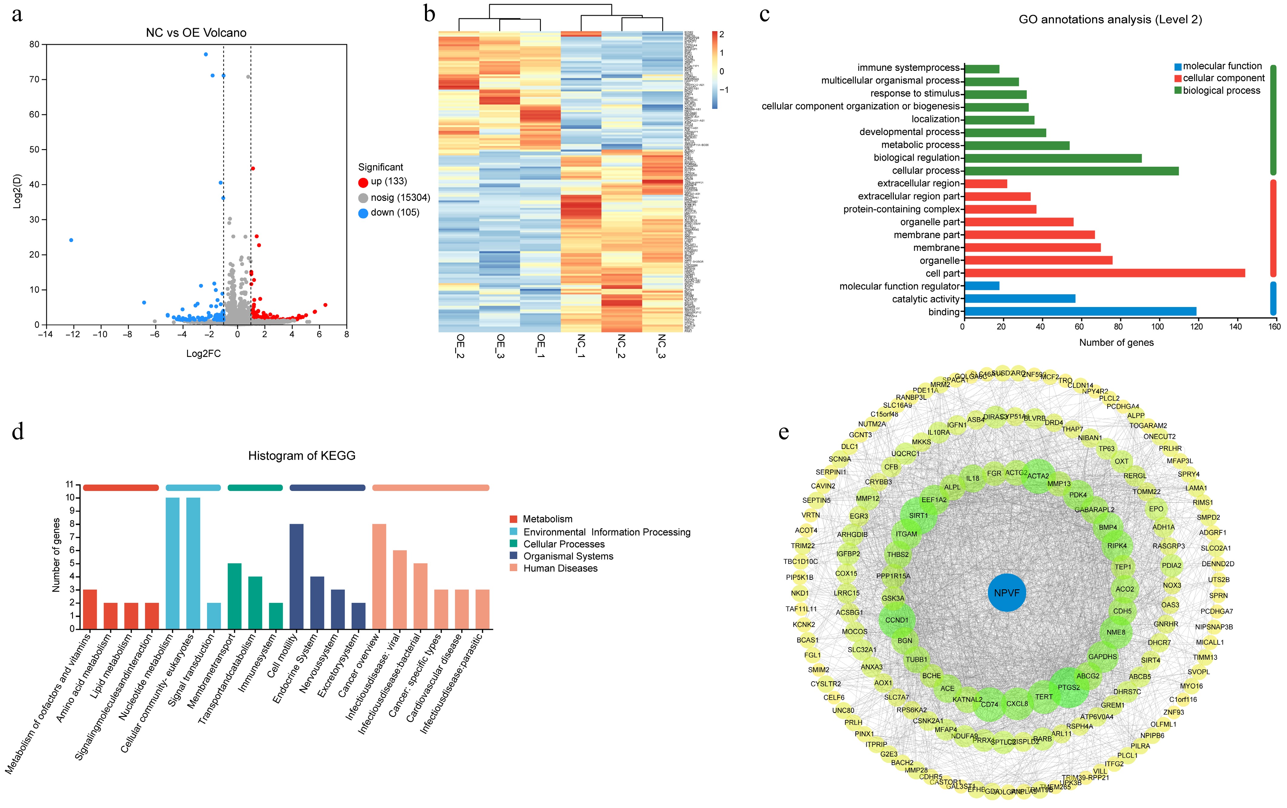
Figure 3.
Alteration of age-related macular degeneration (AMD)-related gene expression by neuropeptide VF (NPVF). (a) Volcano plot showing the differentially expressed genes in NPVF-overexpressing (NPVF-OE), and control (NPVF-NC) retinal pigment epithelium (RPE) cells. The x-axis represents the log2(Fold Change), and the y-axis represents −log10(p-value). A total of 133 upregulated and 105 downregulated genes were identified. (b) Heatmap of differentially expressed genes clearly distinguishing between the NPVF-OE and NPVF-NC groups. (c) Bar graph presenting the enrichment of differential genes, such as 'multicellular organism process', 'response to stimulus,' 'extracellular region'. (d) Bar graph showing the most enriched KEGG pathways. (e) Protein–protein interaction network showing differential genes that may interact with NPVF according to the STRING database with no more than three-step length.
-
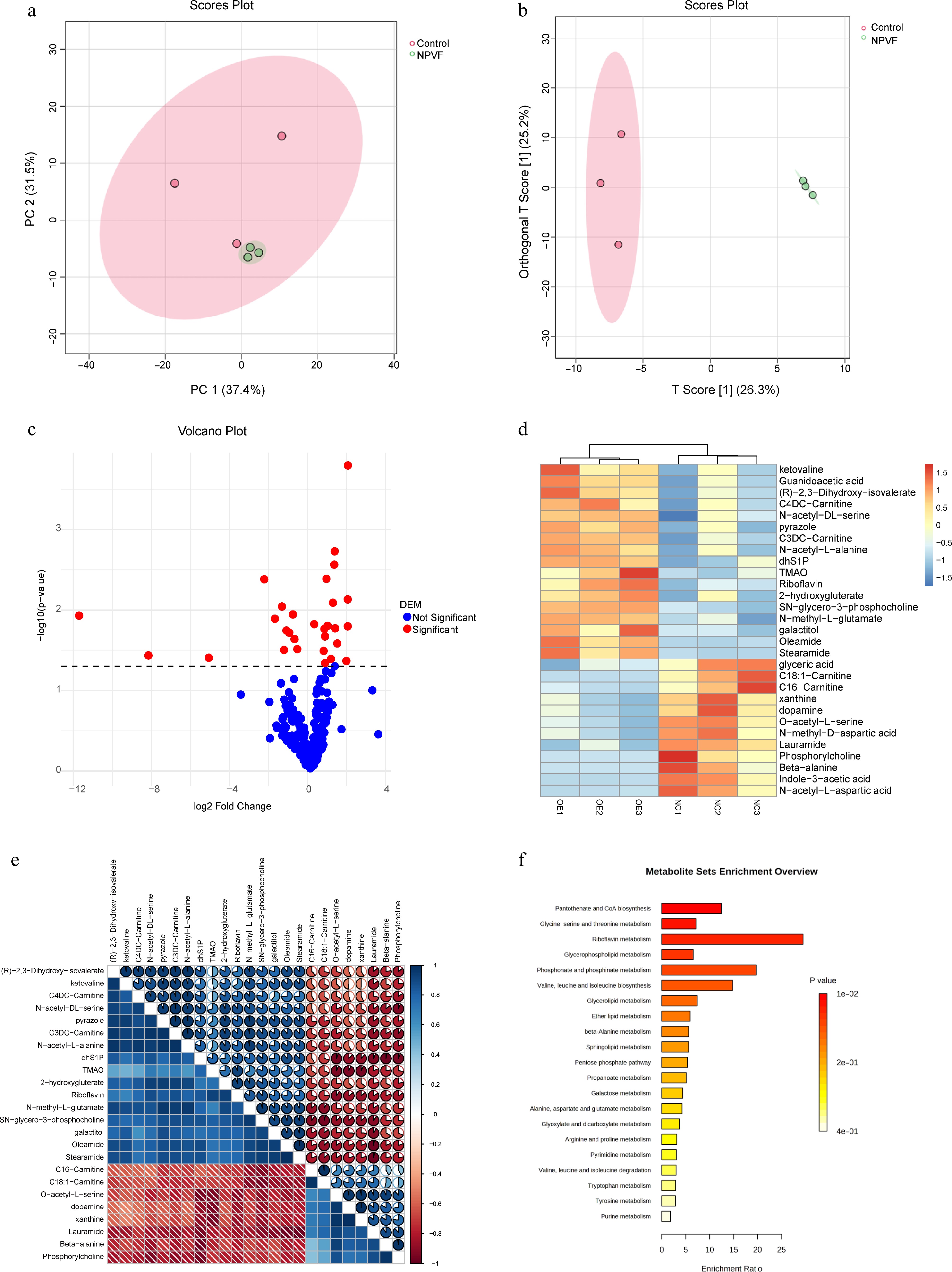
Figure 4.
Alteration of retinal pigment epithelium (RPE) cell line metabolism by neuropeptide VF (NPVF). (a) Scores plot of principal component analyses (PCA) for metabolite quantification in NPVF-overexpressing (NPVF-OE), and control (NPVF-NC) RPE cells. (b) Score plot of OPLS DA analysis, also indicating a clear separation between the NPVF-OE and NPVF-NC groups, consistent with the PCA results and previous transcriptome data. (c) Volcano plot for identifying differential metabolites. (d) Plot showing the separation of the two groups based on differential metabolites. Different colors represent different sample groups. (e) Heatmap showing the correlation of differential metabolites. (f) Bar graph showing enriched metabolic pathways, including amino acid metabolism (such as 'glycine, serine, and threonine metabolism'), and lipid metabolism (such as 'glycerophospholipid metabolism').
-
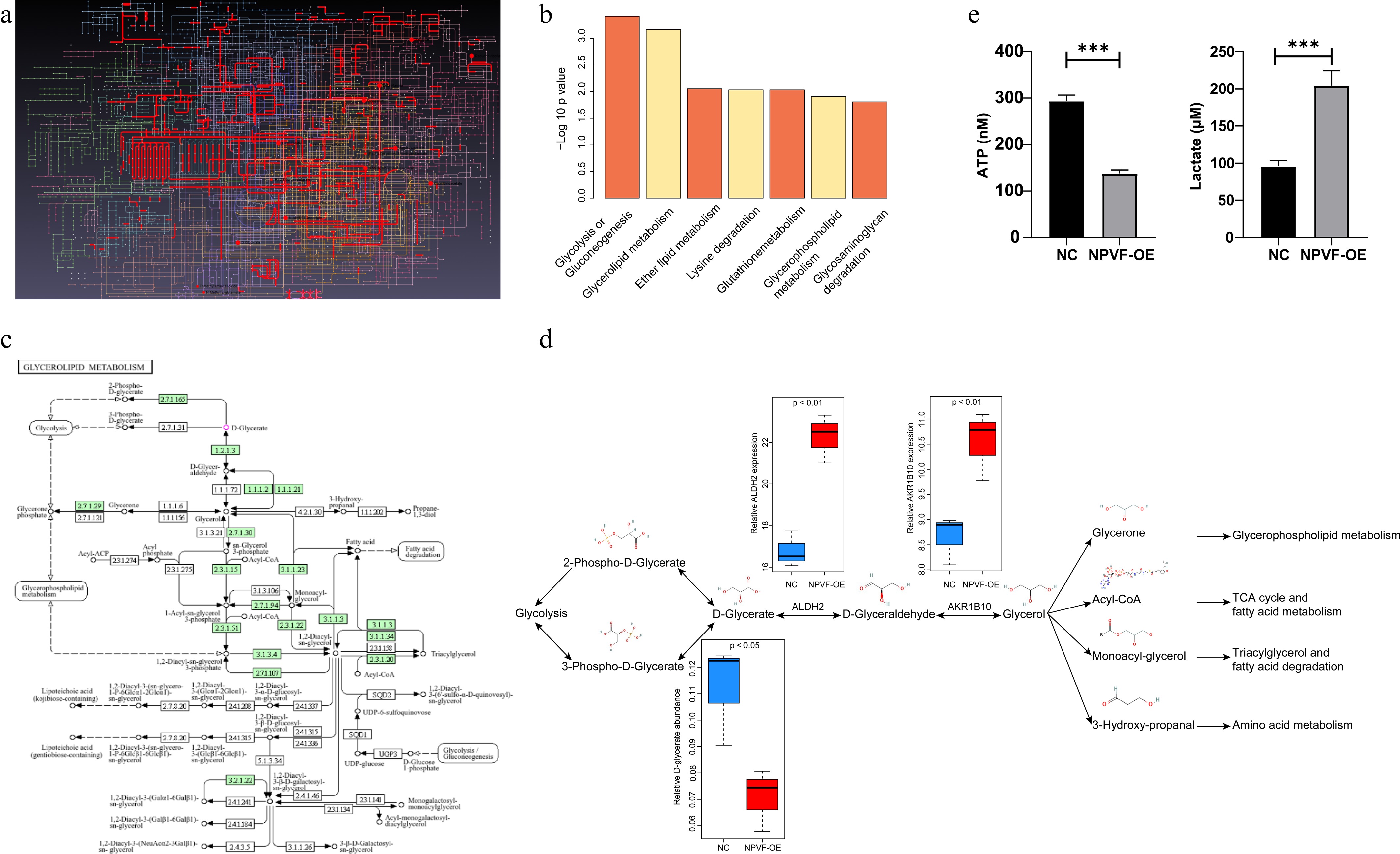
Figure 5.
Integrated analyses of transcriptome and metabolome after neuropeptide VF (NPVF) overexpression. (a) Visualization of the landscape of detected differential metabolites and genes after mapping using the MetaboAnalyst software. Different lines and dots represent different metabolites and genes. (b) Bar graph showing the most enriched pathways, including glycolysis, glycerolipid metabolism, and ether lipid metabolism. The x-axis represents the pathways, and the y-axis shows the significance level [−log10(p-value)[. (c) Schematic diagram highlighting the role of glycolysis and glycerolipid metabolism in age-related macular degeneration (AMD), sourced from the KEGG website (
www.genome.jp/kegg/pathway.html ), and has obtained the necessary permissions from KEGG. (d) Plot illustrating the detailed differences in gene expression and metabolite abundance in glycolysis and glycerolipid metabolism pathways. ALDH2 and AKR1B10 were upregulated, and the metabolite D-glycerate was significantly lower in NPVF-overexpressing (NPVF-OE) than in control (NPVF-NC) cells. (e) ATP and lactate levels in the culture supernatants of NPVF-OE and control RPE cells were measured using an ATP Assay Kit and a colorimetric assay kit (n = three replicates). Two-sided unpaired t-test, *** p < 0.001. -
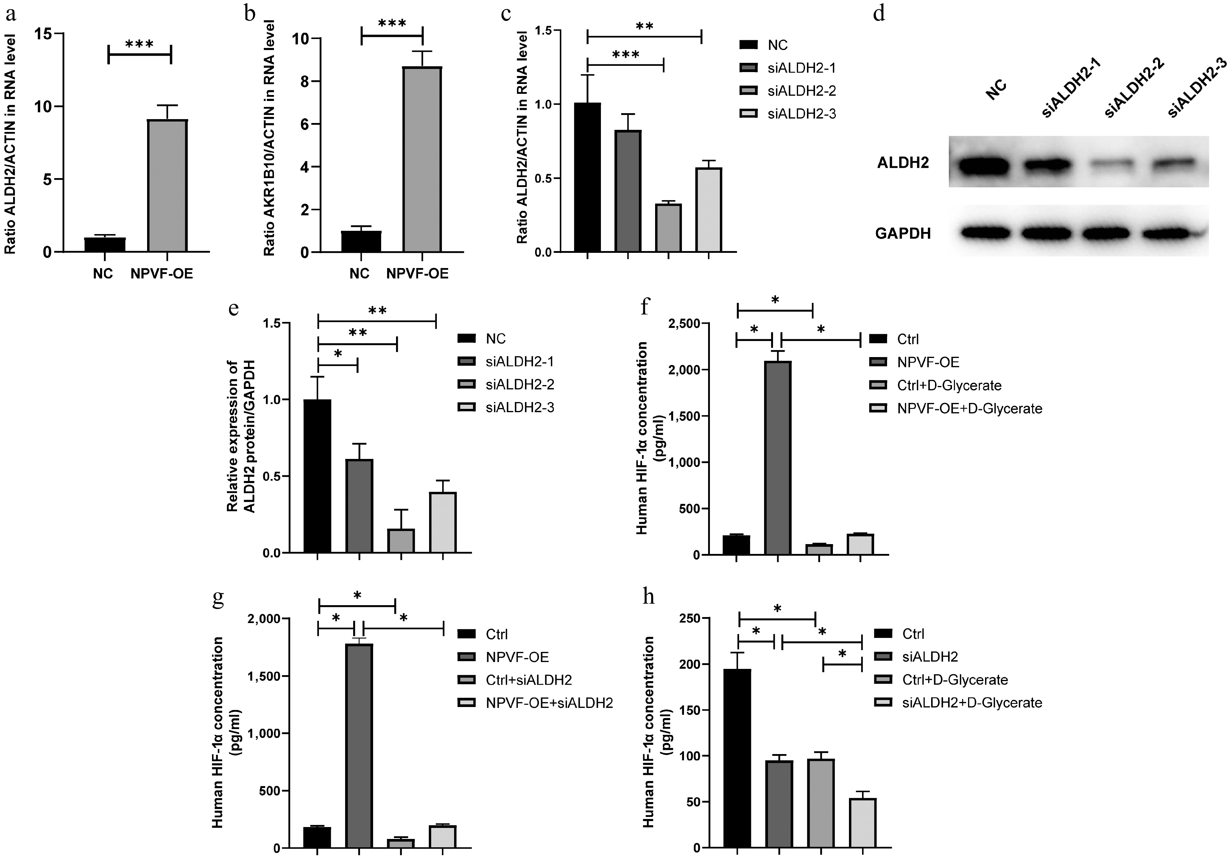
Figure 6.
Neuropeptide VF (NPVF) regulates hypoxia-inducible factor-1α (HIF-1α) via ALDH2/D - glycerate axis. qRT-PCR results showing the expression of (a) ALDH2, and (b) AKR1B10 in NPVF-overexpressing (NPVF-OE), and control (NPVF-NC) retinal pigment epithelium (RPE) cells. (c) qRT-PCR, and (d), (e) western blot results showing the knockdown of ALDH2 gene expression using siRNA in the NPVF-OE RPE cells. (f) Enzyme-linked immunosorbent assay (ELISA) results showing that exogenous D-glycerate inhibited NPVF-induced HIF-1α overexpression. (g) ELISA results showing that ALDH2 knockdown inhibits NPVF-induced HIF-1α overexpression. (h) ELISA results showing that exogenous D-glycerate and ALDH2 significantly reduced HIF-1α. Two-sided unpaired t-test, * p < 0.05, ** p < 0.01, *** p < 0.001.
-
NPVF Dry AMD (n = 16) Wet AMD (n = 9) Total (n = 41) Control (n = 42) log2(Fold Change) = −0.2706, p = 0.4799 log2(Fold Change) = 3.647, p < 0.0001 log2(Fold Change) = 1.268, p = 0.0084 Dry-AMD − log2(Fold Change) = 3.918, p < 0.0001 − Table 1.
NPVF gene expression data in ocular tissues of patients with age-related macular degeneration (AMD), and healthy controls from GSE29801.
-
AMD Controls p-value Sample size 30 30 Age (year) 72.13 ± 9.45 72.37 ± 8.29 0.92 Male 15 (50%) 15 (50%) 1.00 Stage Wet AMD − − Table 2.
Comparison of age-related macular degeneration (AMD), and control group characteristics.
Figures
(6)
Tables
(2)