-
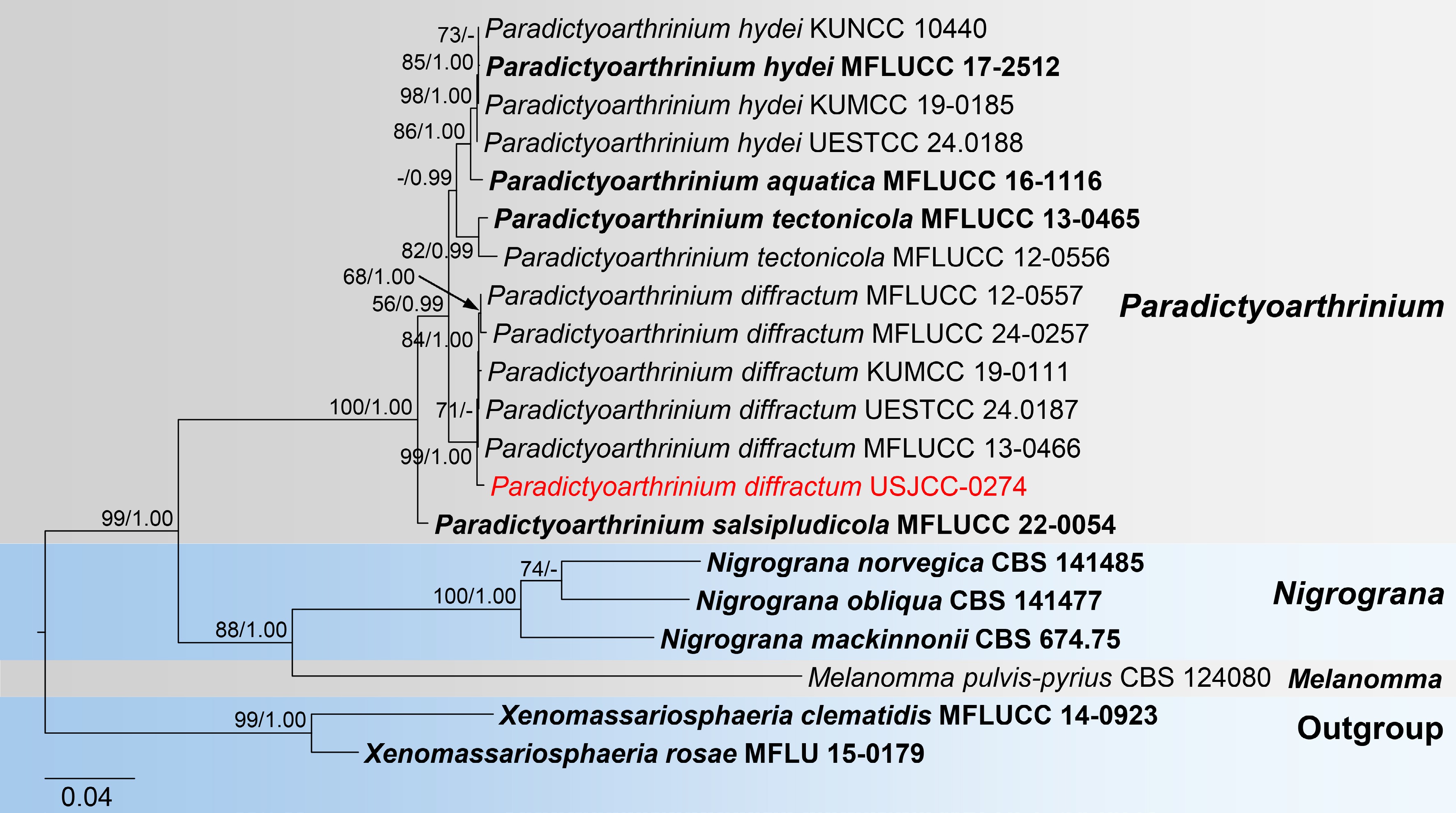
Figure 1.
Maximum likelihood (ML) tree of combined LSU, ITS, and rpb2 sequences from Paradictyoarthrinium species. Xenomassariosphaeria clematidis MFLUCC 14-0923 and X. rosae MFLU 15-0179 were used as the outgroup. Maximum likelihood bootstrap support values (≥ 50%) and Bayesian posterior probability (≥ 0.90) are indicated at nodes. The newly generated strain is indicated in red, while type strains are shown in bold.
-
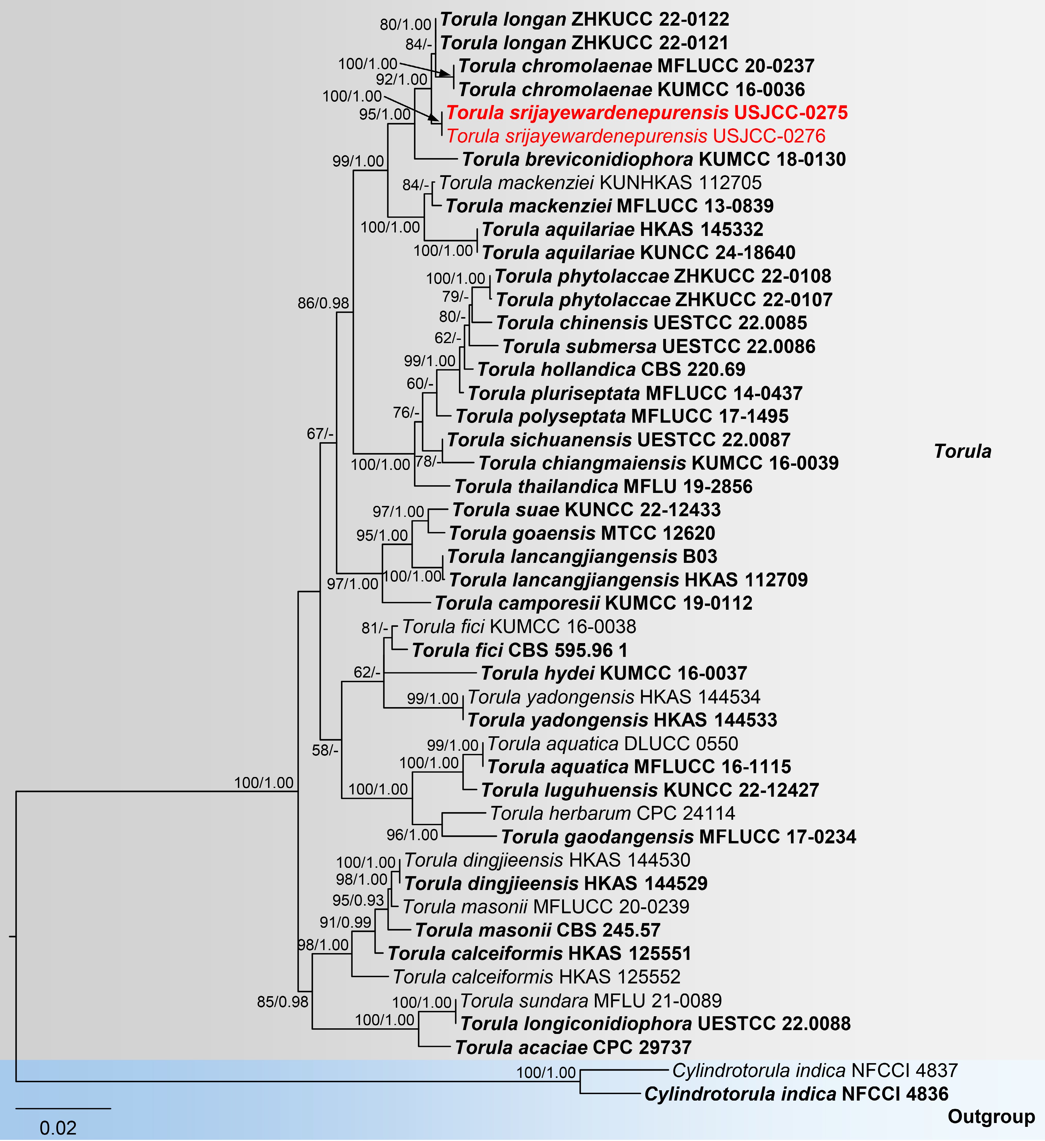
Figure 2.
Maximum likelihood (ML) tree of concatenated ITS, LSU, SSU, and tef1-α sequences from Torula species. Cylindrotorula indica (NFCCI 4836 and NFCCI 4837) was used as the outgroup. Maximum likelihood bootstrap support values (≥ 50%) and Bayesian posterior probability (≥ 0.90) are indicated at nodes. Newly generated strains in this study are highlighted in red, while type strains are shown in bold.
-
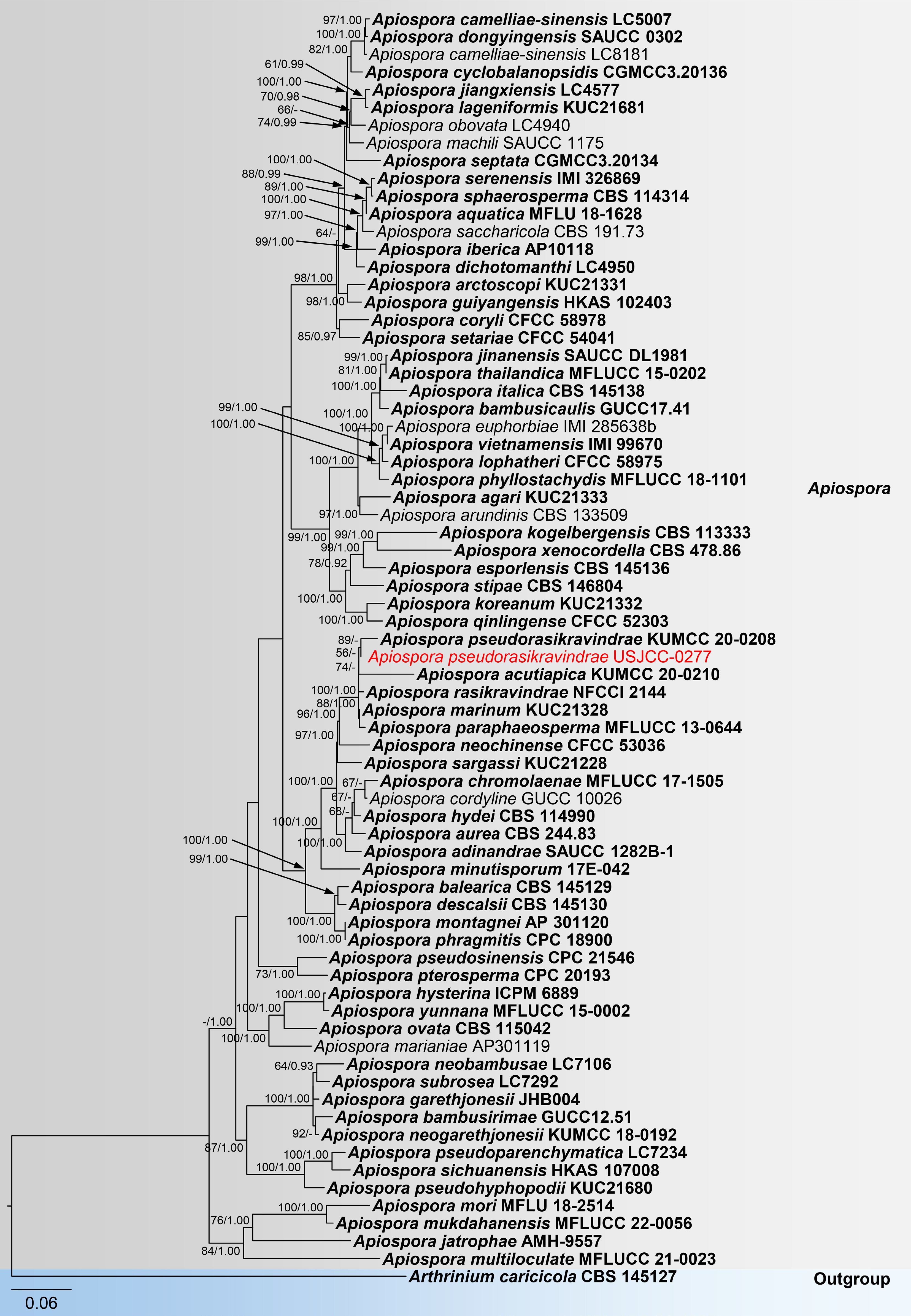
Figure 3.
Maximum likelihood (ML) tree of concatenated ITS, LSU, tef1-α, and tub2 sequences from Apiospora species. Arthrinium caricicola was used as the outgroup. Maximum likelihood bootstrap support values (≥ 50%) and Bayesian posterior probability (≥ 0.90) are indicated at nodes. The newly generated strain is shown in red, while type strains are shown in bold.
-
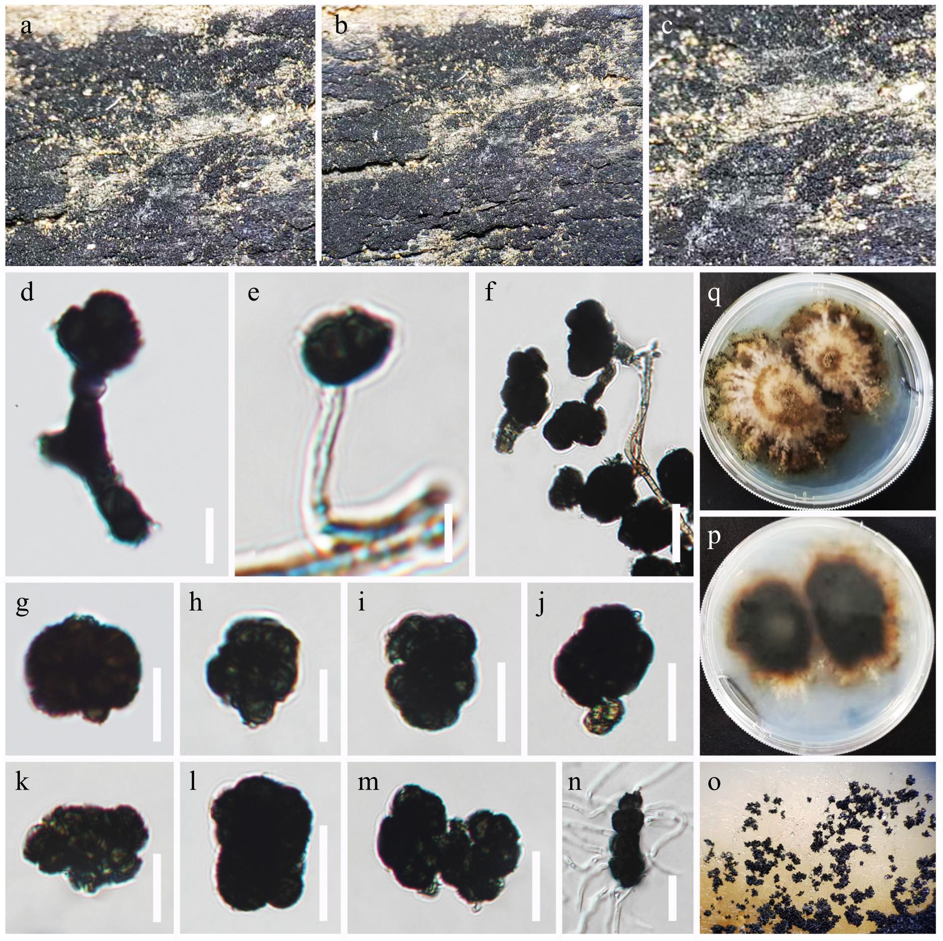
Figure 4.
Paradictyoarthrinium diffractum (USJ-H-255). (a)–(c) Colonies on the host substrate. (d)–(f) Conidiogenous cells and developing conidia. (g)–(m). Conidia. (n) Germinated conidium. (o) Front, and (p) the reverse views of the culture on PDA media. (q) Colonies on PDA media. Scale bars: (d)–(n) = 15 μm.
-
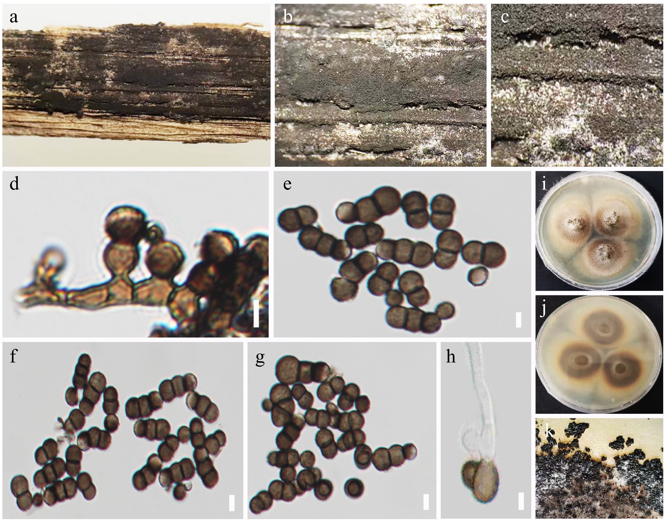
Figure 5.
Torula srijayewardenepurensis (USJ-H-252, holotype). (a)–(c) Colonies on the host substrate. (d) Conidiophores with conidiogenous cells. (e)–(g) Conidia. (h) Germinated conidium. (i) Front, and (j) the reverse view of the culture on PDA medium. (k) Appearance of colonies on PDA. Scale bars: (d)–(h) = 5 μm.
-

Figure 6.
Apiospora pseudorasikravindrae (USJ-H-254). (a)–(c) Colonies on the host substrate. (d), (e) Conidiogenous cells and developing conidia. (f)–(h) Conidia. (i) Germinated conidium. (j) Front, and (k) the reverse views of the culture on PDA media. Scale bars: (d)–(i) 5 μm.
Figures
(6)
Tables
(0)