-
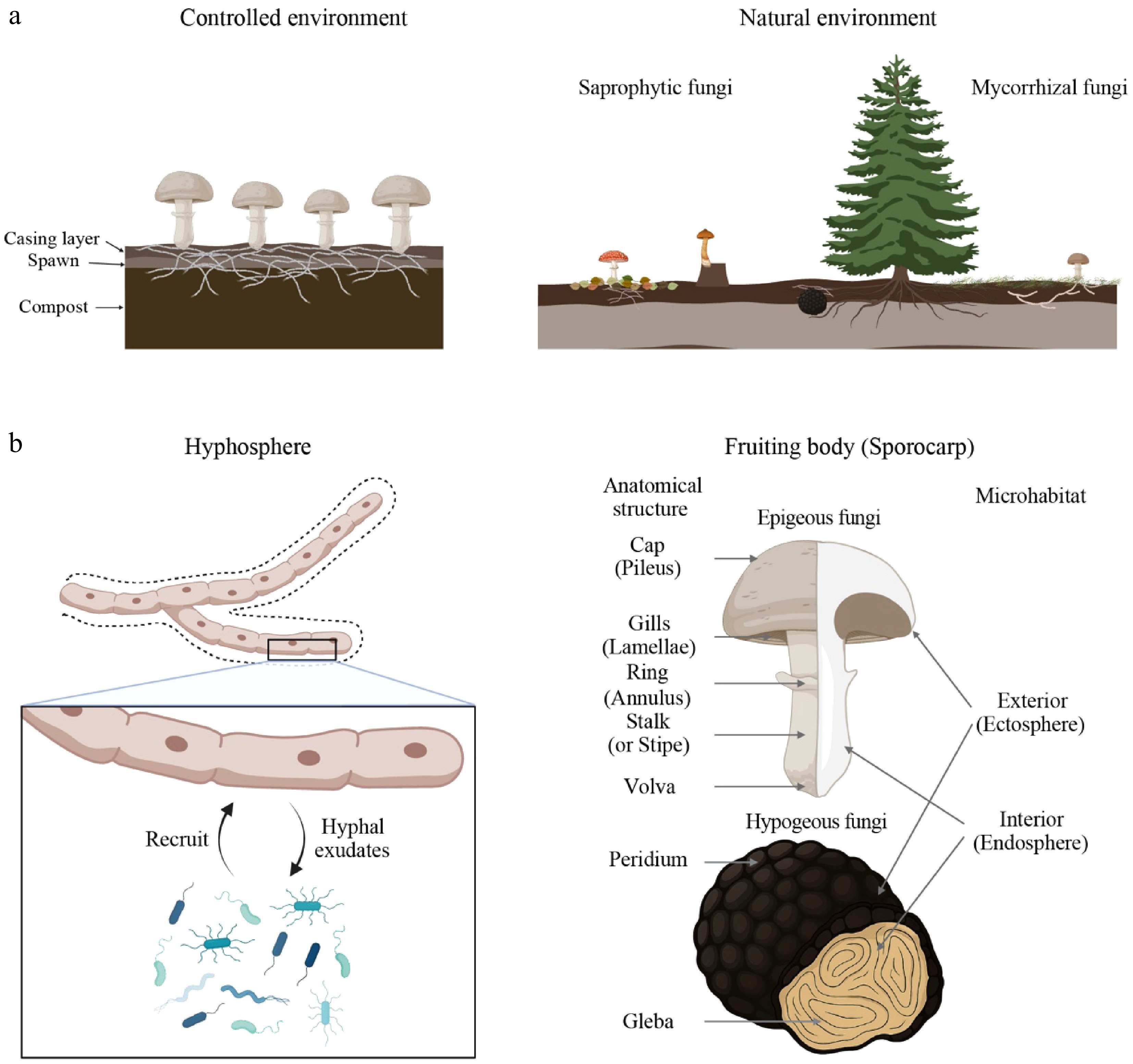
Figure 1.
Macro- and micro-habitat structures of mushroom-forming fungi. (a) Macrohabitats in which mushroom-forming fungi reside. Cultivated mushroom fungi, such as Agaricus bisporus, are generally grown under the standardized conditions consisting of compost soils, spawn, and casing layers (left panel). In contrast, most wild mushroom-forming fungi naturally inhabit forest environments, where saprotrophic species are found in the litter and humus layers, and mycorrhizal species associate with the root zones of host plants (right panel). (b) Microhabitats where mushroom fungi-associated microbiomes are generally found. The hyphosphere is the soil region affected by fungal hyphae (left panel; space indicated with the dashed line). Mushroom fungi secrete hyphal exudates acting as nutrient sources or signal molecules to attract microbes that can provide nutrients or protect fungi from other harmful microbes, similar to the plant microbiomes in the rhizosphere soils. Another microhabitat is the fruiting body (sporocarp). The distinguished anatomical structure of the fruiting bodies is depicted in the right panel. Mushroom-associated microbiomes could be found on the outer layer of the fruiting bodies (ectosphere) as epiphytes or inside them (endosphere) as endophytes. The figure was created in BioRender (
https://biorender.com ). -
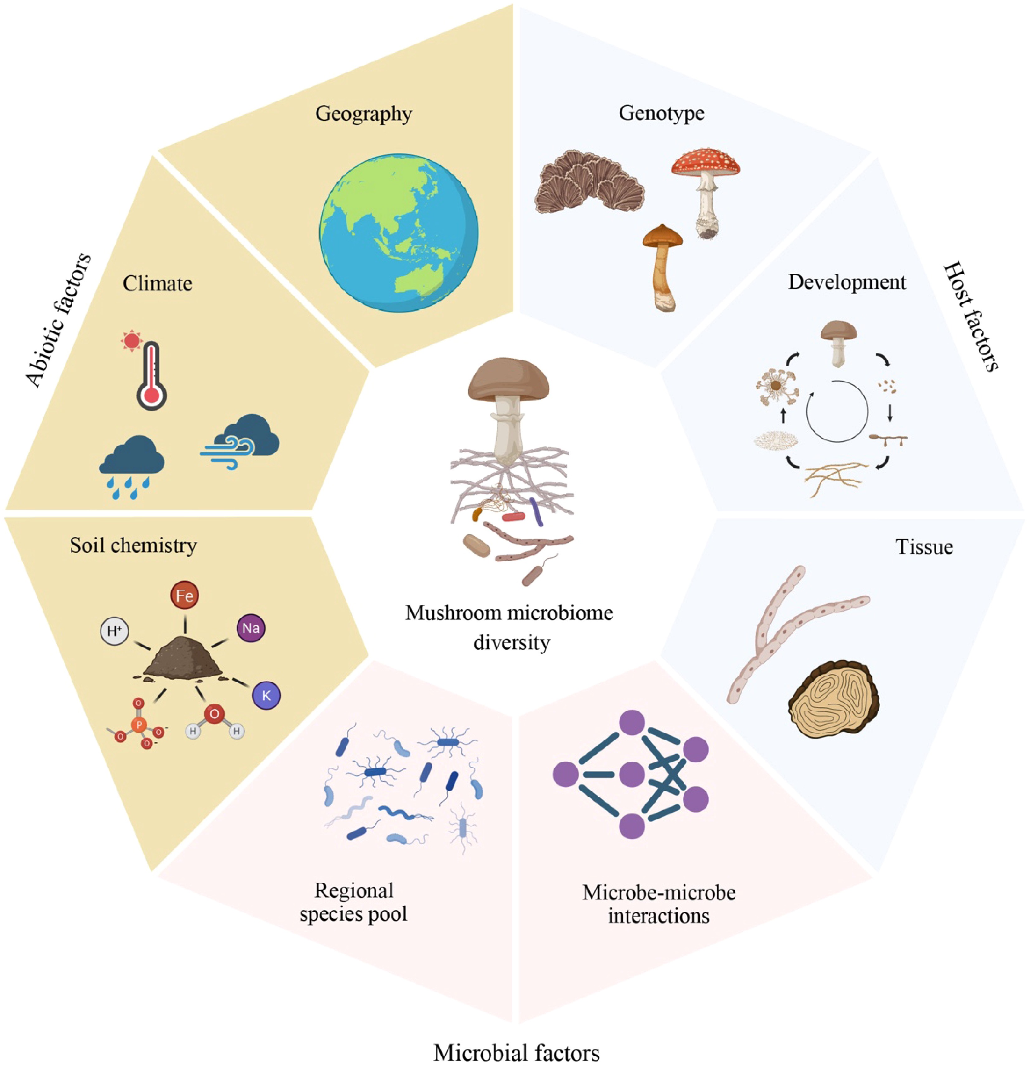
Figure 2.
Abiotic and biotic factors influencing mushroom microbiome diversity. Microbiomes associated with mushroom-forming fungi are shaped by abiotic (brown-filled cells), host-related (blue-filled cells), and microbe-associated factors (red-filled cells). Although each factor is depicted separately, these factors are tightly linked to each other. The figure was created in BioRender.
-
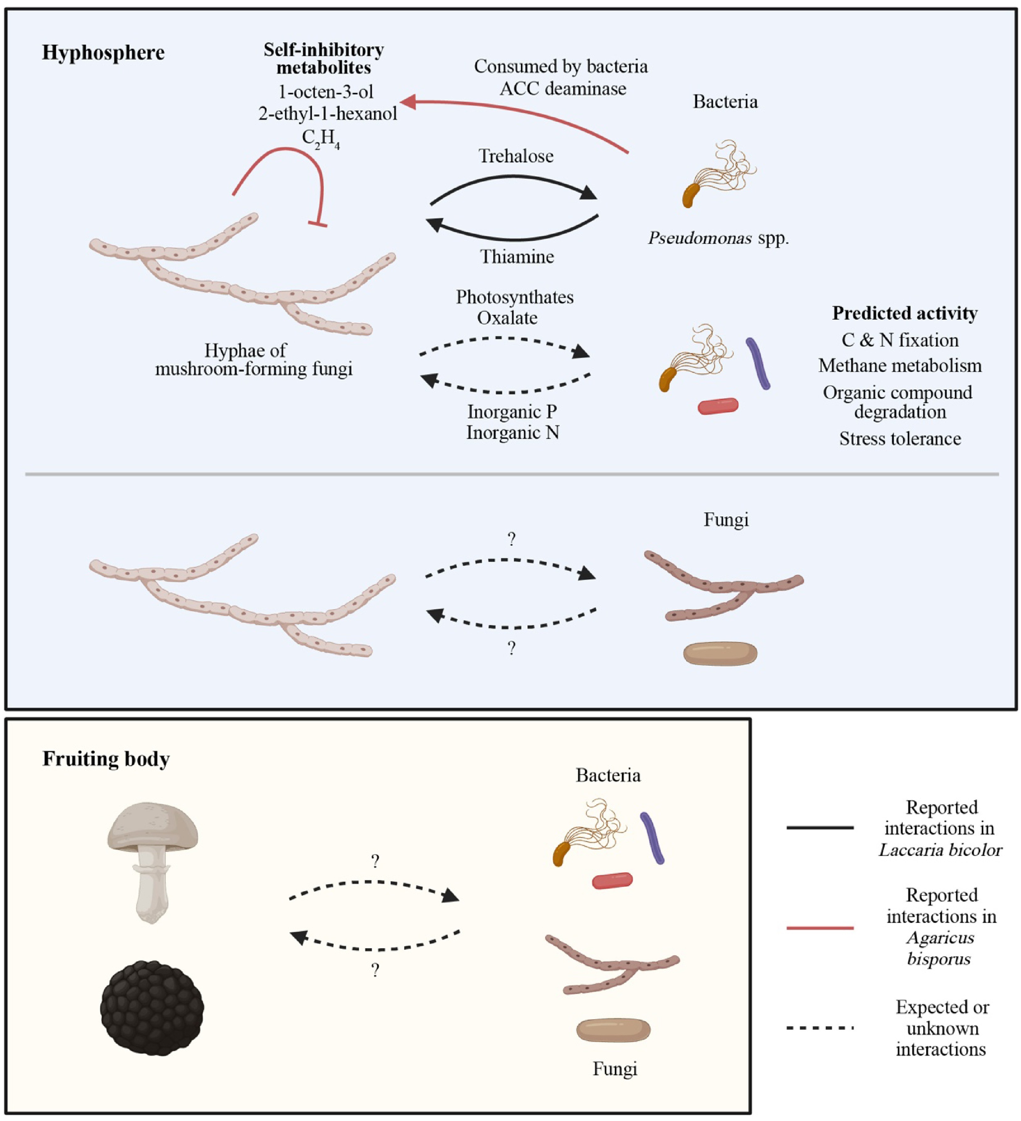
Figure 3.
Functions of mushroom-forming fungi-associated microbiomes. The functions of mushroom microbiomes have been investigated using culture-dependent and independent methods. Culture-dependent studies revealed that some bacteria, including Pseudomonas fluorescens, remove self-inhibitory metabolites produced by mushroom-forming fungi, such as Agaricus bisporus, improving the fungal growth and fruiting body formation. In addition, the Laccaria bicolor-P. fluorescens model demonstrated that fungal trehalose attracts the bacterium, which can produce thiamine required for fungal growth. Shotgun metagenomics, marker gene-based functional prediction, and metabolomics have further expanded our knowledge of the roles of mushroom microbiomes. A metabolic approach showed that mushroom-forming ectomycorrhizal fungi can provide plant-derived carbohydrates (photosynthates) to nearby soil microbes. The general functionality of the hyposphere bacterial microbiomes, including C and N fixation, methane metabolism, degradation of organic compounds (chitin, cellulose, peptidoglycan, aromatic compounds, etc.), and stress tolerance, has been reported. In particular, from the findings of arbuscular mycorrhizal fungi-associated hyphosphere microbiomes (the provision of inorganic P by hyphosphere microbiomes), it is expected that microbes associated with mushroom-forming fungi may show similar functional roles. On the other hand, the knowledge of biological mechanisms governing the interactions between a mushroom fungus and other fungi in the hyphosphere or adjacent bacteria and fungi inhabiting the fungal fruiting bodies remains lacking, although some studies showed that they promote host fungal growth under in vitro culture conditions. The figure was created in BioRender.
-
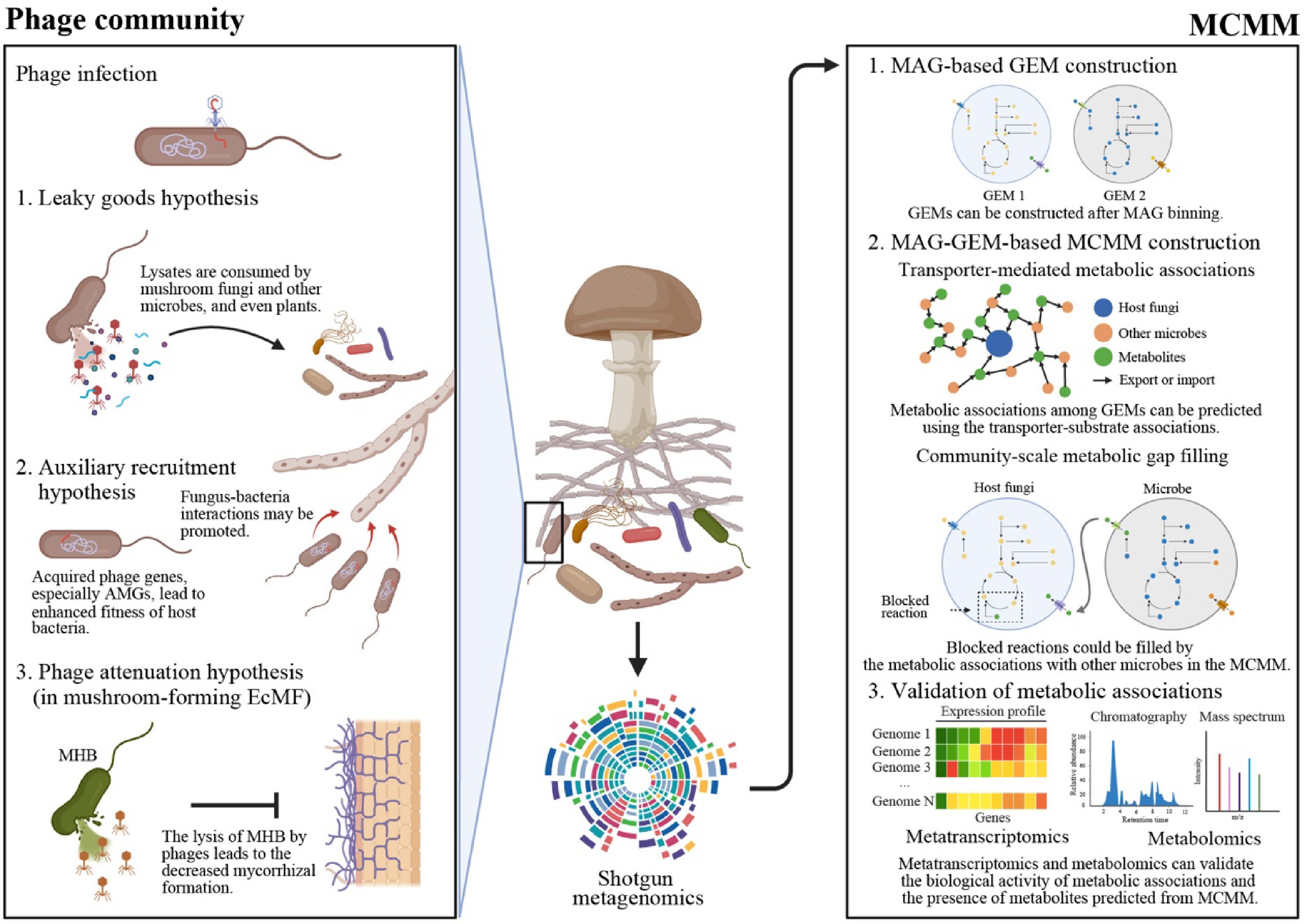
Figure 4.
The proposed holistic framework for a comprehensive understanding of mushroom-microbiome associations. To expand our knowledge of mushroom microbiomes, we proposed two agendas: phage communities (left panel) and microbial community-scale metabolic model (MCMM) (right panel). The phage communities are expected to contribute to the ecological functions of mushroom microbiomes by directly regulating bacterial populations and introducing auxiliary metabolic genes (AMGs) to bacterial populations. Phage-induced bacterial cell lysis can improve soil nutrient conditions by viral shunt (Leaky goods hypothesis). Furthermore, during lysogenic interactions, phage's AMGs can be introduced to bacterial hosts, leading to increased bacterial fitness and promoted associations between mushroom-forming fungi and bacteria (Auxiliary recruitment hypothesis). In case of mushroom-forming mycorrhizal fungi, bacterial lysis can lead to decreased mycorrhizal formation or diminished mycorrhizal activity (phage attenuation hypothesis). These putative phages' roles imply the necessity for researching phage communities in mushroom microbiomes, although thorough experimental approaches should be performed to prove or disprove the suggested hypotheses. Please note that the figure describing the phage-associated hypotheses (left panel) is modified from the artwork in by Berrios[115]. Please see this latest insightful forum article for more information on mycorrhizal fungi-bacteria-phage interactions. Another experimental approach is MCMM, a powerful tool for examining metabolic associations among microbes at the community level (right panel). From metagenome data, genome-scale metabolic models (GEMs) of each metagenome-assembled genome (MAG) can be reconstructed. Using these GEMs, potential cross-feeding interactions and transporter-mediated metabolic exchanges can be inferred, enabling the identification of putative key metabolites required for mushroom-forming fungi. Furthermore, community-scale metabolic gap-filling of GEMs, including host fungi, enables a mechanistic understanding of the observed microbial associations by revealing essential metabolic interdependencies and complementary pathways in the examined mushroom microbiomes. Finally, metatranscriptomics and metabolomics can help researchers assess whether the predicted metabolic interactions are biologically active and whether the predicted key metabolites are present in the samples. For more information of MCMM, please see a cutting-edge review article addressed by Quinn-Bohmann et al.[119]. The figure was created in BioRender.
Figures
(4)
Tables
(0)