-
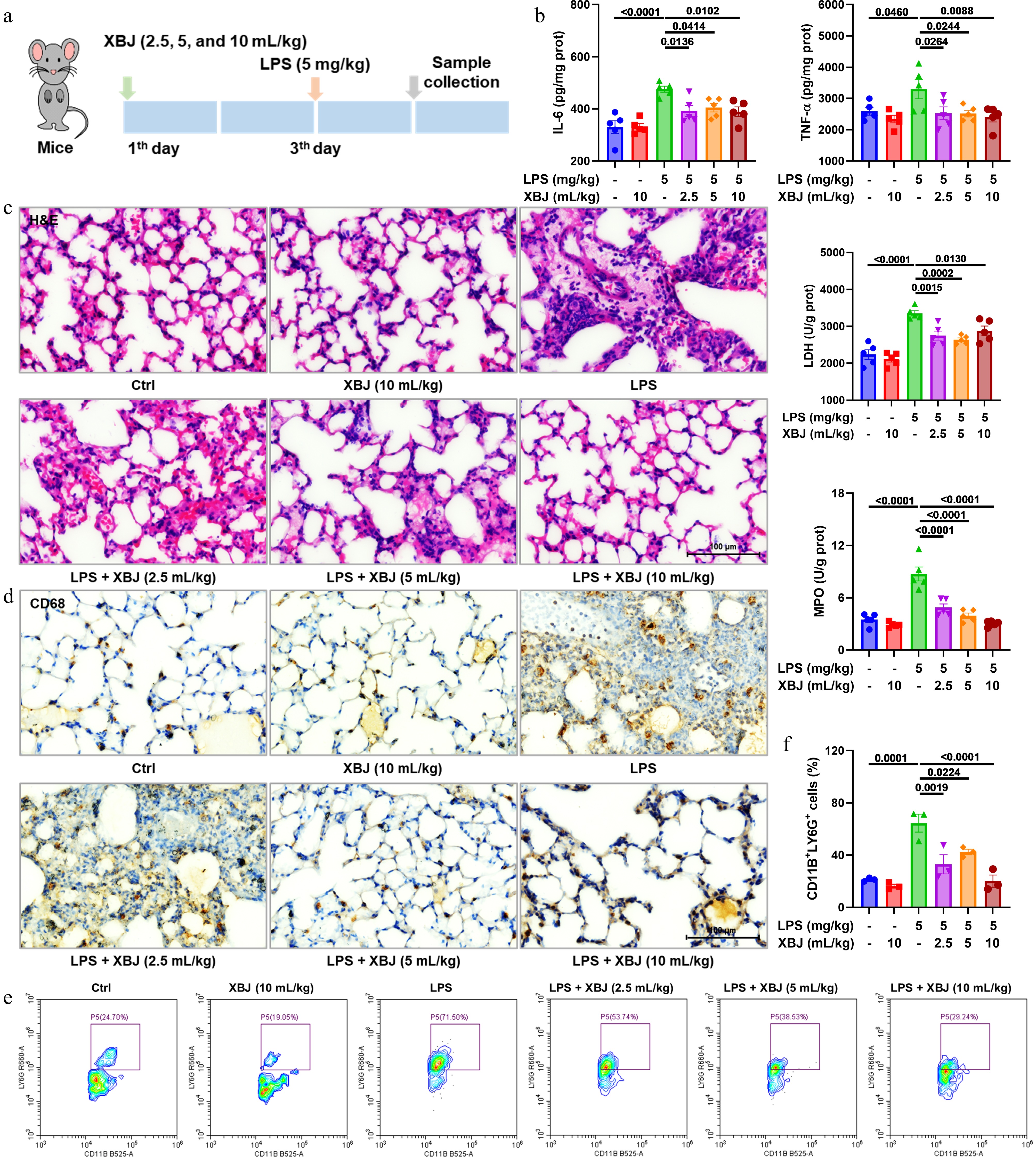
Figure 1.
XBJ suppressed LPS-induced pulmonary inflammation and tissue injury. (a) The experimental protocol; (b) effects of XBJ toward IL-6, TNF-α, LDH, and MPO in ALI (n = 5); (c) H&E staining plots; (d) representative CD68 staining plots; (e), (f) flow cytometric profiling of CD11B+LY6G+ cells (n = 3).
-
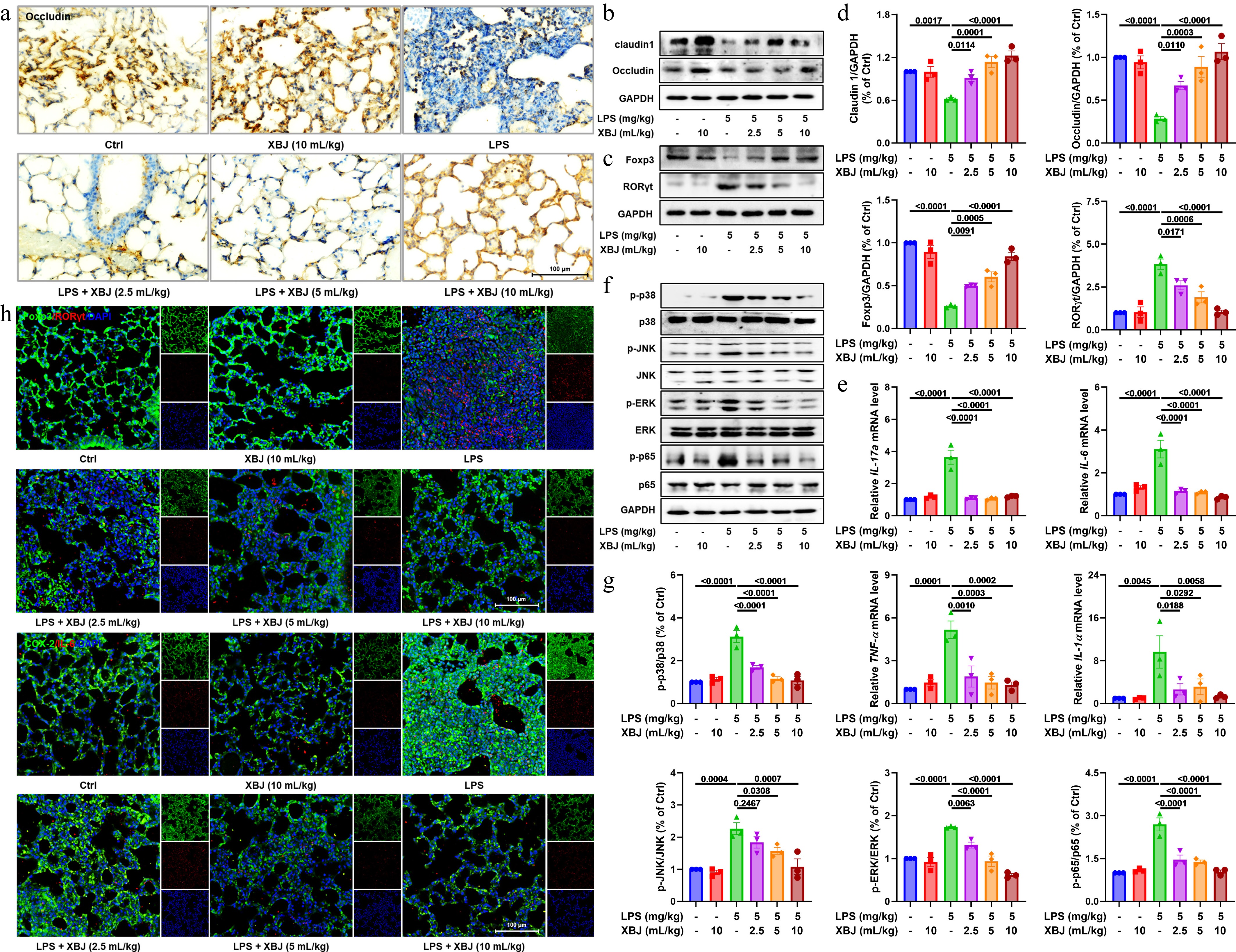
Figure 2.
Protective effects of XBJ against ALI via barrier repair, immune regulation, and MAPK and NF-κB pathway inhibition. (a) Representative occludin staining plots; (b)–(d) expressions of claudin1, occludin, Foxp3, and RORγt proteins (n = 3); (e) mRNA expressions of IL-17a, IL-6, TNF-α, and IL-1α (n = 3); (f), (g) expressions of key proteins in the MAPK and NF-κB pathways (n = 3); (h) immunofluorescence staining of Foxp3, RORγt, COX-2, and IL-6.
-
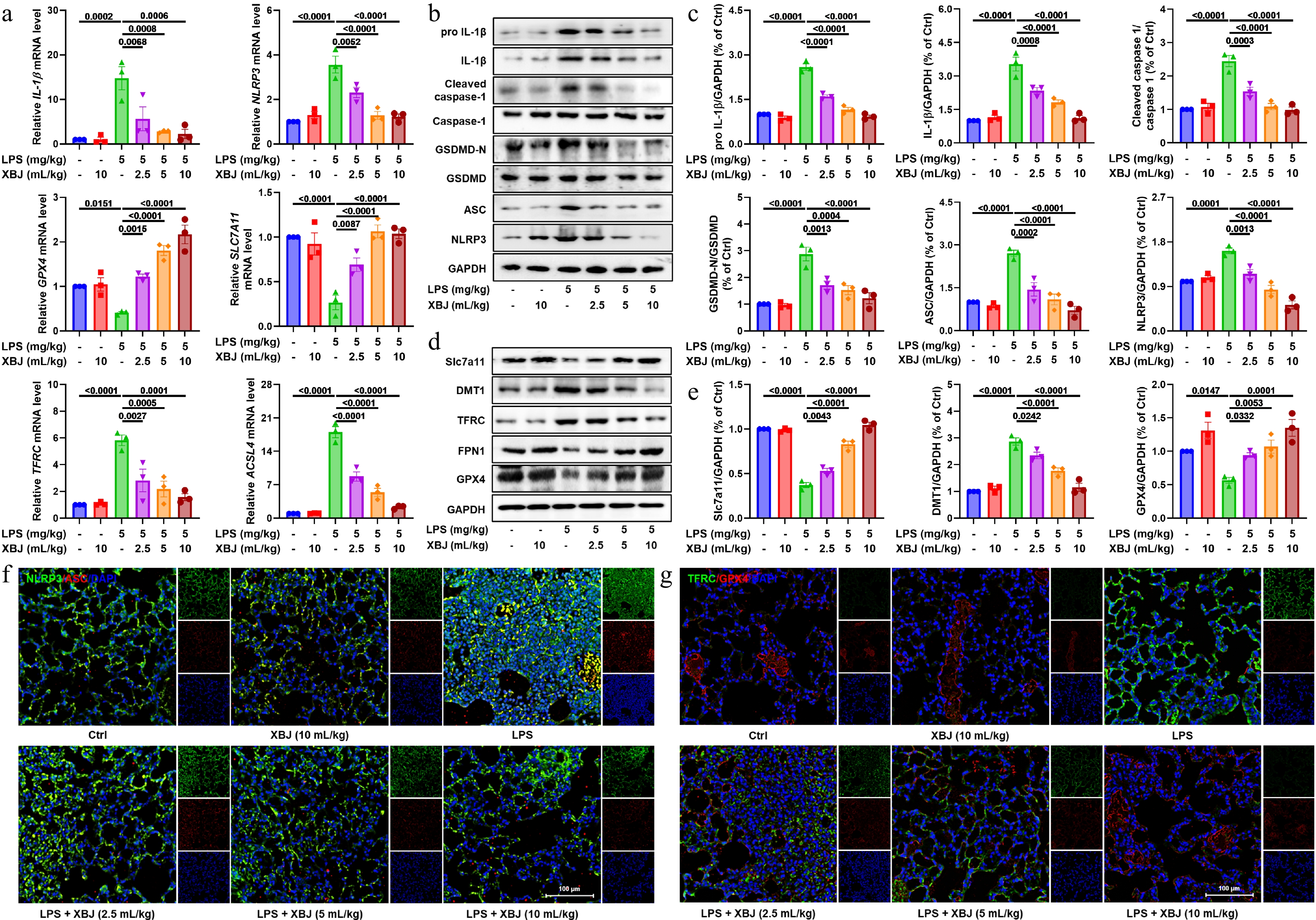
Figure 3.
XBJ alleviated ALI by inhibiting the NLRP3 pathway and ferroptosis. (a) mRNA expressions of GPX4, TFRC, ACSL4, Slc7a11, IL-1β, and NLRP3 (n = 3); (b), (c) effects of XBJ toward NLRP3 pathways in ALI (n = 3); (d), (e) Western blotting analysis of Slc7a11, DMT1, TFRC, FPN1, and GPX4 in ALI (n = 3); (f), (g) representative image of immunofluorescence of TFRC, GPX4, ASC, and NLRP3.
-
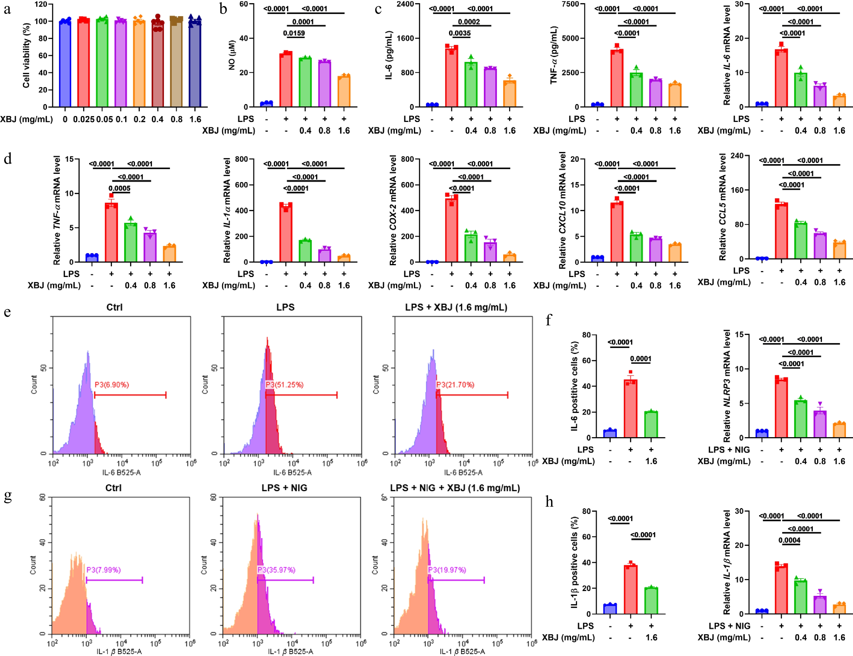
Figure 4.
XBJ suppressed LPS-induced inflammation in MH-S cells. (a) CCK-8 assay of MH-S cells treated with XBJ (n = 5); (b) XBJ inhibited NO production in MH-S cells (n = 3); (c) effects of XBJ toward IL-6 and TNF-α (n = 3); (d) effects of XBJ on mRNA expression levels of IL-6, TNF-α, COX-2, IL-1α, IL-1β, CCL5, CXCL10, and NLRP3 in LPS-mediated MH-S cells (n = 3); (e)–(h) flow cytometry of IL-6 and IL-1β (n = 3).
-
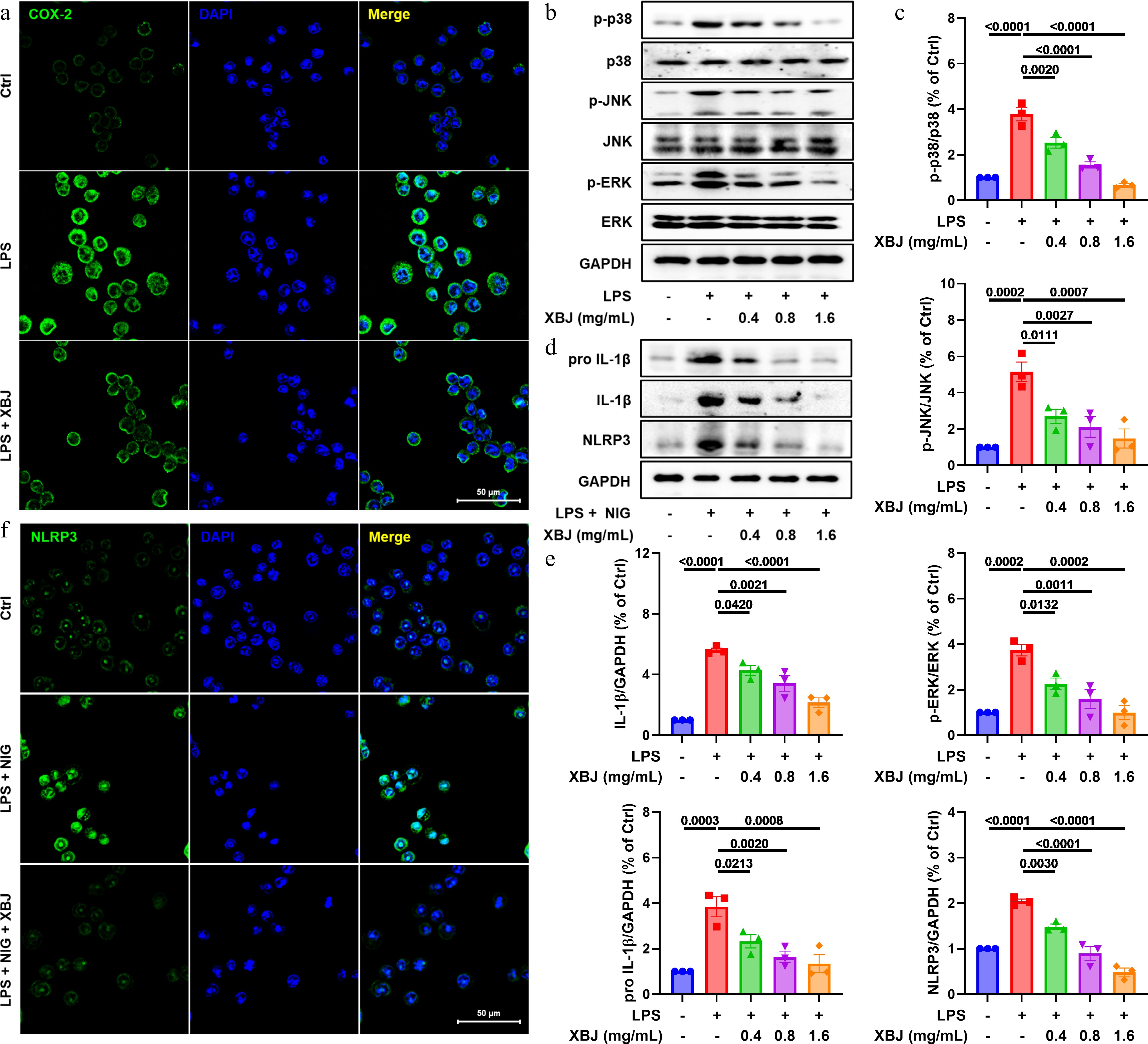
Figure 5.
XBJ suppressed LPS-induced inflammation via MAPK/NLRP3 pathways in vitro. (a), (f) Representative image of COX-2 and NLRP3 staining; (b), (d) XBJ attenuated expression of p-p38, p38, p-JNK, JNK, p-ERK, ERK, pro IL-1β, IL-1β, and NLRP3 (n = 3); (c), (e) quantification of p-p38/p38, p-JNK/JNK, p-ERK/ERK, pro IL-1β, IL-1β, and NLRP3 (n = 3).
-
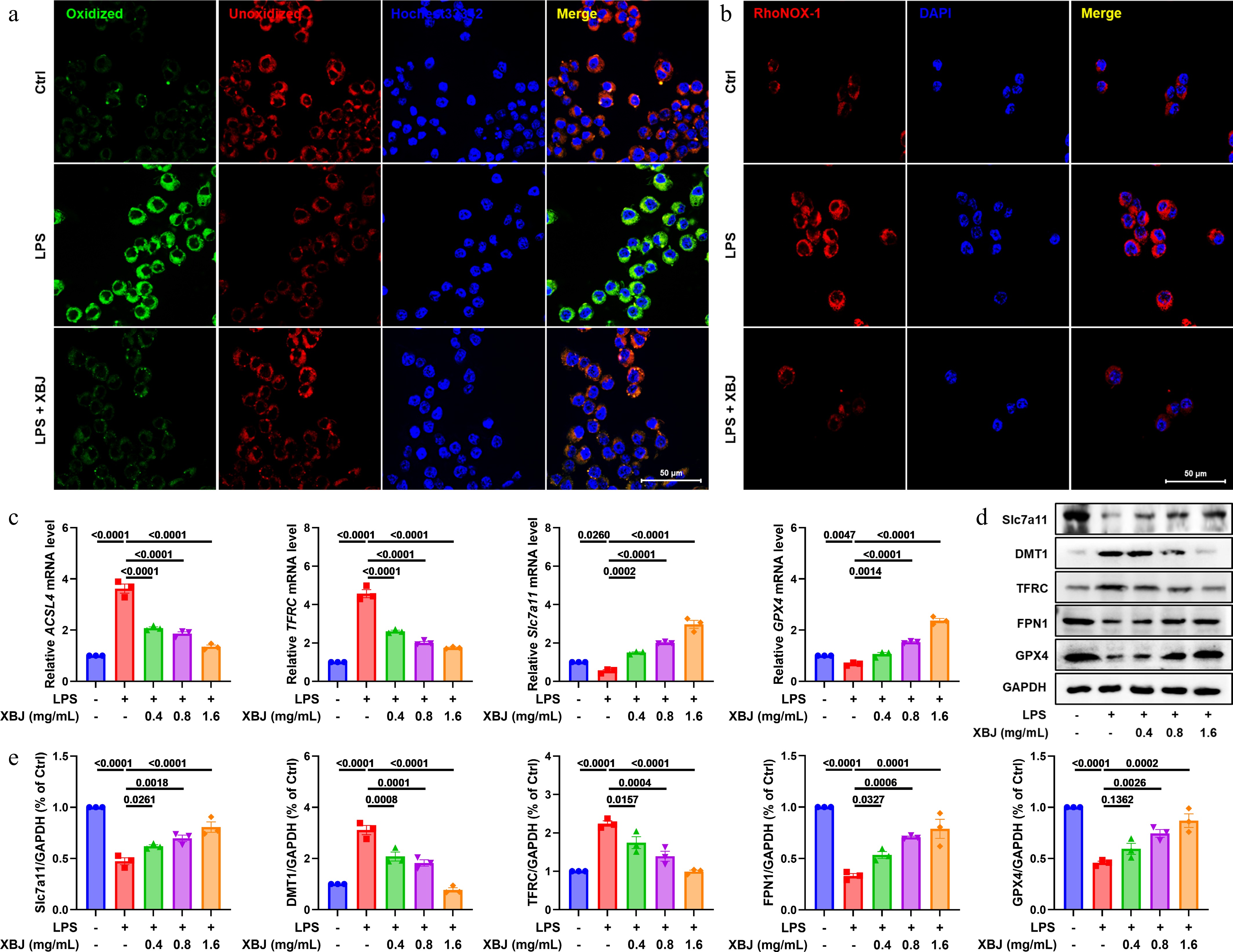
Figure 6.
XBJ inhibited ferroptosis in vitro. (a) Representative images of oxidized and unoxidized staining; (b) representative images of RhoNOX-1 staining; (c) mRNA levels of ACSL4, Slc7a11, TFRC, and GPX4 (n = 3); (d), (e) XBJ decreased expression levels of Slc7a11, DMT1, TFRC, FPN1, and GPX4 in LPS-mediated MH-S cells (n = 3).
-
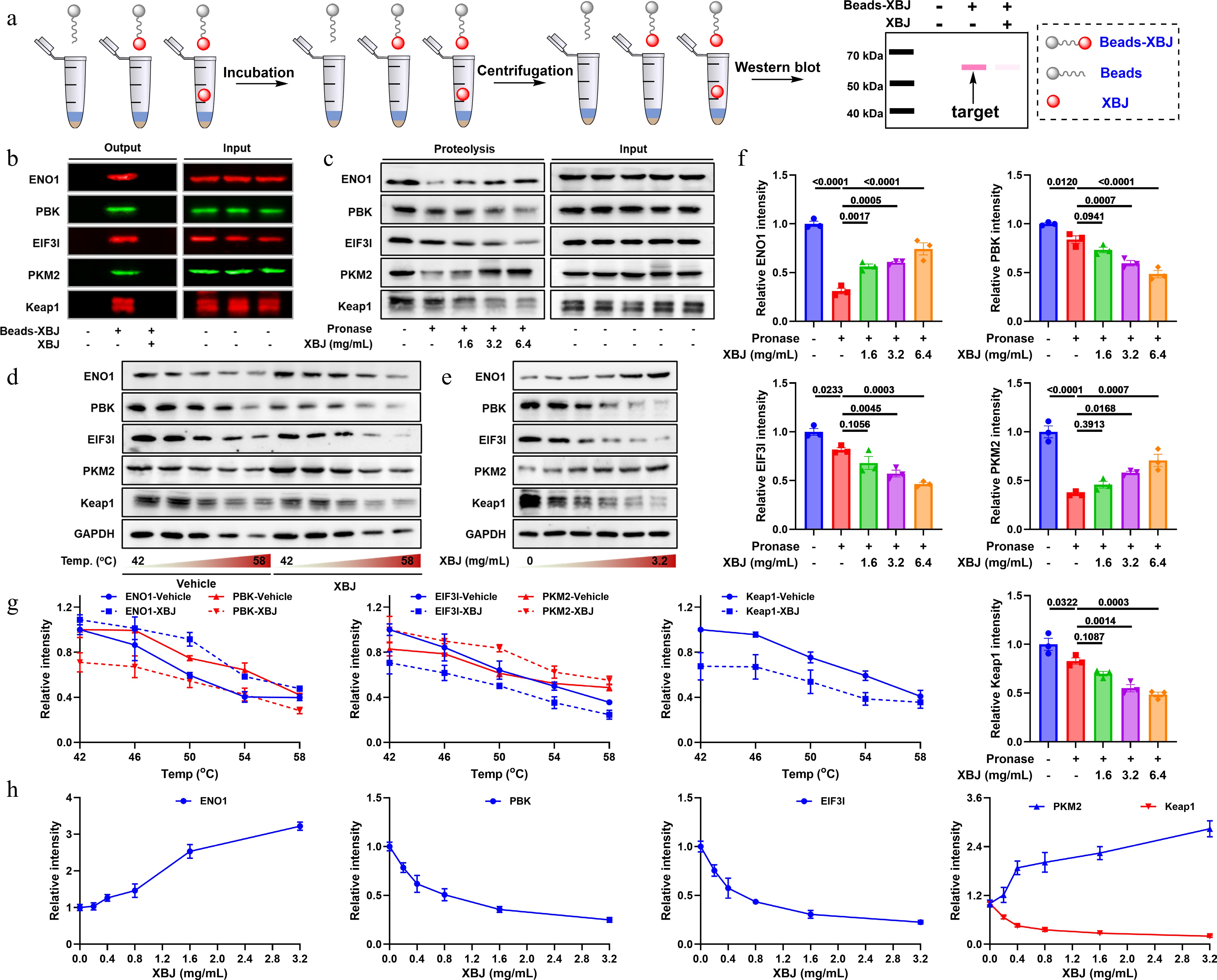
Figure 7.
ENO1, PBK, EIF3I, PKM2, and Keap1 were potential targets of XBJ. (a) Flowchart of affinity chromatography; (b) pull-down result; (c) DARTS result; (d), (e) CETSA result; (f)–(h) quantitative results of DARTS and CETSA.
-
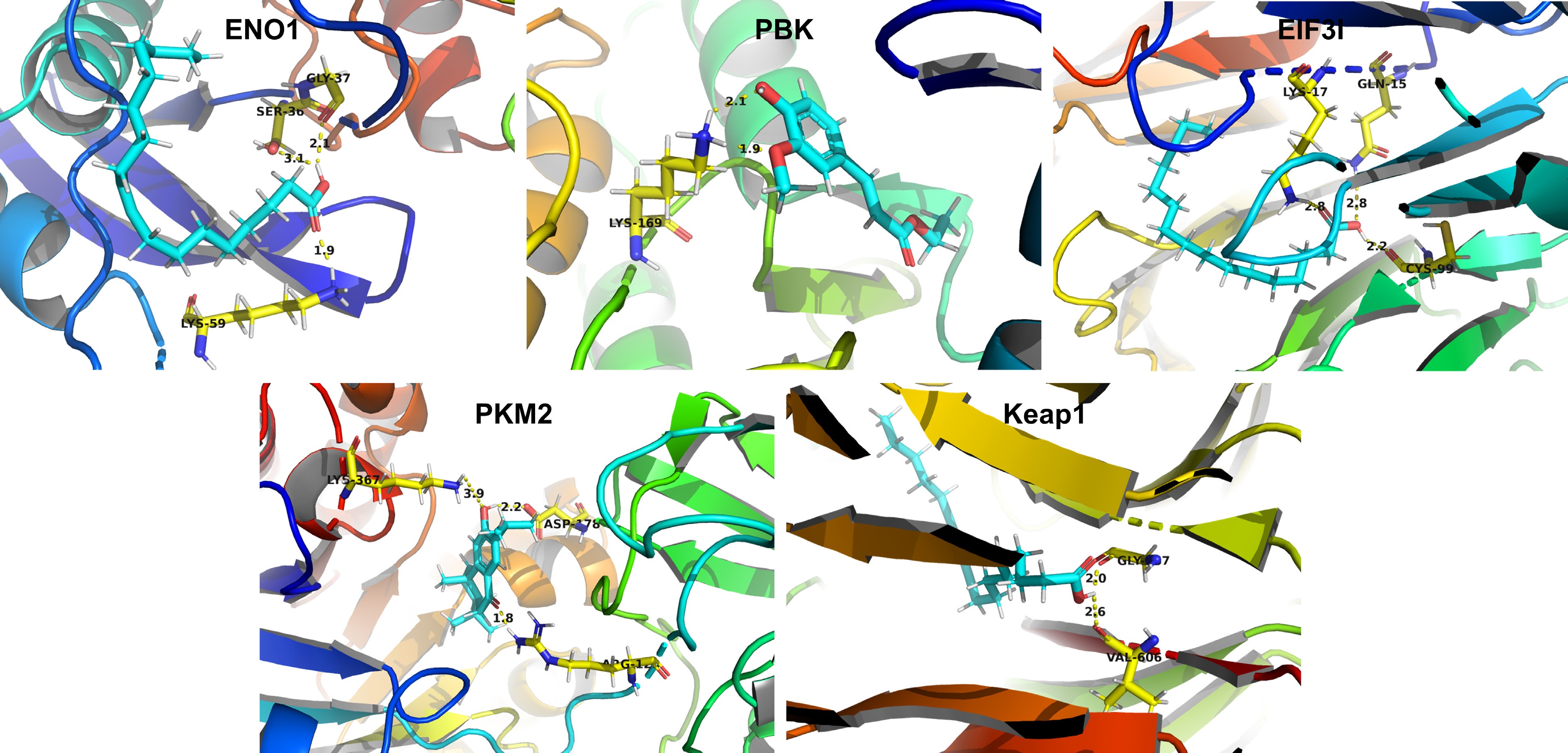
Figure 8.
Molecular docking results.
-
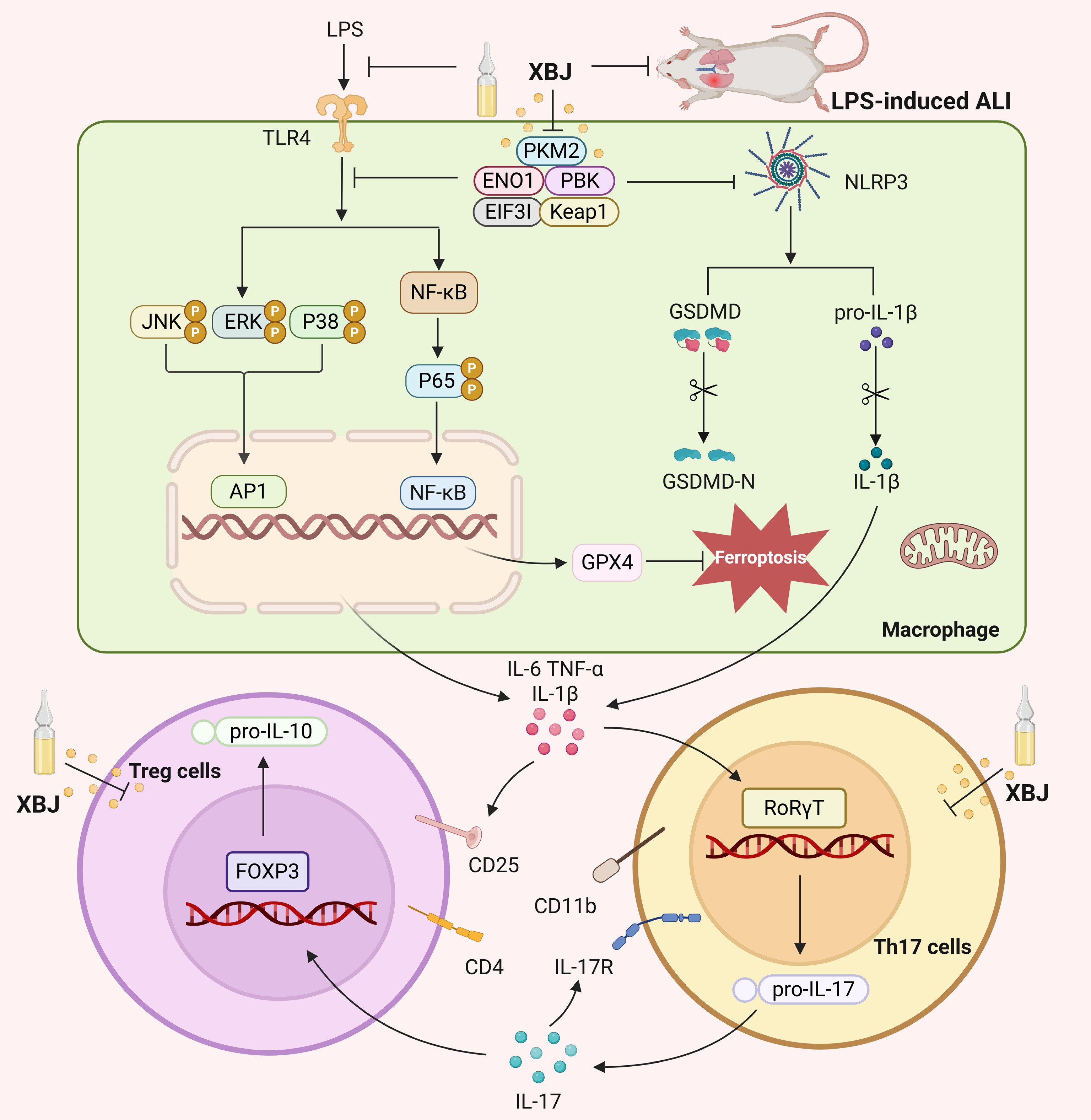
Figure 9.
Mechanism of XBJ for treating ALI.
Figures
(9)
Tables
(0)