-
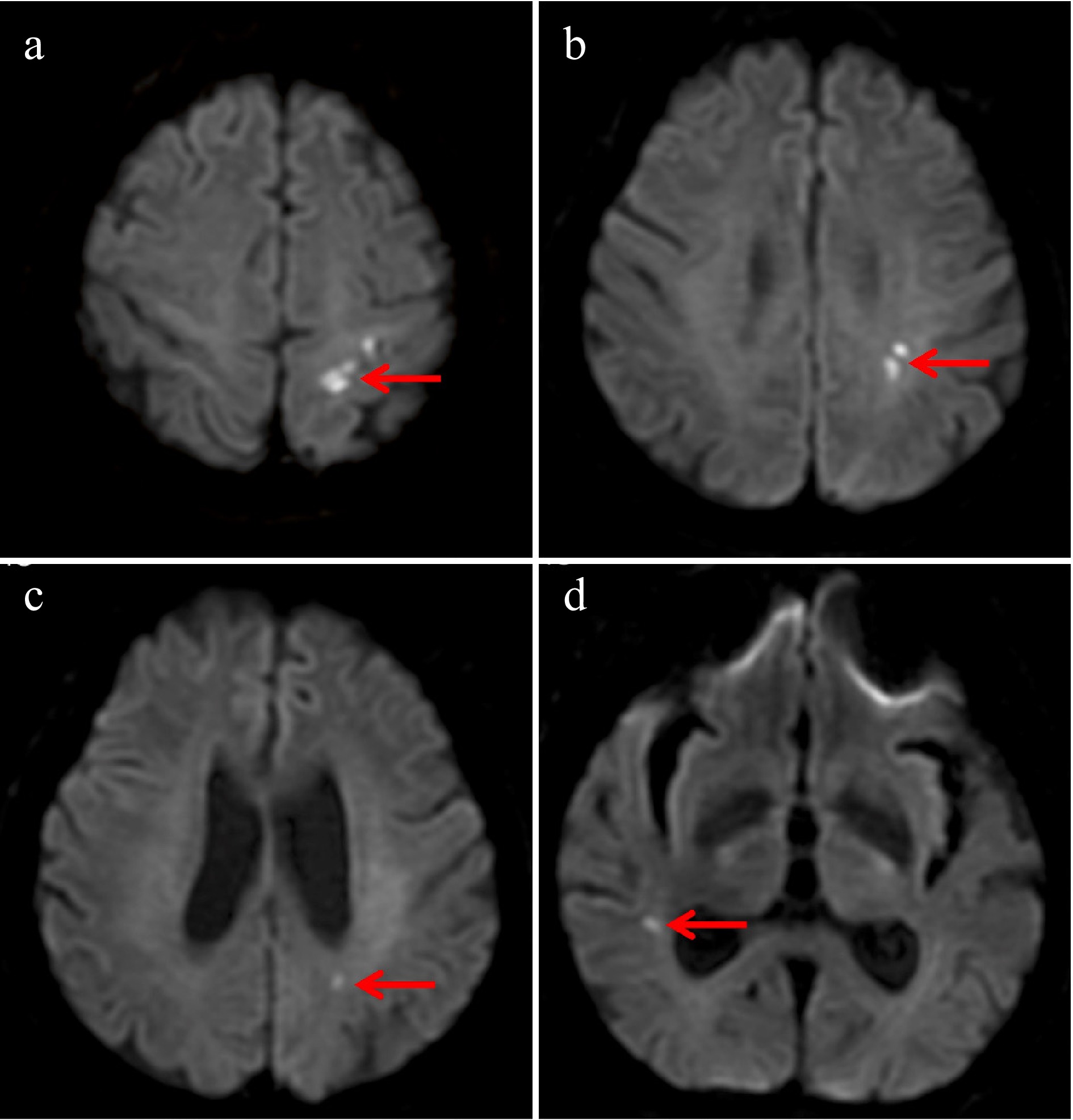
Figure 1.
Brain magnetic resonance imaging–diffusion-weighted imaging (MRI-DWI) demonstrated multiple cerebral infarctions involving both the anterior and posterior circulations. Acute infarction in (a) the left anterior cerebral artery (ACA) region, (b) the left middle cerebral artery (MCA) region, (c) the left posterior cerebral artery (PCA) region, and (d) the right MCA region.
-
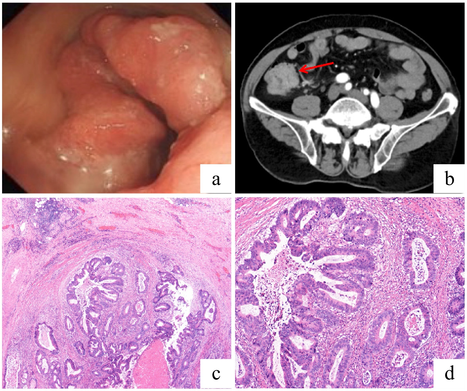
Figure 2.
Diagnostic and histopathological findings of ileocecal adenocarcinoma. (a) Colonoscopic view of a large, irregular mass in the ileocecal region. (b) Contrast-enhanced abdominal CT scan (axial view) showing irregular wall thickening with a marked enhancement in the ascending colon and ileocecal region. (c) Low-power photomicrograph (hematoxylin and eosin [HE] staining) of the resected specimen revealing an ulcerated, moderately differentiated adenocarcinoma with transmural invasion into the muscular layer. (d) High-power photomicrograph (HE staining) demonstrating the neoplastic glands with enlarged, hyperchromatic nuclei and evident mitotic activity. No significant perineural or lymphovascular invasion was identified.
-
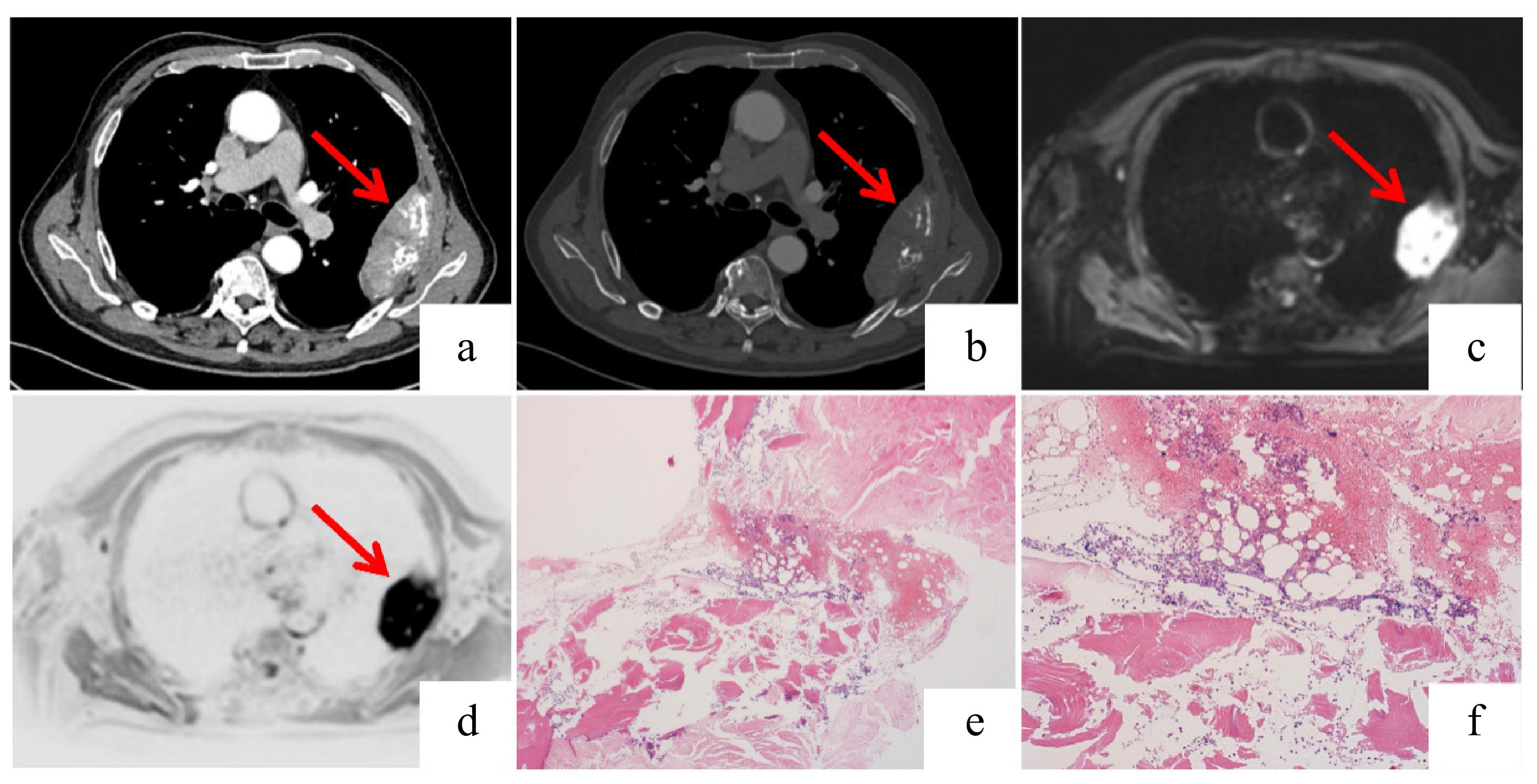
Figure 3.
Imaging and histopathological findings of the plasmacytoma. (a), (b) Axial and coronal contrast-enhanced chest CT scans demonstrating irregular left pleural thickening (measuring approximately 9.8 × 3.1 cm in the maximum cross-section) with associated destruction of the adjacent rib. (c), (d) Fusion positron emission tomography (PET)-MRI images reveal a rounded mass in the left chest wall showing hyperintensity on DWI with corresponding hypointensity on the apparent diffusion coefficient (ADC) map, consistent with restricted diffusion. Increased signal intensity is also noted in the adjacent rib on the MRI sequence. (e), (f) The bone marrow biopsy specimens show scattered plasma cells with lambda light chain restriction indicated by immunohistochemistry: (f) lambda-positive; (e) kappa-negative. This confirmed the presence of monoclonal plasma cells. The overall cellularity and number of plasma cells were not significantly increased.
-
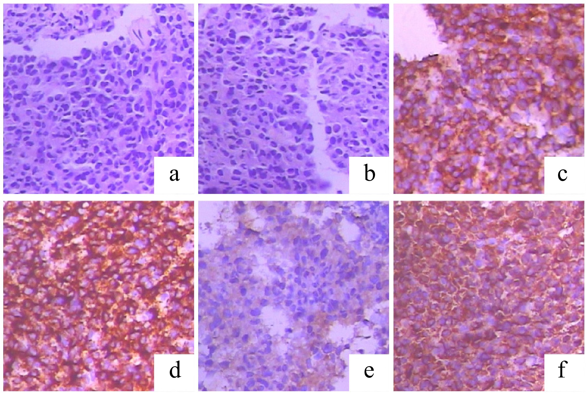
Figure 4.
Histopathological and immunohistochemical (IHC) features of the left rib mass, diagnostic of plasmacytoma. (a), (b) Hematoxylin and eosin (H&E) staining reveals sheets of monomorphic plasma cells. (c), (d) The neoplastic cells show strong and diffuse positivity for the plasma cell markers (c) CD38 and (d) CD138. (e), (f) IHC staining demonstrates lambda light chain restriction, supporting a monoclonal plasma cell proliferation: (f) positive for lambda light chains; (e) negative for kappa light chains.
Figures
(4)
Tables
(0)