-
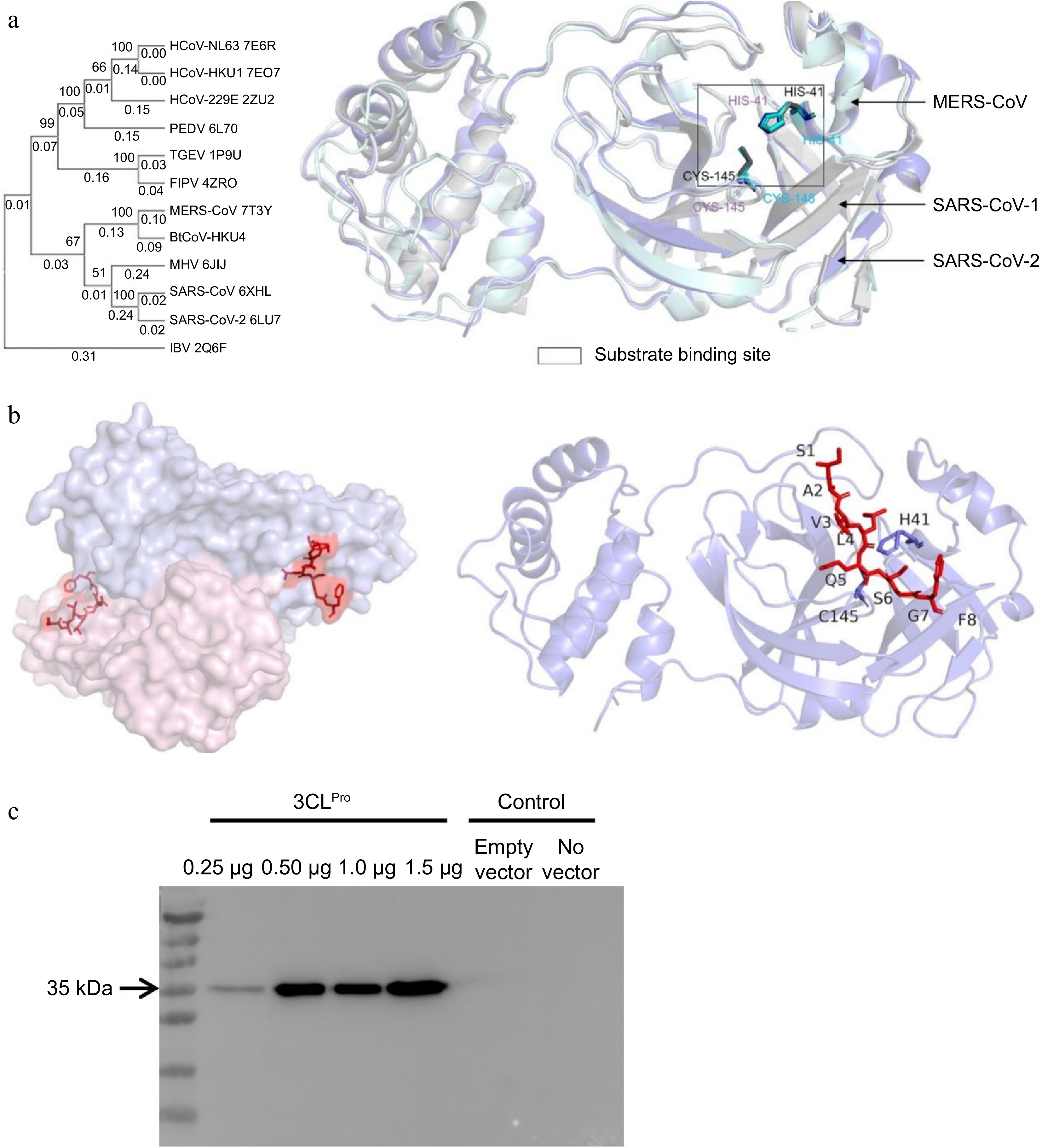
Figure 1.
The substrate binding pocket of 3CLPro. (a) Structural alignment of three coronaviruses 3CLPro (SARS-CoV-1, SARS-CoV-2, and MERS-CoV). The catalytic dyad was indicated in the black box. (b) The substrate peptide (red sticks) bound to 3CLPro. The protease was a homodimer in native conformations. In the design process, only one protomer (right panel) was used as the input model. (c) 3CLPro expression in HEK-293T cells. The cells were transfected with different amounts of pCMV vectors encoding 3CLPro (33.8 kD, without the Flag tag) and the protein expression levels were detected by Western blot using anti-Flag antibodies.
-
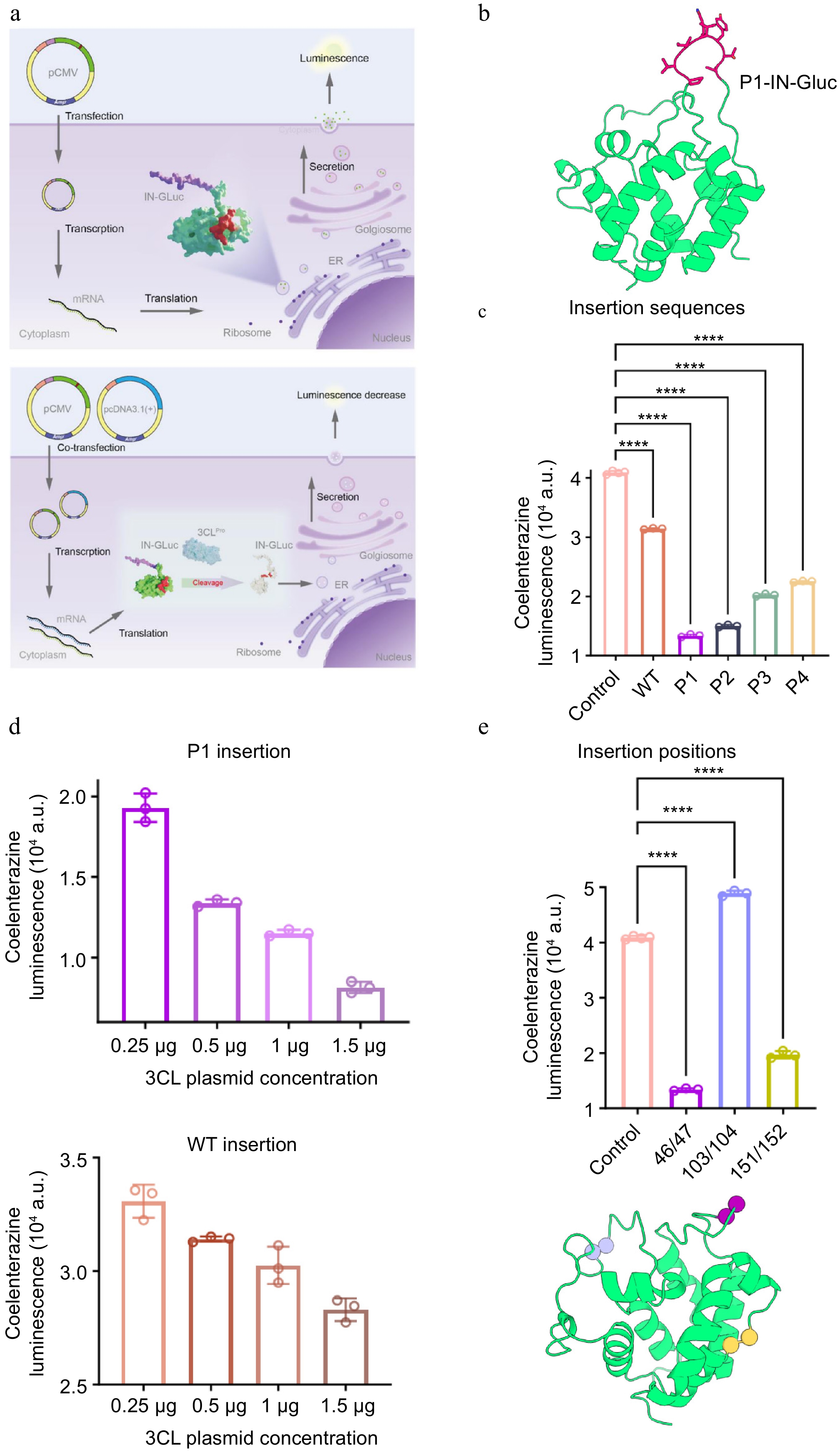
Figure 2.
The design of IN-Gluc. (a) The design principle of IN-Gluc. 3CLPro cleaved the peptide sequence inserted in the middle of Gluc, a secreted enzyme catalyzing the oxidation of coelenterazine to a luminescent molecule, leading to a reduction of luminescence (lower panel). In the absence of 3CLPro or infections, the Gluc was readily secreted out of the cytosol (upper panel). (b) The AlphaFold3 predicted structural model of P1-IN-Gluc. (c) The effects of different designed sequences when inserted into Gluc. The control was deactivated 3CLPro H41A/C145A expressed at the same level. (d) The 3CLPro dose-dependent signal change when P1 or WT peptide was inserted into Gluc. (e) The effects of different insertion positions and the locations of 46/47, 103/104, and 151/152 positions indicated as spheres. n = 3 as the number of replications (n = 4 for the control sample).
-
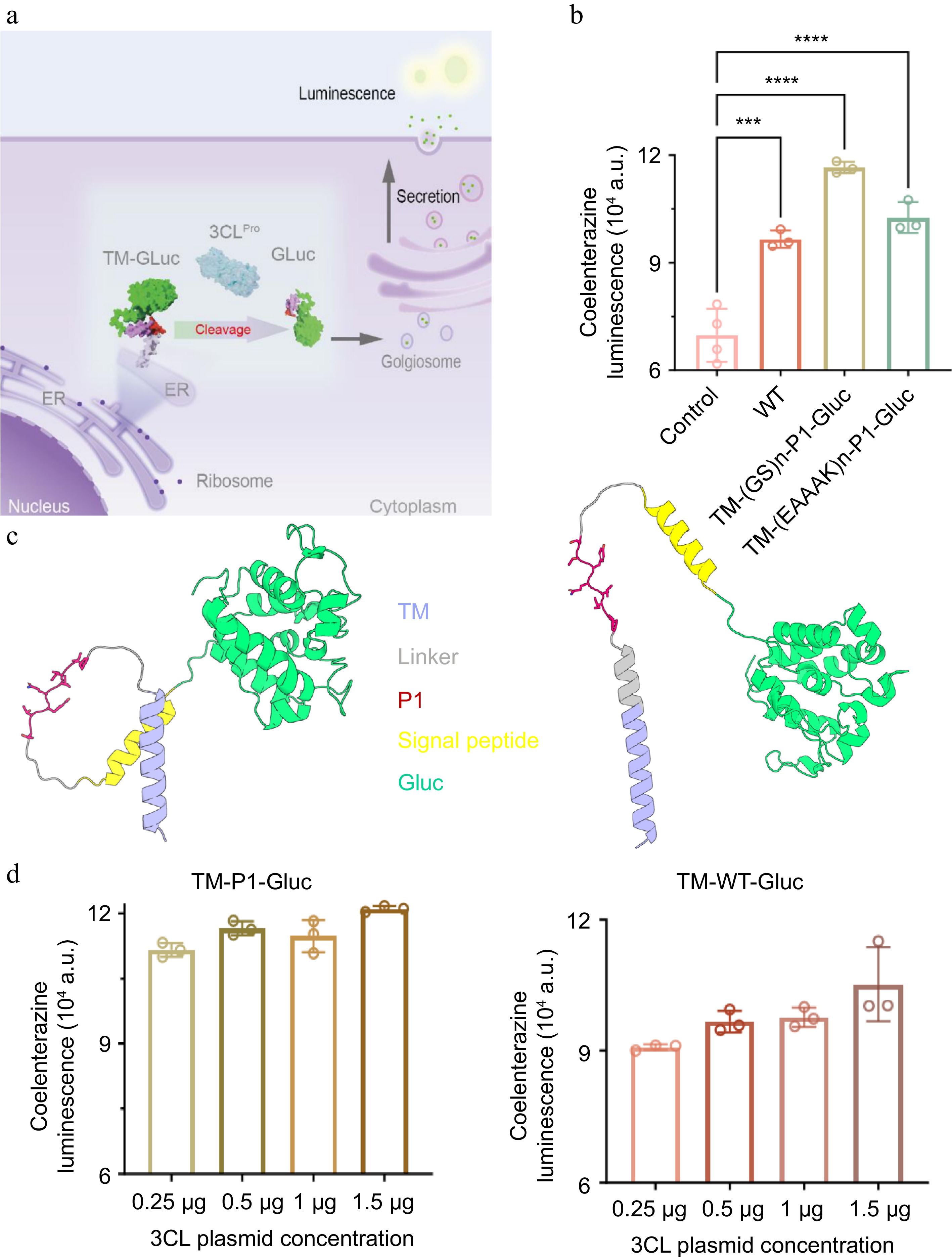
Figure 3.
The design of TM-Gluc. (a) The design principle of TM-Gluc. 3CLPro-mediated cleavage released Gluc for extracellular secretion, leading to elevation of luminescence. (b) The effects of flexible linker and rigid linker when fusing the P1 peptide with the membrane anchoring sequence TM. (c) The AlphaFold3 predicted structural model of P1-TM-Gluc (left: flexible linker, right: rigid linker). (d) The 3CLPro dose-dependent signal change when P1 or WT peptide was fused at the N-terminal of Gluc. n = 3 as the number of replications (n = 4 for the control sample).
-
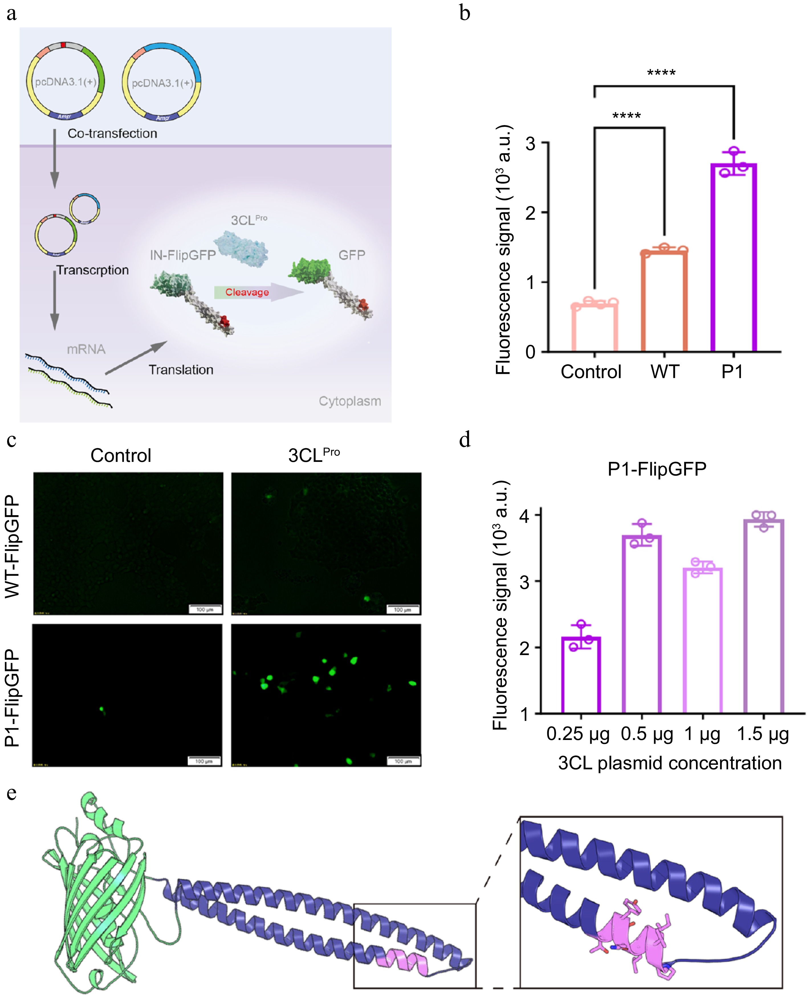
Figure 4.
The design of IN-FlipGFP. (a) The design principle of IN-FlipGFP. 3CLPro-mediated cleavage released conformational constraint of FlipGFP and restored the green fluorescence. (b) The comparison between WT peptide insertion and P1 peptide insertion in the FlipGFP construct. (c) The image of cells expressing 3CLPro and IN-FlipGFP indicating the brightest signal came from P1-IN-FlipGFP, with 3CLPro H41A/C145A expression as the control (left panel). (d) The 3CLPro dose-dependent signal change of the P1-IN-FlipGFP construct. (e) The AlphaFold3 predicted structural model of P1-IN-FlipGFP with the insertion site highlighted. n = 3 as the number of replications (n = 4 for the control sample).
-
ID Sequence AlphaFold3 predictions Rosetta energy (REU) WT SAVLQSGF ipTM = 0.8 −56.1 P1 PVILQYTT ipTM = 0.72 −75.8 P2 MSRLQTSN ipTM = 0.73 −64.3 P3 PVILNYTH ipTM = 0.64 −71.5 P4 KPRLQAGN ipTM = 0.69 −47.5 Table 1.
Computational characterization of WT and designed sequences.
Figures
(4)
Tables
(1)