-
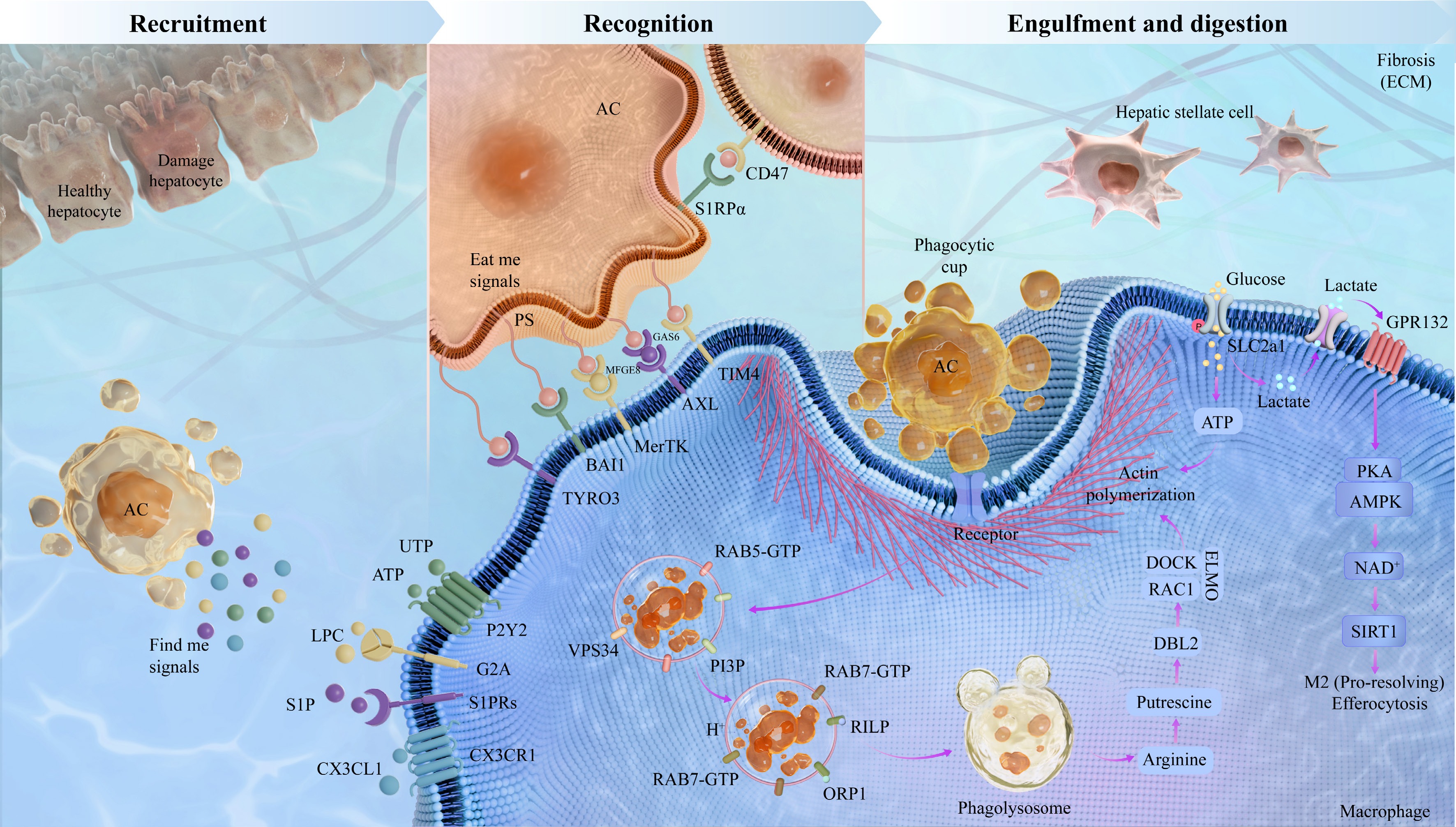
Figure 1.
The sequential stages of efferocytosis in liver fibrosis. Efferocytosis is a multifaceted process divided into four phases: the recruitment stage, the recognition stage, and the engulfment and digestion stage. In the recruitment stage, apoptotic hepatocytes release soluble mediators LPC, S1P, ATP, UTP, and CX3CL1 to recruit macrophages. During the recognition stage, macrophages recognize apoptotic hepatocytes through interactions between 'eat me' signals, PS, and phagocyte surface receptors including MerTK, AXL, TIM, BAI1, TYRO3, and STABILIN. Healthy cells display the 'don't-eat-me' signal CD47; this classical protective signal is often overexpressed under conditions associated with defective clearance of apoptotic cells, and blockade of the CD47-SIRPα axis has been exploited in cancer therapies and atherosclerosis. In the engulfment and digestion stage, macrophages efficiently digest and degrade engulfed apoptotic hepatocytes along with cellular contents. This stage exerts anti-inflammatory effects through phagolysosomal degradation of ACs, a process involving RAB7-GTP, RILP, and VPS34 that generates metabolites and pro-resolving IL10 and TGFβ signals. Such factors promote M2 macrophage polarization and TGFβ release, subsequently suppressing HSCs activation, enhancing ECM degradation, and mitigating fibrosis.
-
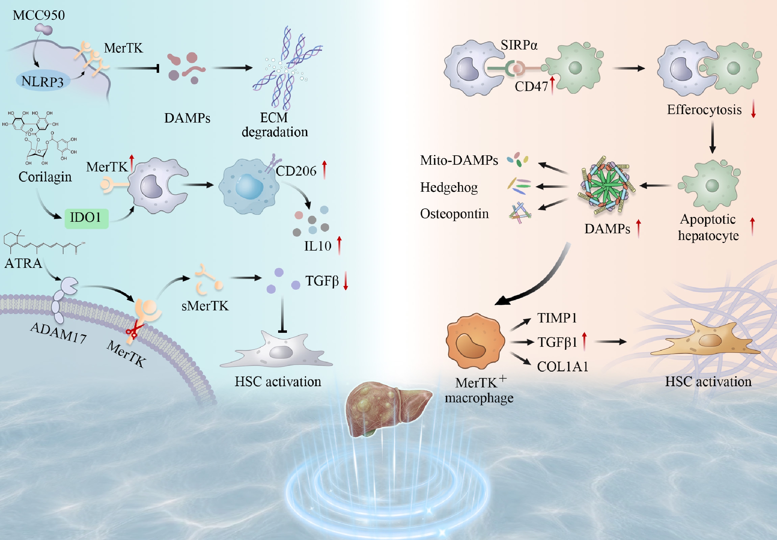
Figure 2.
The dual role of efferocytosis-related molecules in liver fibrosis. In the normal liver, macrophages efficiently clear apoptotic hepatocytes by upregulating MerTK and related molecules, promoting downregulation of TGFβ and TIMP1, thereby exerting anti-fibrotic and pro-resolution effects. In contrast, during liver fibrosis progression, efferocytosis becomes dysfunctional, accompanied by abnormalities in the CD47-SIRPα signaling axis, activation of the NLRP3 inflammasome (which can be inhibited by MCC950), release of DAMPs and mitochondrial DAMPs, ADAM17-mediated shedding of sMerTK, as well as activation of multiple pathways involving Hedgehog, osteopontin, VEGF, IDO1, and IL10. These changes collectively promote HSCs activation and ECM deposition (e.g., COL1A1), ultimately driving fibrosis progression.
-
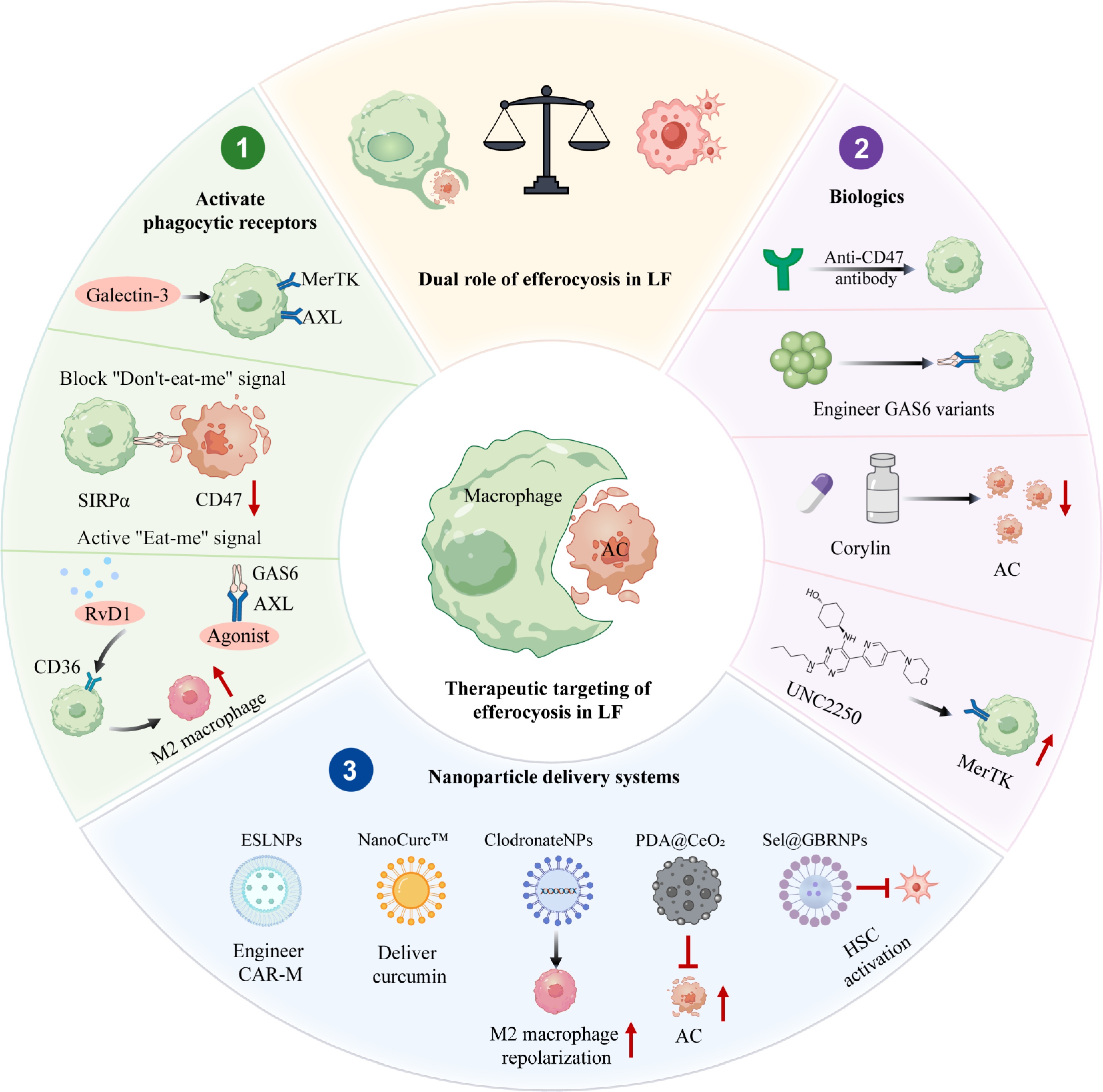
Figure 3.
Therapeutic strategies targeting efferocytosis. This figure outlines efferocytosis-targeted strategies for liver fibrosis, grouped into three main approaches: (1) enhancing protective efferocytosis by blocking CD47-SIRPα, supplementing bridging molecules (e.g., MFGE8), restoring MerTK function, or using engineered GAS6 ligands, (2) inhibiting pathological efferocytosis via AXL/MerTK inhibition, Galectin-3 blockade, or Corylin treatment, and (3) nanoparticle delivery systems including ESLNPs, NanoCurc™, Sel@GBRNPs plus Riociguat, and PDA@CeO2, which enable targeted modulation and promote resolution.
-
Regulator Targets Mechanisms Key effects Intervention strategies Ref. Protective role MerTK NLRP3 activation inhibits MerTK-mediated efferocytosis Reduces DAMPs release and promote
fibrosis resolutionMCC950 [57,58] GPNMB Ly6C+ monocytes switch to restorative macrophages via ERK for MMP resolution Matrix degradation, HSCs inactivation, scar remodeling, and fibrosis resolution Liposome-induced phagocytosis [59] MerTK Suppresses pro-fibrotic factors, upregulates MMPs, and promotes ECM degradation Promotes matrix degradation and reduces inflammation Enhance MerTK function [60] TREM2 TREM2 expression in LAMs promotes efferocytosis and collagen degradation Apoptotic hepatocytes clearance and HSCs inactivation, fibrosis resolution TREM2 agonists [67] CD47, SIRPα Blocks CD47-SIRPα to promote macrophage clearance of necroptotic hepatocytes Inhibits HSCs activation and reduces liver fibrosis Anti-CD47 antibody [70] MerTK, GAS6 ADAM17-mediated MerTK cleavage releases soluble fragment that blocks GAS6 Inhibits HSCs activation and
reduces liver fibrosisEnhance MerTK function [71] CD68 Macrophage engulfment of apoptotic cells upregulate MMP3, MMP8, and MMP9 Fibrosis reversal, septa fragmentation, and liver architecture restoration Roux-en-Y anastomosis [72] TIM4 Restores TIM4 mediated efferocytosis and increases IL10 Suppresses HSCs activation, and reverses fibrosis progression TIM4 restoration [73] TIM4 Inhibits AKT1 and ROS mediated mitophagy axis in KCs, reducing TGFβ1 secretion Reverses fibrosis, restores architecture, and attenuates post-LT fibrosis TIM4 mAb [74] Pathological role αvβ3 CCN1 via αvβ3 bridges ACs to macrophages, triggering efferocytosis and TGFβ1 Activates HSCs and induces myofibroblast transdifferentiation Block CCN1-αvβ3 binding [27] MerTK Increases MerTK expression via rs4374383 GG/GA, and enhances HSCs procollagen Activates HSCs and fibrosis progression MerTK inhibitors [81] TREM2+ macrophage TREM2+ SAMs highly express SPP1 promotes type I collagen production Promotes scar formation and exacerbates liver fibrosis Osteopontin neutralization [82] AXL Phosphorylates SMAD3 via AXL, and upregulates PAI1, MMP9, SNAIL, and TGFβ1 Activates HSCs and ECM deposition AXL inhibitors [85,86] GAS6 GAS6 impairs β-oxidation and upregulates inflammatory cytokines Recruitments of macrophages drives liver fibrosis progression BGB324 [87] NADPH oxidase Induces apoptotic body phagocytosis, NADPH oxidase activation, and ROS production Activates HSCs and extracellular matrix deposition ROS scavenger [89] FasL Apoptotic body engulfment by KCs upregulate FasL and TNFα Induces inflammation and activates HSCs Gadolinium chloride [90] αvβ3 Enhances apoptotic body phagocytosis and activates NF-κB pathway Activates HSCs and increase production of TGFβ and collagen I Galectin-3 inhibition [91] CD36 CD36 regulated by FABP7 controls KCs phagocytosis and MCP1, TGFβ production Activates HSCs and fibrosis progression FABP7 modulation [92] Table 1.
Core regulatory factors and mechanisms of efferocytosis in liver fibrosis.
-
Identifiers Start date Liver fibrosis type Time frame Phase Status NCT04197479 November 2019 NASH with liver fibrosis (stage F2–F3) 52 weeks Phase 3 Active, not recruiting NCT03900429 April 2019 NASH with liver fibrosis (stage F1–F3) 36 weeks Phase 3 Completed NCT04951219 July 2021 NASH with compensated cirrhosis (stage F4) 54 weeks Phase 3 Recruiting − − TIM4 engineered macrophages for MASH-associated
liver fibrosis− Preclinical Preclinical stage − − Anti-CD47 antibody for liver fibrosis − Preclinical Liver fibrosis-specific
trial pendingTable 2.
The ongoing clinical trials of associated drugs or therapies in liver fibrosis.
Figures
(3)
Tables
(2)