-
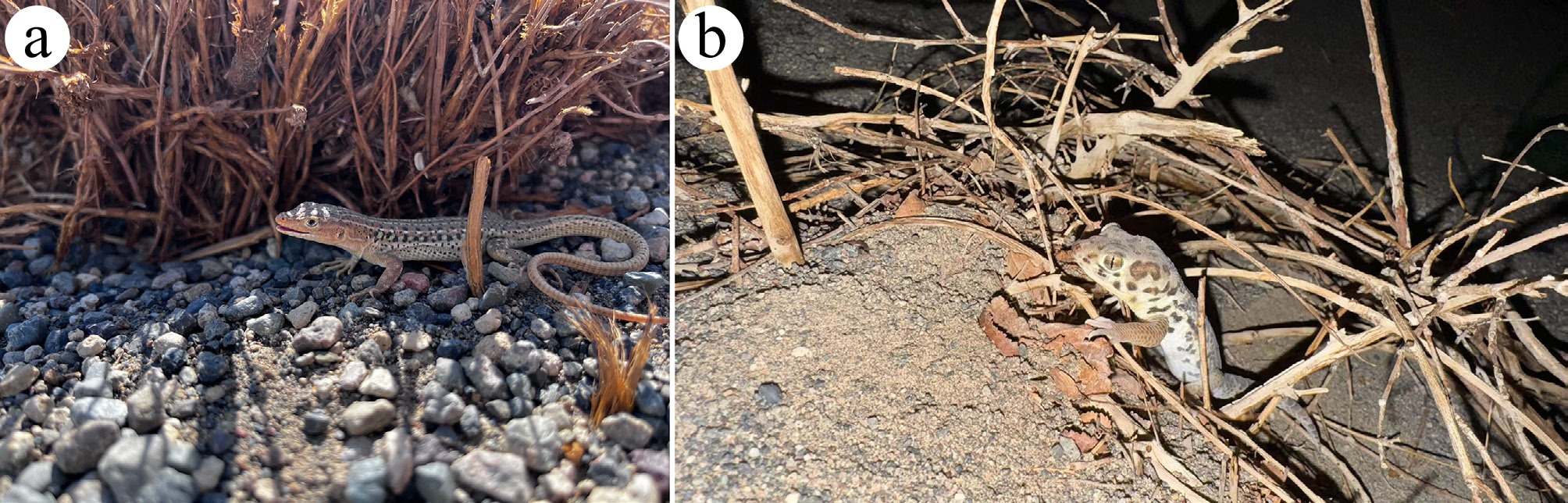
Figure 1.
Wild foraging habitats of the two lizard species in the Turpan Basin, Xinjiang Uygur Autonomous Region, China. (a) Eremias roborowskii inhabiting an area beneath Alhagi camelorum in the wild. (b) Teratoscincus roborowskii crawling in the wild.
-
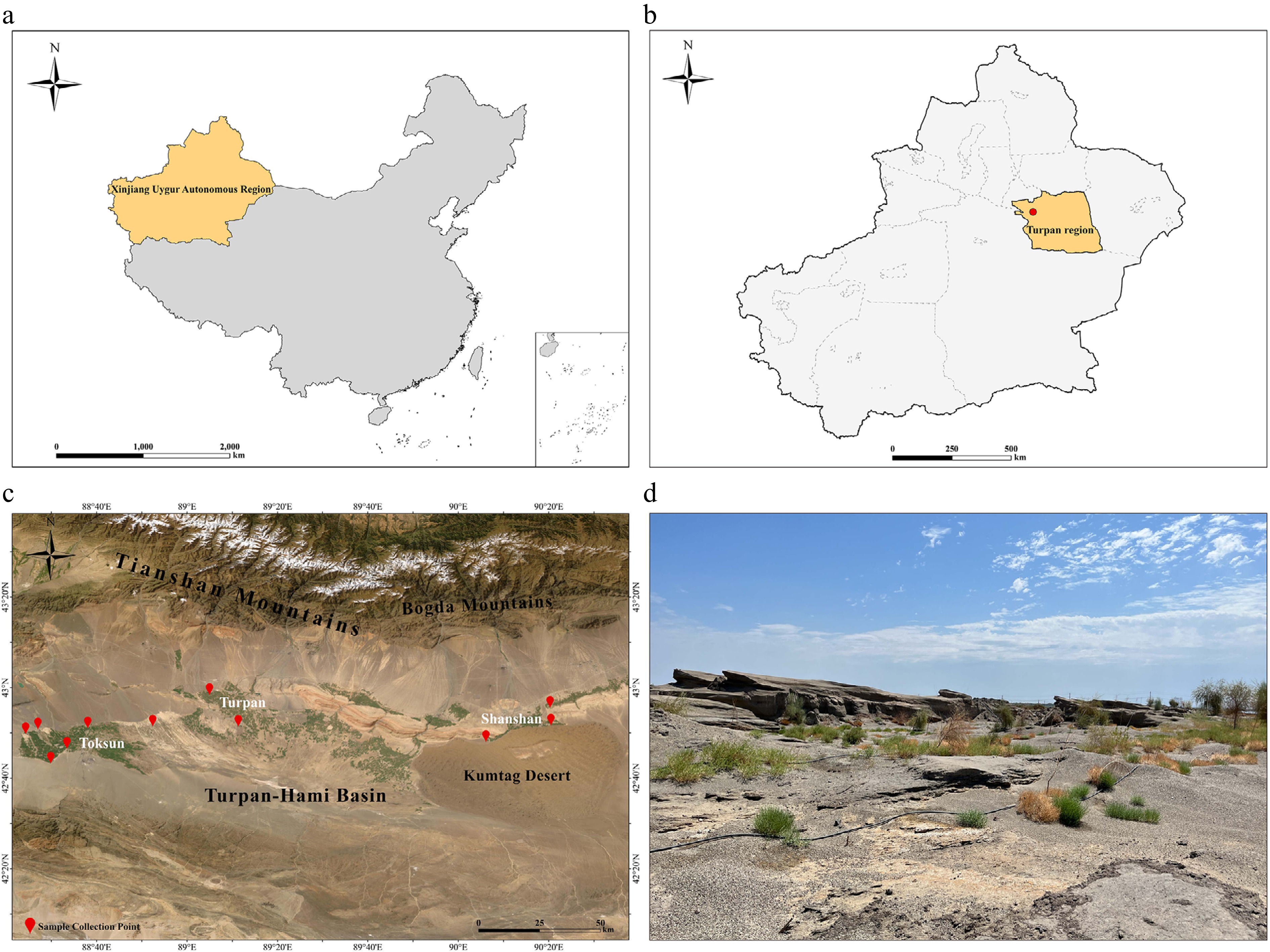
Figure 2.
Sampling sites and geographic distribution of the two lizard species collected during 2024−2025 field surveys in the Turpan Basin, Xinjiang, China. (a) Map of the People's Republic of China, highlighting the location of the Xinjiang Uygur Autonomous Region. (b) Map of the Xinjiang Uygur Autonomous Region, indicating the Turpan research area. (c) Topographic map of the Turpan Basin, with red markers denoting sampling sites in Toksun, Turpan, and Shanshan. (d) Representative arid desert habitat of the research region, illustrating the typical environment inhabited by the two lizard species. (The provincial administrative boundary data of China and the geographic map were obtained from the Resource and Environmental Science Data Registration and Publication System and the built-in geographic base map service of ArcGIS Pro software, respectively).
-
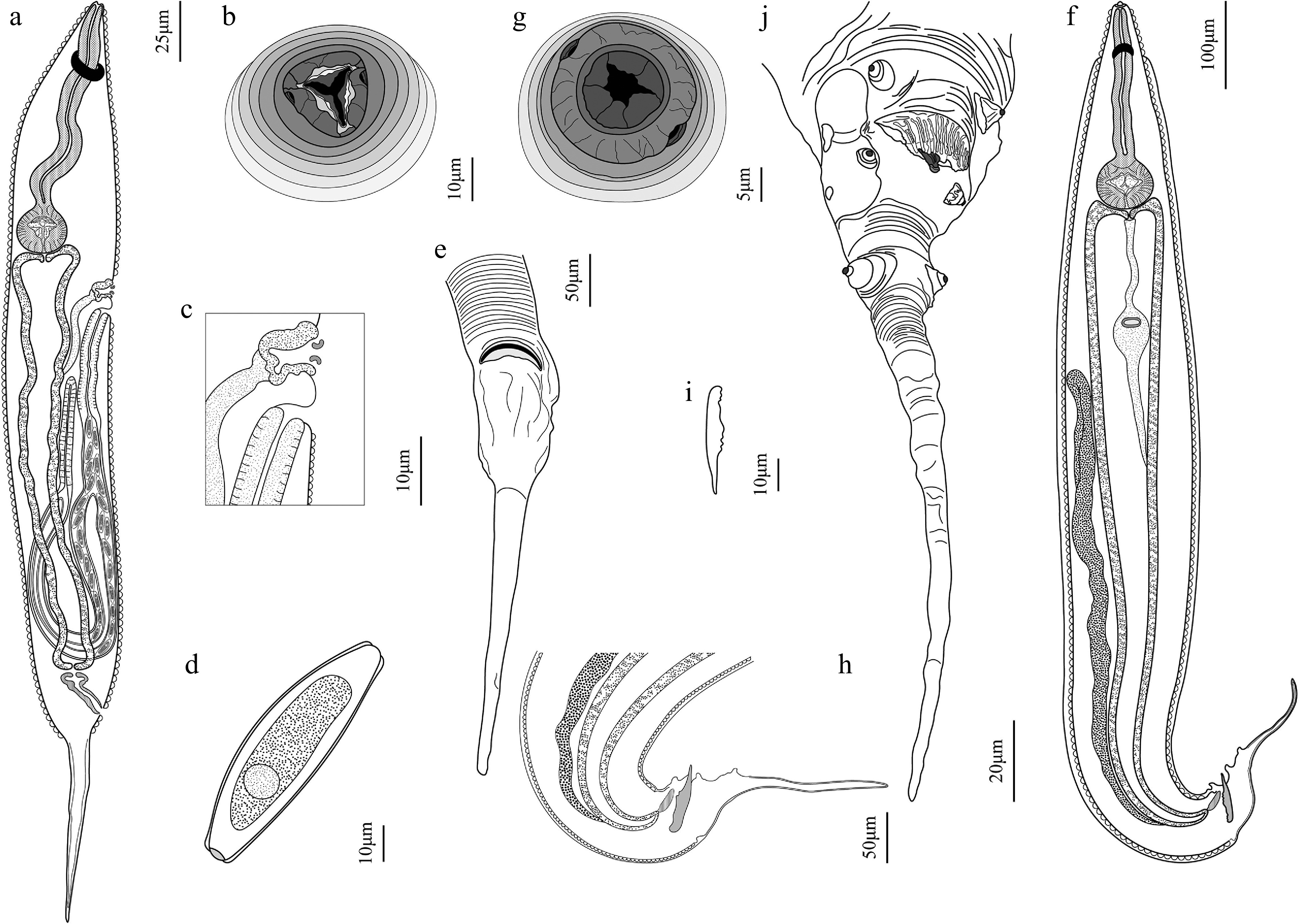
Figure 3.
Spauligodon turpanoeremius sp. nov. (a)–(e) female, and (f)–(j) male. (a) Female, gravid, entire, ventral view; (b) en-face view; (c) excretory pore and vulva region; (d) egg; and (e) posterior end, ventral view. (f) Male, entire, lateral view; (g) en-face view; (h) posterior end with spicule, lateral view; (i) spicule; and (j) caudal extremity, ventral view.
-
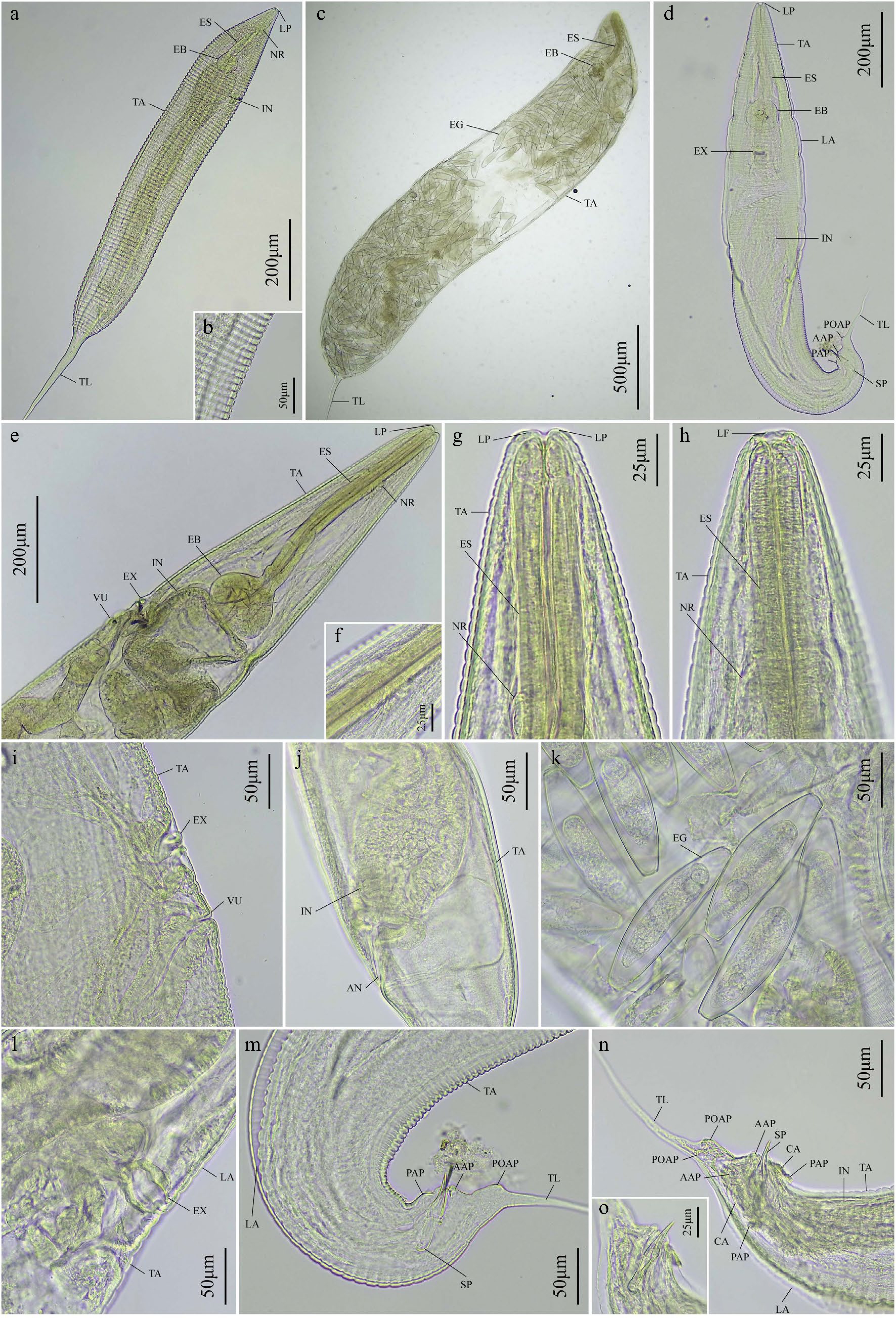
Figure 4.
Digital micrographs of Spauligodon turpanoeremius sp. nov. isolated from Eremias roborowskii. (a)–(c), (e)–(g), (i)–(k) Female. (d), (h), (l), (m)–(o) Male. (a) Female, whole mount, lateral view; (b) transverse striation of cuticle; (c) female gravid, whole mount, lateral view; (d) male, whole mount, lateral view; (e) female, anterior end, lateral view; (f) nerve ring; (g) cephalic end, ventral view; (h) male, cephalic end, ventral view; (i) female, excretory pore and vulva, lateral view; (j) female, posterior end, ventral view; (k) eggs; (l) male, excretory pore, lateral view; (m) male, posterior end, lateral view; (n) male, caudal morphology, highlighting the spicule, caudal alae, and papillae, ventral view; (o) spicule. AAP, adanal papillae; AN, anus; CA, caudal alae; EB, esophageal bulb; EG, eggs; ES, esophagus; EX, excretory pore; IN, intestine; LA, lateral alae; LF, labial flaps; LP, lips; NR, nerve ring; PAP, preanal papillae; POAP, postanal papillae; SP, spicule; TA, transverse annulations; TL, tail; VU, vulva.
-
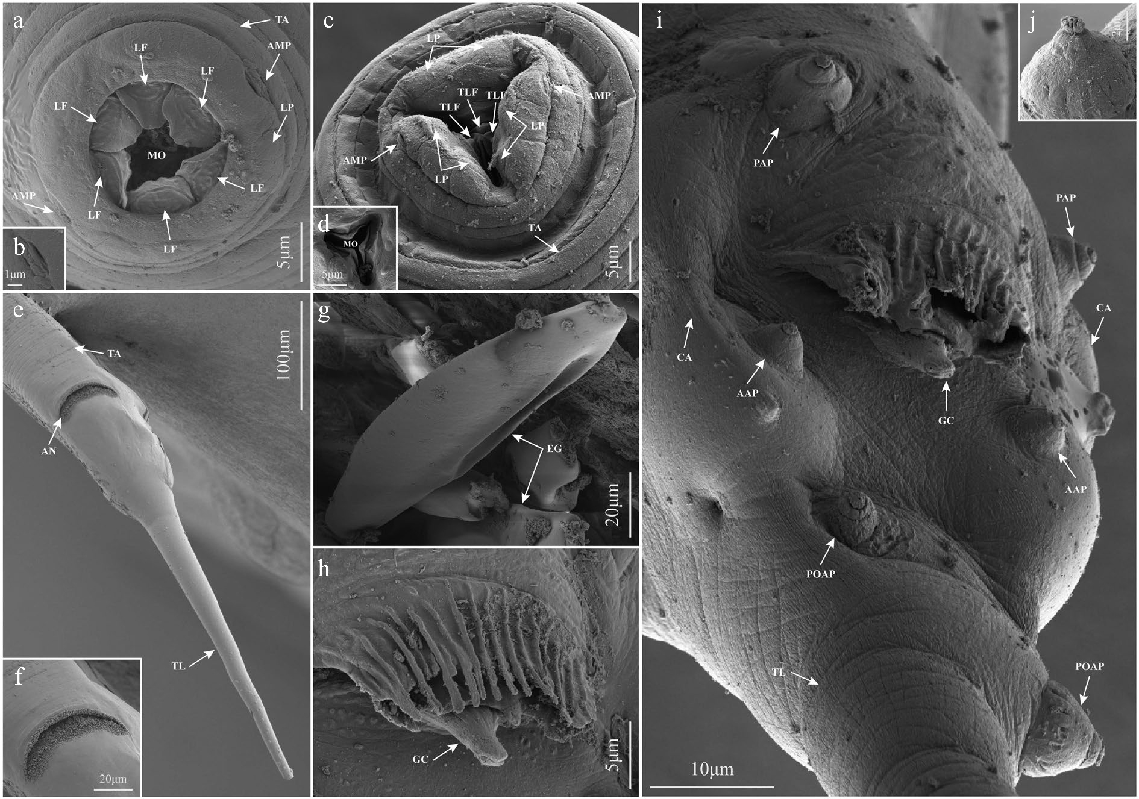
Figure 5.
Scanning electron micrographs of Spauligodon turpanoeremius sp. nov. isolated from Eremias roborowskii. (a), (b), (h)–(j) Male. (c)–(g) Female. (a) Cephalic end of the male; (b) amphid at the cephalic end; (c) cephalic end of the female; (d) three equal triangular labial flaps and the mouth opening at the cephalic end; (e) smooth caudal region in the female; (f) anus at the caudal region; (g) egg morphology in the female; (h) complex membranous curtain and genital cone in the male anal region; (i) ventral view of the caudal extremity of the male; (j) caudal papillaes. AAP, adanal papillae; AMP, amphid; AN, anus; CA, caudal alae; EG, eggs; GC, genital cone; LF, labial flaps; LP, lips; MO, mouth opening; PAP, preanal papillae; POAP, postanal papillae; TA, transverse annulations; TL, tail; TLF, triangular labial flaps.
-
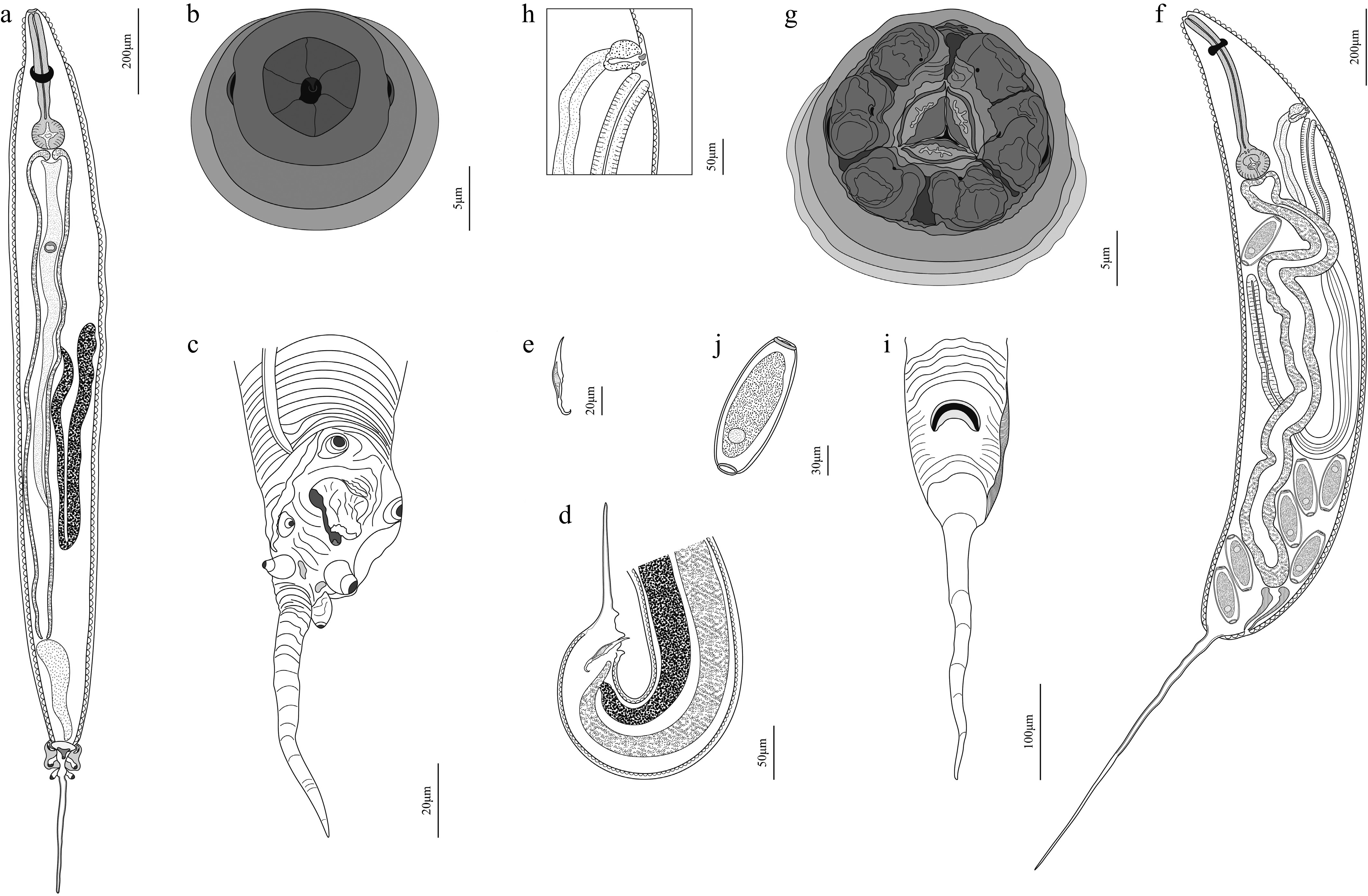
Figure 6.
Spauligodon turpanoscincus sp. nov. (a)–(e) Male. (f)–(j) Female: (a) male, entire, ventral view; (b) male, en-face view; (c) male, caudal extremity, lateral view; (d) male, posterior end with spicule, lateral view; (e) spicule. (f) female, gravid, entire, ventral view; (g) female, en-face view; (h) female, excretory pore and vulva region; (i) female, posterior end with lateral alae, ventral view; (j) egg.
-

Figure 7.
Digital micrographs of Spauligodon turpanoscincus sp. nov. isolated from Teratoscincus roborowskii. (a)–(c), (f), (h), (j) Female. (d), (e), (g), (i), (k), (l), (m) Male: (a) female, whole mount, lateral view; (b) anus, lateral view; (c) female, gravid, whole mount, lateral view; (d) male, whole mount, ventral view; (e) transverse striation of cuticle; (f) female, cephalic end, ventral view; (g) male, cephalic end, ventral view; (h) female, excretory pore and vulva, lateral view; (i) male, esophageal bulb, intestine and excretory pore, ventral veiw; (j) eggs; (k) male, caudal morphology, caudal alae and papillaes, ventral view; (l) highlighting the genital cone, lateral view; (m) male, posterior end, lateral view. AAP, adanal papillae; AN, anus; CA, caudal alae; EB, esophageal bulb; EG, eggs; ES, esophagus; EX, excretory pore; GC, genital cone; IN, intestine; LA, lateral alae; LP, lips; NR, nerve ring; PAP, preanal papillae; POAP, postanal papillae; SP, spicule; TA, transverse annulations; TL, tail; VU, vulva.
-
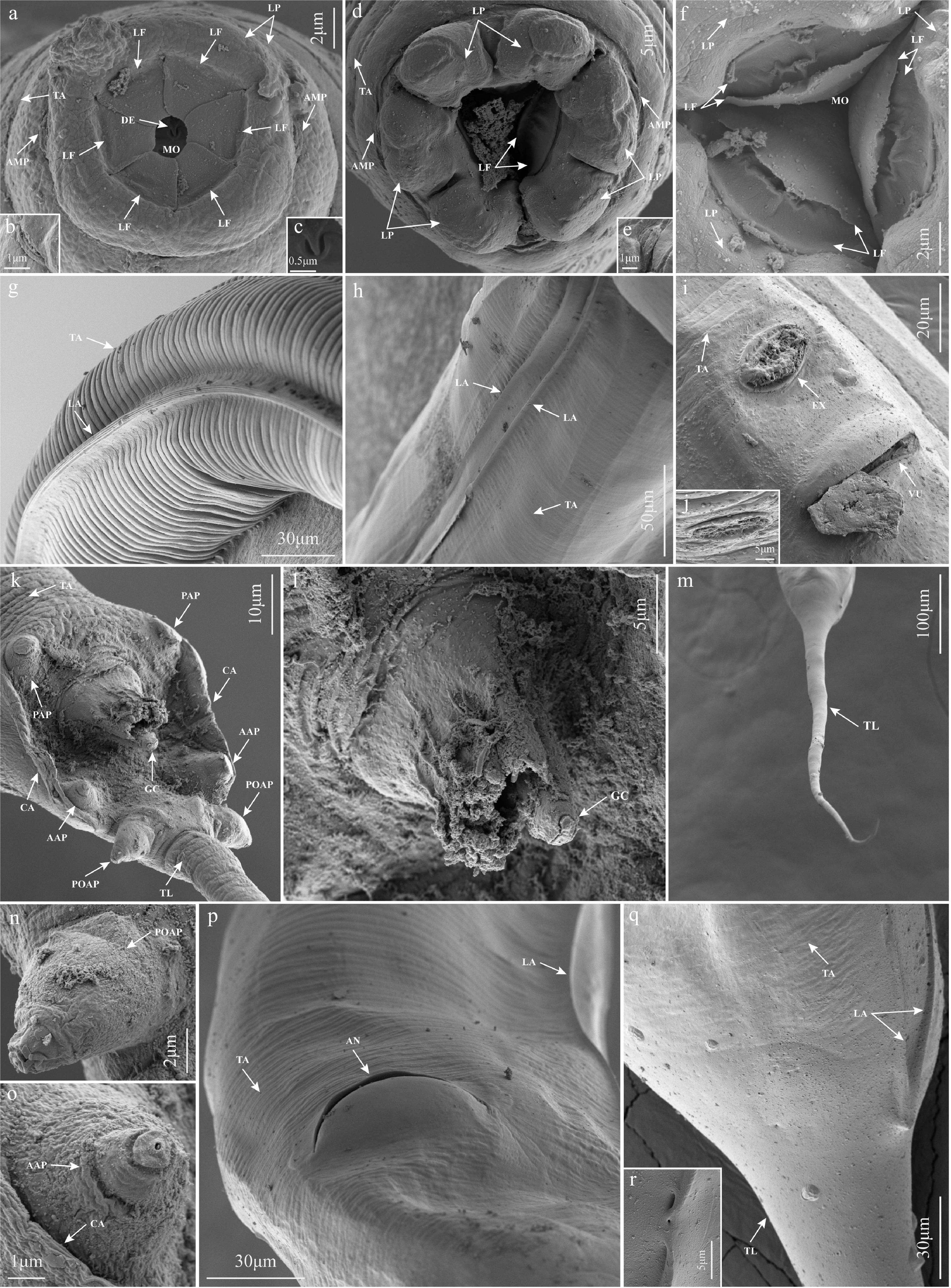
Figure 8.
Scanning electron micrographs of Spauligodon turpanoscincus sp. nov. isolated from Teratoscincus roborowskii. (a)–(c), (g), (k), (l), (n), (o) Male. (d)–(f), (h)–(j), (m), (p)–(r) Female: (a) cephalic end of the male; (b) amphid at the cephalic end; (c) denticle at the cephalic end in the male; (d) cephalic end of the female; (e) amphid at the cephalic end in the female; (f) three well-developed bilobed labial flaps and the mouth opening at the cephalic end in the female; (g) lateral view of the midbody of the male; (h) lateral view of the midbody of the female; (i) ventral view of the excretory pore and vulva in the anterior region of the female; (j) excretory pore in the female; (k) ventral view of the caudal extremity of the male; (l) complex membranous curtain and genital cone in the male anal region; (m) caudal filament of the female; (n) postanal papillae; (o) adanal papillae; (p) ventral view of the anus in the caudal region of the female; (q) smooth caudal region in the female; (r) termination point of the lateral alae and cuticular pit in the caudal region of the female. AAP, adanal papillae; AMP, amphid; AN, anus; CA, caudal alae; DE, denticle; EX, excretory pore; GC, genital cone; LA, lateral alae; LF, labial flaps; LP, lips; MO, mouth opening; PAP, preanal papillae; POAP, postanal papillae; TA, transverse annulations; TL, tail; VU, vulva.
-
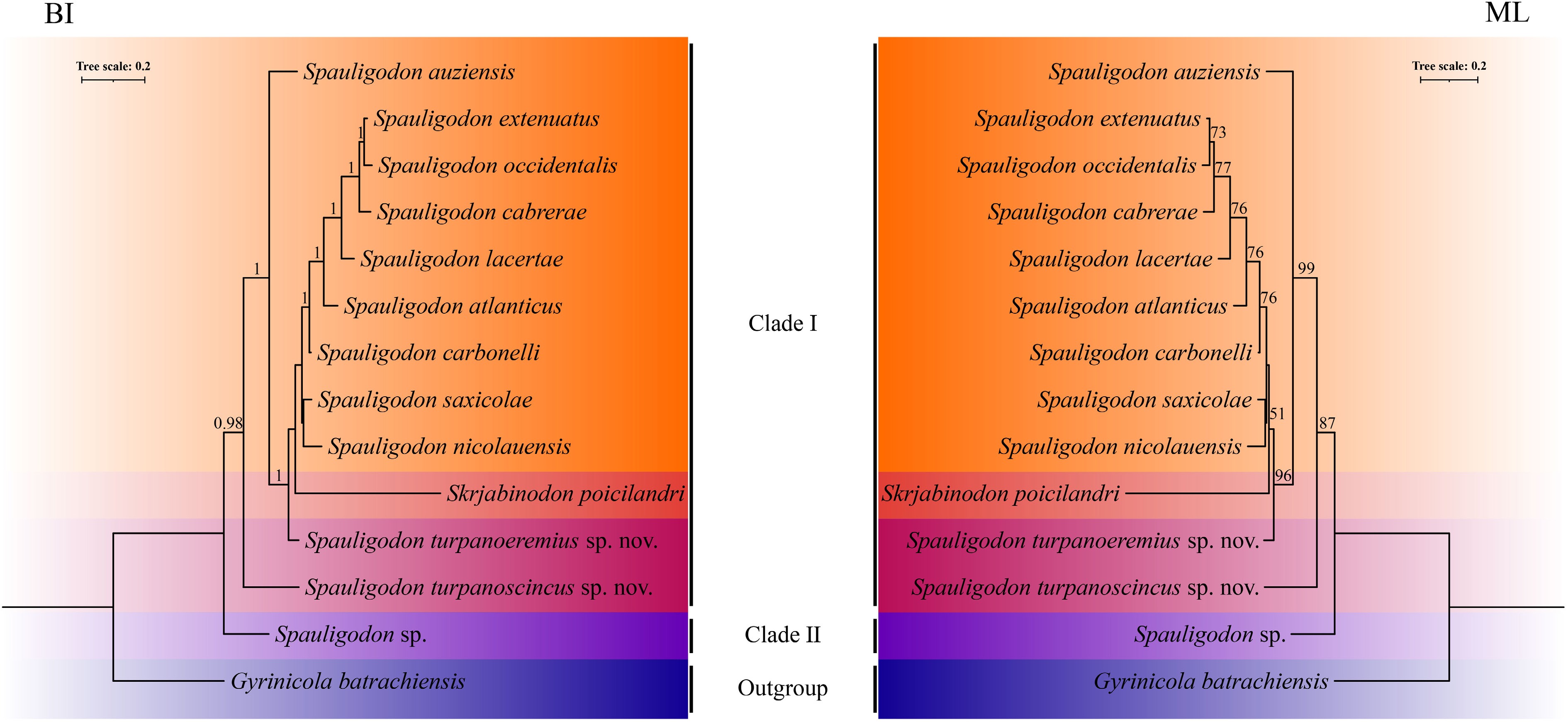
Figure 9.
Bayesian inference (BI) and maximum likelihood (ML) inference based on the concatenated (18S + 28S + COI) sequence data shows the phylogenetic relationships among the representatives of Pharyngodonidae, Spauligodon turpanoeremiussp. nov. and S. turpanoscincussp. nov.
-
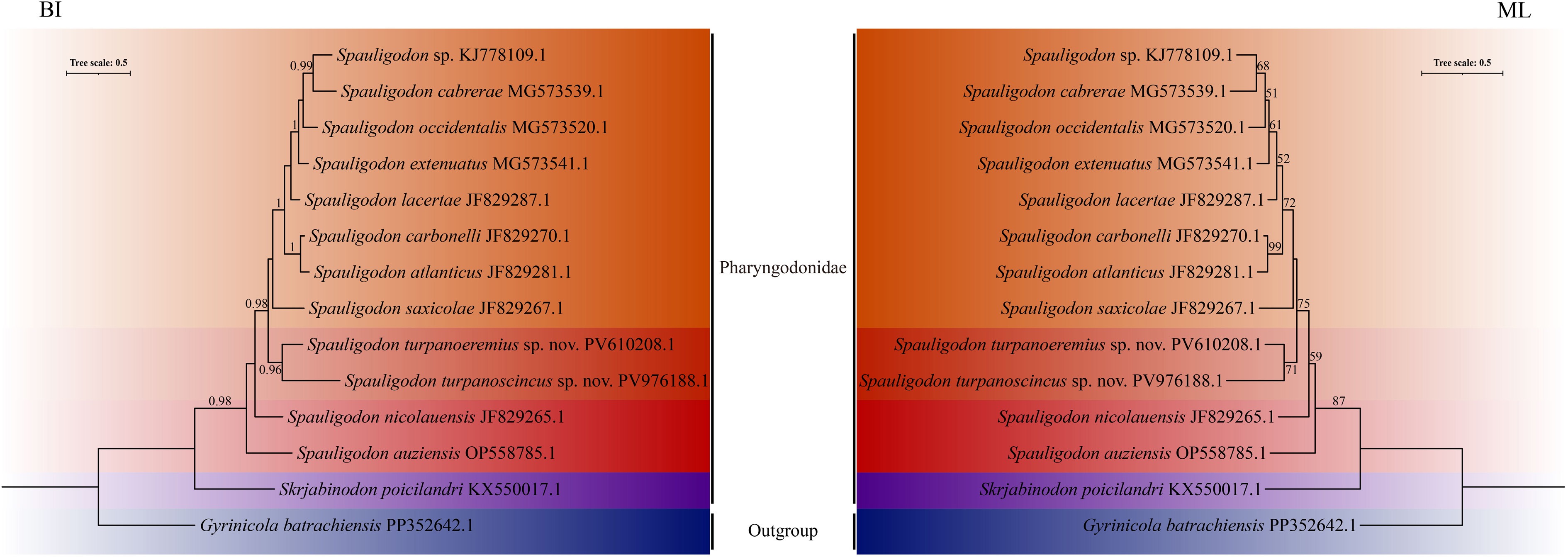
Figure 10.
Phylogenetic relationships between Spauligodon turpanoeremius sp. nov. and S. turpanoscincus sp. nov. inferred from COI sequences using Bayesian inference (BI) and maximum likelihood (ML) methods.
-
Table 1.
Detailed information on primers used for amplifying different target regions by PCR in the present study.
-
1) Double lateral alae in males Spauligodon sharpiloi 1.1) Single lateral alae in males 2 2) Tail filament with 1−5 spines in males Spauligodon carbonelli 2.1) Caudal filament in males lack spines 3 3) Mouth opening with a cephalic papillae on each of six labial flaps, two symmetrically arranged amphids present lateral to the lips in males Spauligodon auziensis 3.1) Mouth opening without cephalic papillaes on each of six labial flaps, two symmetrically arranged amphids present lateral to the lips in males 4 4) Mouth opening with a pair of setiform erect cephalic papillae on each of three labial flaps, two symmetrically arranged amphids present lateral to the lips in females; a pair of short-stem papillae at the base of tail filament in males Spauligodon extenuatus 4.1) Mouth opening without cephalic papillaes on each of three labial flaps, two symmetrically arranged amphids present lateral to the lips in females; a pair of prominent and pedunculated papillae at the base of tail filament in males 5 5) Cephalic end with three slightly bilobed lips in females 6 5.1) Cephalic end with three conspicuously bilobed lips in females 7 6) Vulva situated in the anterior of the esophageal bulb just behind the excretory pore in females Spauligodon atlanticus 6.1) Vulva situated in the posterior of the esophageal bulb just behind the excretory pore in females Spauligodon nicolauensis 7) Males approximately half the length of females; lateral alae reaching maximum width on the anterior of the anus; a pair of pedunculated papillae present the anterior to the anus, postanal papillae larger than the first pair Spauligodon occidentalis 7.1) Males approximately one-third the length of females; lateral alae terminating the anterior to the anus; a pair of short-stem papillae present the anterior to the anus, postanal papillae larger than the first pair 8 8) Parallel double lateral alae in females, broad caudal alae in males Spauligodon turpanoscincus 8.1) Absence of lateral alae in females; inconspicuous caudal alae in males 9 9) Tail filament with 5−9 spines in females; eggs oval Spauligodon cabrerae 9.1) Caudal filament in females lack spines; eggs barrel-shaped 10 10) Presence of genital cone and single spicule in males Spauligodon turpanoeremius 10.1) Presence of genital cone but absence of spicule in males 11 11) Parasites primarily inhabiting the large intestine Spauligodon saxicolae 11.1) Parasites primarily inhabiting the small intestine Spauligodon lacerate -
1 2 3 4 5 6 7 8 9 10 11 12 13 14 1. S. turpanoeremius sp. nov. 13.75 16.15 21.24 17.81 13.05 15.30 14.38 15.04 16.44 15.04 15.27 26.83 31.42 2. S. turpanoscincus sp. nov. 8.46 15.96 19.51 17.62 13.53 16.02 16.41 16.63 16.02 17.74 15.08 27.91 30.60 3. S. atlanticus 7.50 8.54 18.86 15.88 5.95 12.04 12.38 19.61 13.79 15.43 12.38 29.00 30.39 4. S. auziensis 9.34 9.92 9.35 21.36 18.17 19.15 18.17 19.03 20.23 21.11 17.82 30.89 29.93 5. S. cabrerae 7.85 9.30 3.78 9.58 14.49 13.96 13.96 18.15 14.66 17.98 14.14 29.27 31.76 6. S. carbonelli 6.31 7.67 2.75 8.46 4.69 12.91 10.61 18.49 13.61 15.27 11.74 29.27 30.71 7. S. extenuatus 7.09 8.89 3.37 9.64 3.25 4.22 8.73 17.80 10.82 15.88 12.22 29.81 30.37 8. S. lacertae 7.03 9.01 3.25 9.40 3.08 3.93 1.92 17.36 10.65 15.11 12.38 28.46 28.78 9. S. nicolauensis 7.81 8.59 8.27 8.75 8.22 7.54 7.86 7.86 19.37 18.81 17.20 26.56 30.23 10. S. occidentalis 7.32 9.01 3.31 9.63 3.60 4.51 2.56 2.61 7.92 17.10 12.39 28.46 30.19 11. S. saxicolae 5.84 8.43 5.39 8.75 5.98 4.98 5.51 5.22 7.07 5.92 17.36 28.46 29.42 12. Spauligodon sp. 7.75 8.75 7.42 5.61 7.48 6.82 7.07 7.25 8.16 7.07 7.05 27.64 29.26 13. Skrjabinodon poicilandri 16.81 17.77 18.05 18.33 18.05 17.32 18.35 17.99 17.45 18.04 16.97 17.75 34.96 14. Gyrinicola batrachiensis 20.01 20.70 20.30 20.53 20.60 19.83 20.48 19.88 19.70 20.77 19.28 20.24 21.79 Table 2.
Uncorrected p-distance (%) of Spauligodon turpanoeremius sp. nov. and S. turpanoscincus sp. nov. sequences (COI above diagonal; concatenated sequences below diagonal).
Figures
(10)
Tables
(3)