-
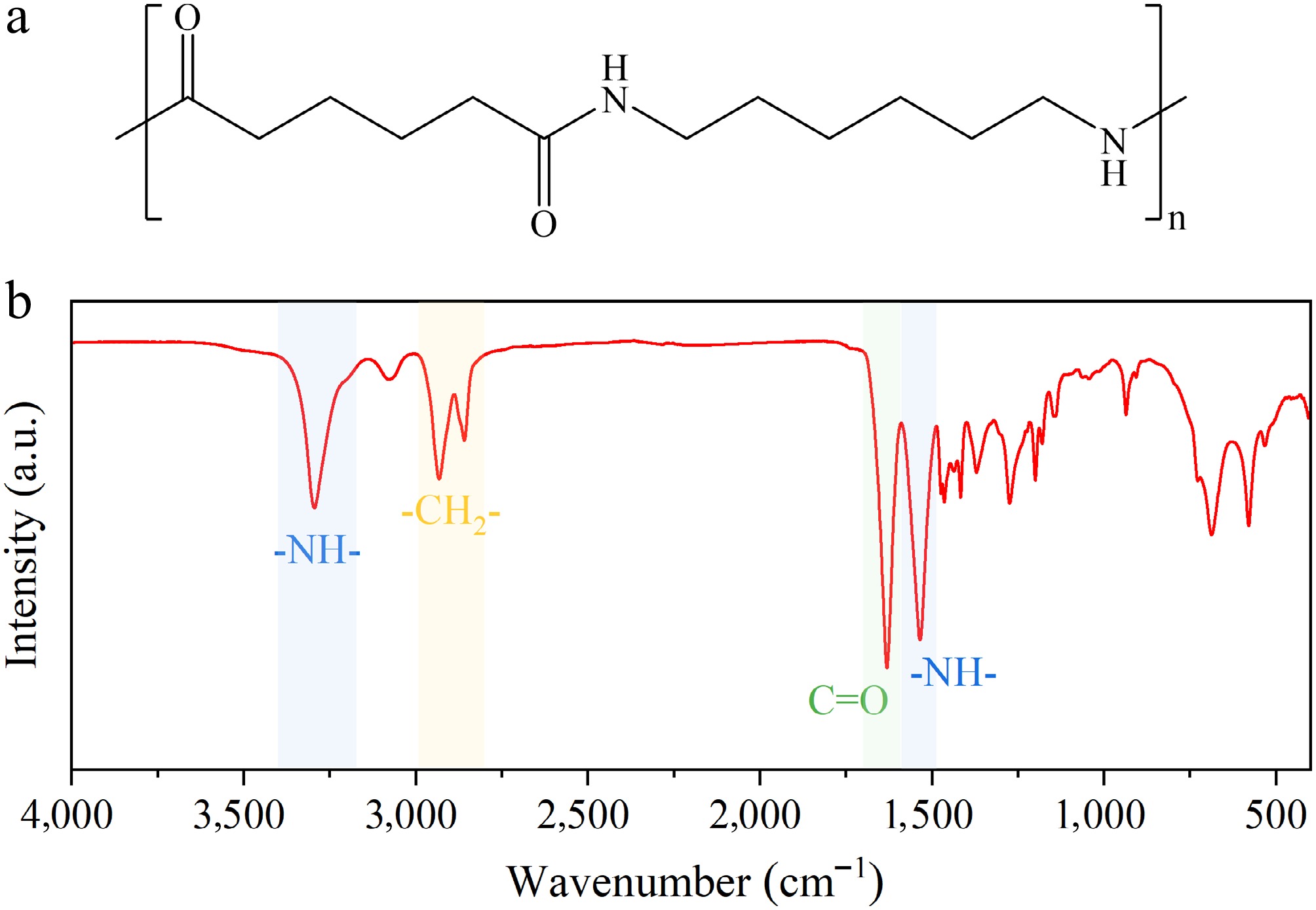
Figure 1.
(a) Chemical structure. (b) Infrared spectrum of nylon 6/6.
-
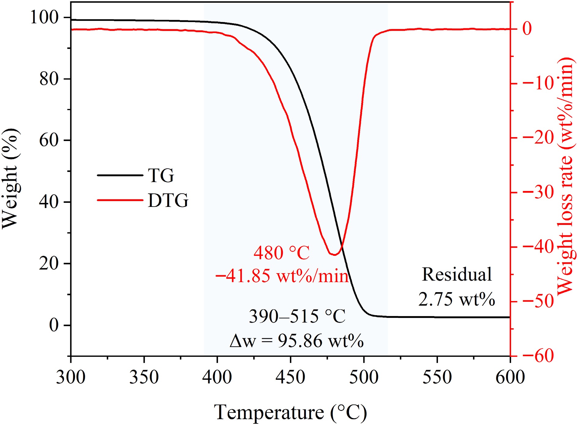
Figure 2.
TG and DTG curves of nylon 6/6.
-
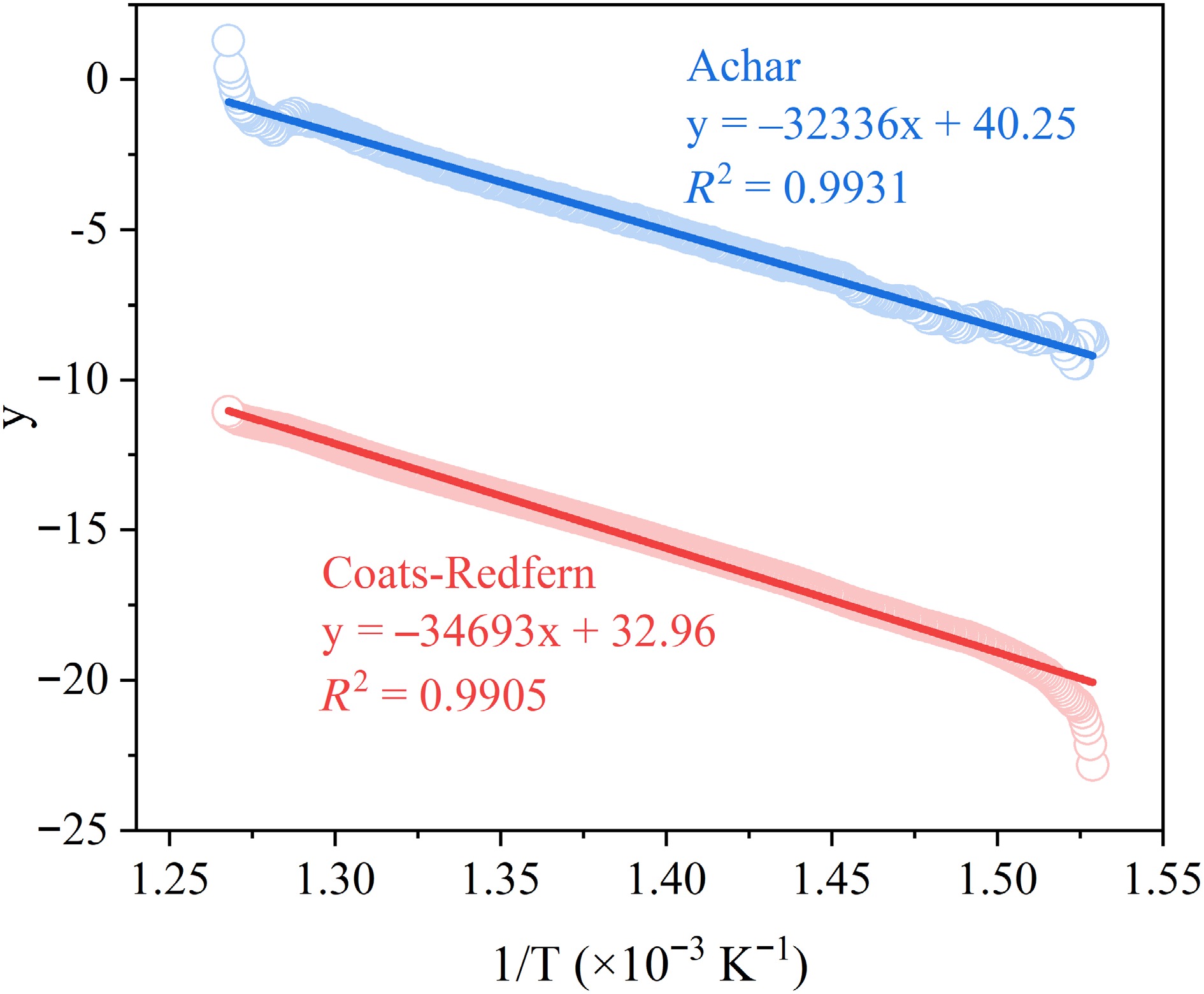
Figure 3.
Linear fitting curves using the F1 kinetic model for nylon 6/6 pyrolysis.
-
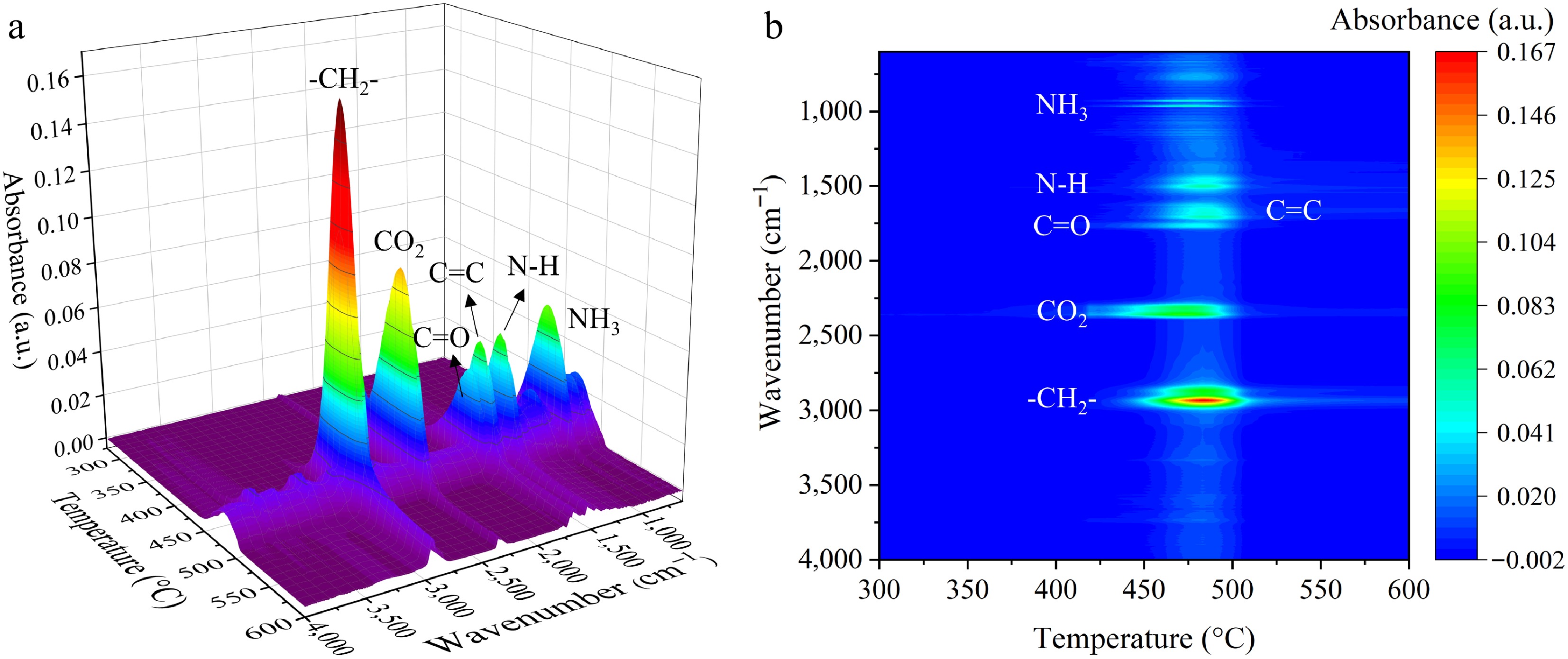
Figure 4.
(a) 3D FTIR spectrum. (b) Corresponding 2D projection of nylon 6/6 pyrolysis volatiles.
-
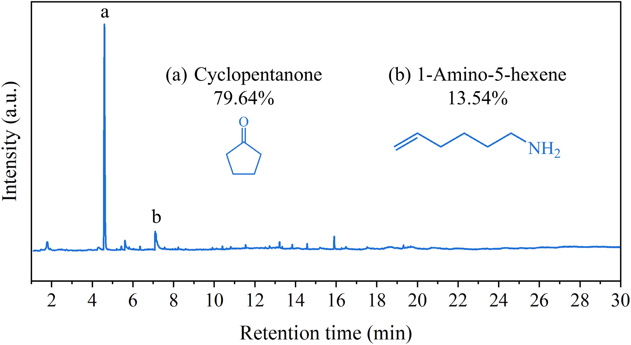
Figure 5.
GC/MS analysis results of nylon 6/6 pyrolysis volatiles.
-
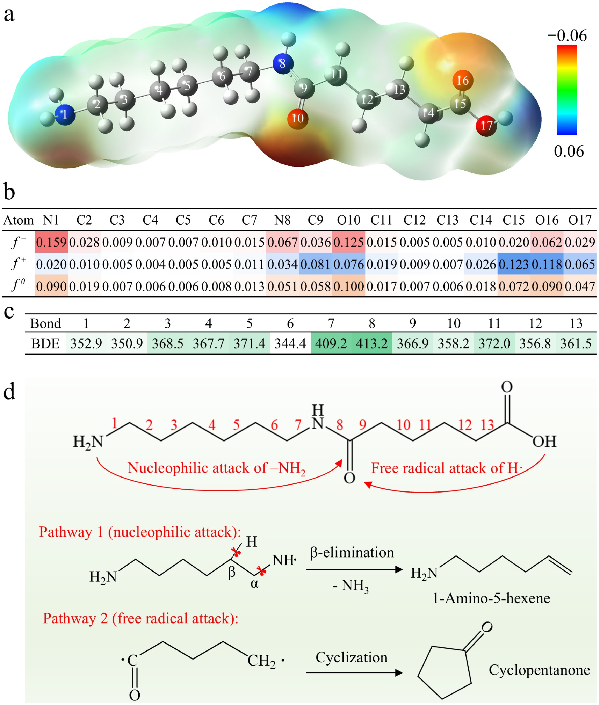
Figure 6.
(a) Electrostatic potential (ESP)-mapped surface of nylon 6/6 model molecule. (b) Fukui functions of key atom sites. (c) Bond dissociation energies (BDEs) of backbone chemical bonds. (d) Two possible decomposition pathways of the nylon 6/6 model molecule.
-
Model
codeCoats-Redfern Achar Ea (kJ/mol) lnA (min−1) R2 Ea (kJ/mol) lnA (min−1) R2 F1 288 46.4 0.9905 268 43.2 0.9931 F2 396 65.4 0.9296 428 71.0 0.8789 F3 529 89.0 0.8195 587 98.7 0.7832 F4 672 114.0 0.7428 746 126.5 0.7252 F5 810 139.8 0.6932 905 154.2 0.6877 D1 485 76.9 0.9480 358 55.2 0.7733 D2 508 80.2 0.9612 410 63.6 0.8744 D3 543 85.0 0.9784 493 76.6 0.9752 D4 519 80.7 0.9672 440 67.4 0.9242 A3/2 188 30.0 0.9890 169 26.8 0.9755 A2 138 21.6 0.9884 110 18.5 0.9401 A3 88 13.2 0.9872 69 10.1 0.8068 A4 63 8.8 0.9859 44 5.8 0.6011 R1 237 37.1 0.9453 110 15.5 0.3624 R2 257 40.1 0.9692 189 28.7 0.8738 R3 266 41.3 0.9773 216 32.9 0.9519 P2 112 16.9 0.9391 -14 -4.7 0.0133 P3 71 9.9 0.9320 -56 -11.6 0.1840 P4 50 6.3 0.9236 -77 -15.2 0.3089 Table 1.
Kinetic analysis results using different mechanism functions.
Figures
(6)
Tables
(1)