-

Figure 1.
Maximum likelihood tree from the concatenated nrSSU, EF-1α, and mtSSU sequences, showing phylogeny of Stemonitidales. Only nodes with UBS/PP/TBE ≥ 70/70/70 are shown. The voucher numbers are indicated after the species names. Newly obtained sequences in this study are indicated in bold, new species described in this study are indicated in red, and holotype or isotype specimens are marked with red stars. The scale bar represents the number of substitutions per site.
-
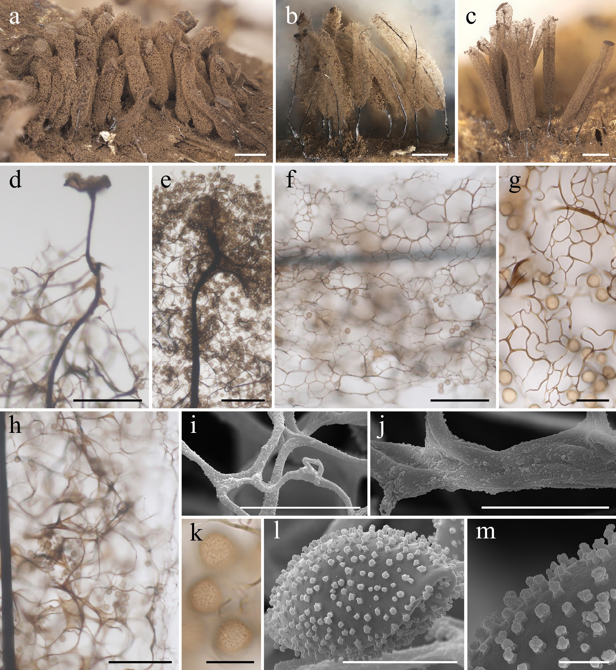
Figure 2.
Morphological characteristics of Corallosporopsis gabrieliana (a), (b), (d), (f), (h)–(j), (l), (m) LE306548; (c), (e), (g), (k). LE306833. (a)–(c) Mature sporocarps (DM). (d), (e) Apical view of the sporotheca (LM). (f), (g) Surface net (LM). (h) Capillitium in the middle part of the sporotheca (LM). (i) Detail of the surface net (SEM). (j) Detail of the capillitium threads (SEM). (k) Spores (LM). (l) Spore (SEM). (m) Detail of the spore ornamentation (SEM). Scale bars: (a)–(c) = 1 mm; (d)–(f), (h) = 100 μm; (g) = 20 μm; (i)–(k) = 10 μm; (l) = 5 μm; (m) = 1 μm.
-
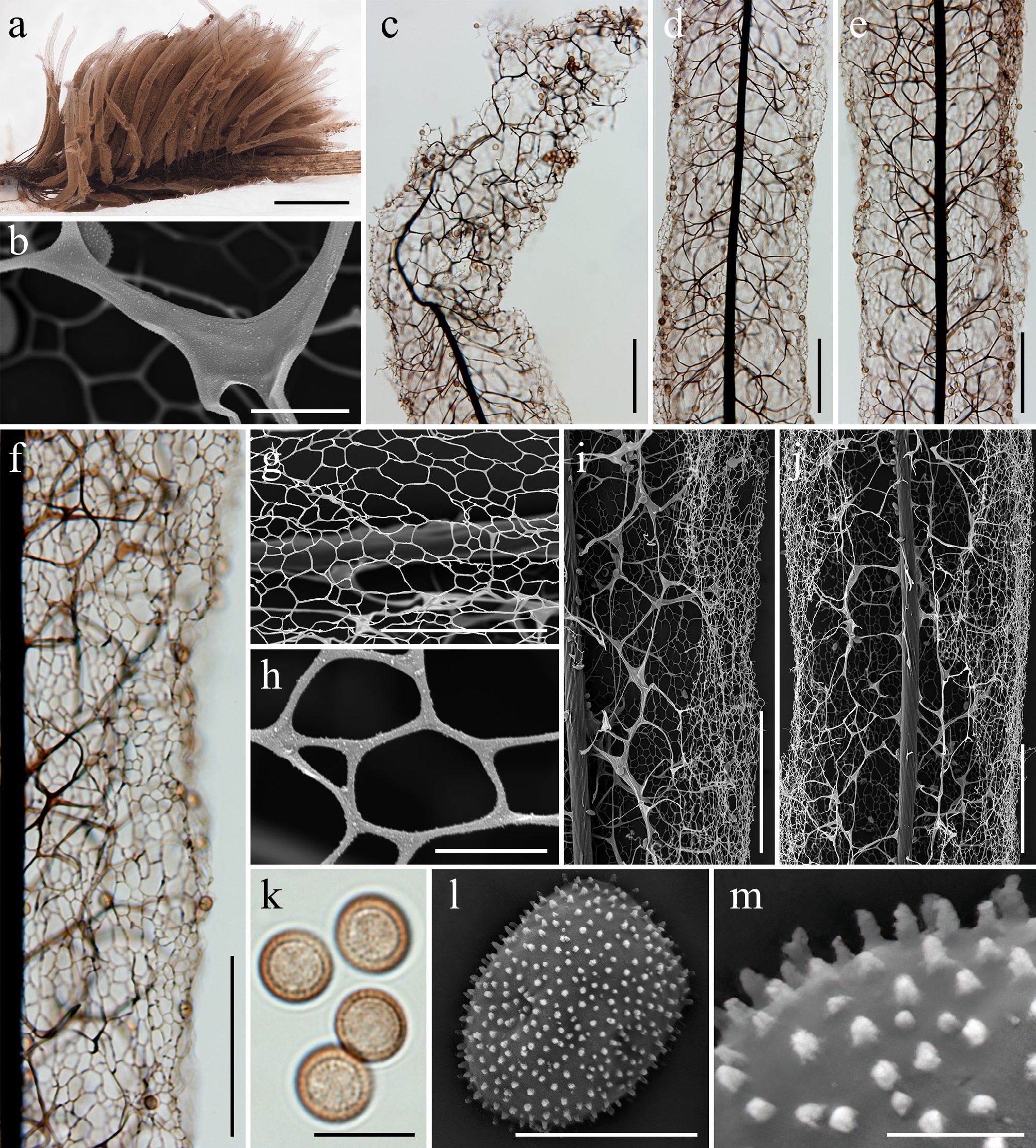
Figure 3.
Morphological characteristics of Corallosporopsis herbatica (HMAS 98461). (a) Mature sporocarps (DM). (b) Detail of the capillitium threads (SEM). (c) Apical view of the sporotheca (LM). (d), (e) Capillitium in the middle part of the sporotheca (LM). (f) Surface net (LM). (g), (h) Detail of the surface net (SEM). (i), (j) Capillitium in the middle part of the sporotheca (SEM). (k) Spores (LM). (l) Spore (SEM). (m) Detail of the spore ornamentation (SEM). Scale bars: (a) = 2 mm; (c)–(f), (i), (j) = 100 μm; (b), (g), (k) = 10 μm; (h), (l) = 5 μm; (m) = 1 μm.
-
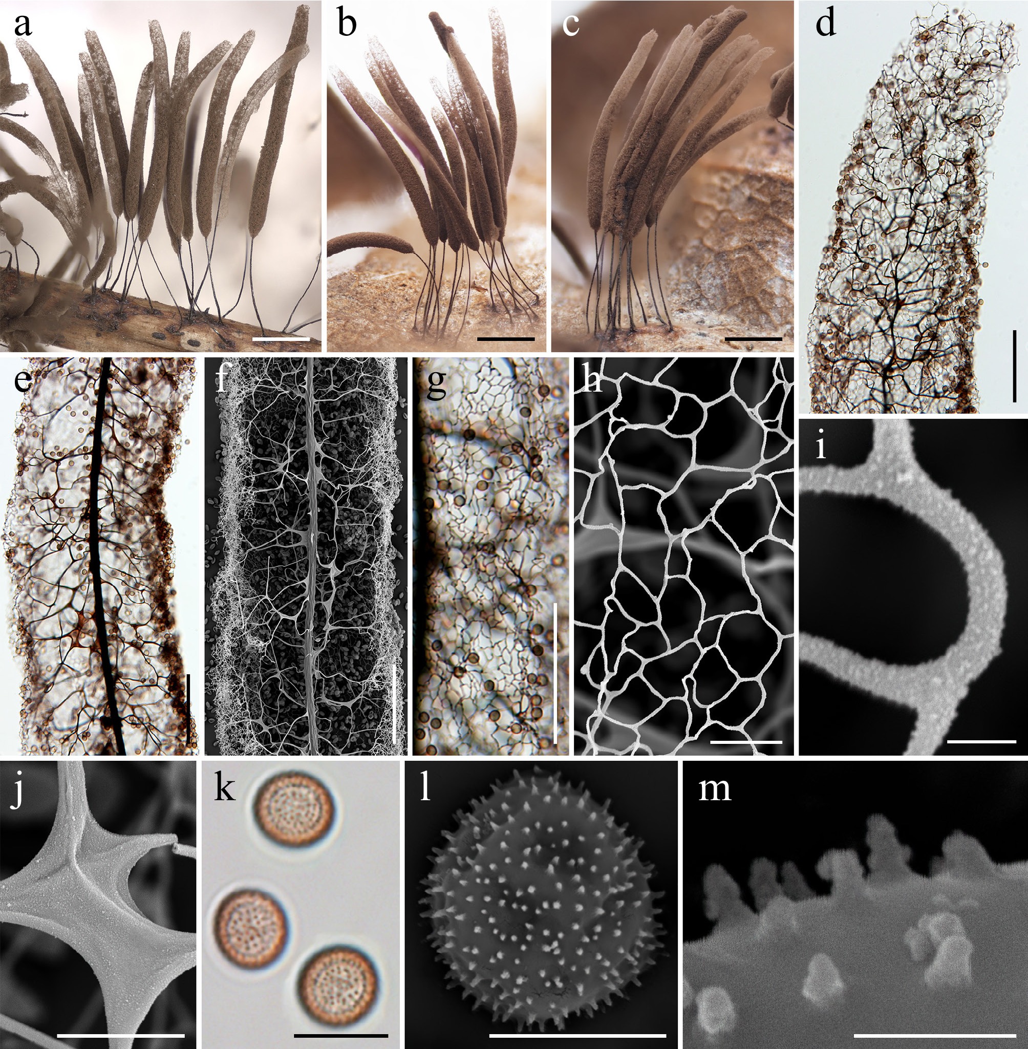
Figure 4.
Morphological characteristics of Corallosporopsis herbatica (LE348810). (a)–(c) Mature sporocarps (DM). (d) Apical view of the sporotheca (LM). (e) Capillitium in the middle part of the sporotheca (LM). (f) Capillitium in the middle part of the sporotheca (SEM). (g) Surface net (LM). (h), (i) Detail of the surface net (SEM). (j) Detail of the capillitium threads (SEM). (k) Spores (LM). (l) Spore (SEM). (m) Detail of the spore ornamentation (SEM). Scale bars: (a)–(c) = 1 mm; (d)–(g) = 100 μm; (h), (j), (k) = 10 μm; (l) = 5 μm; (i), (m) = 1 μm.
-
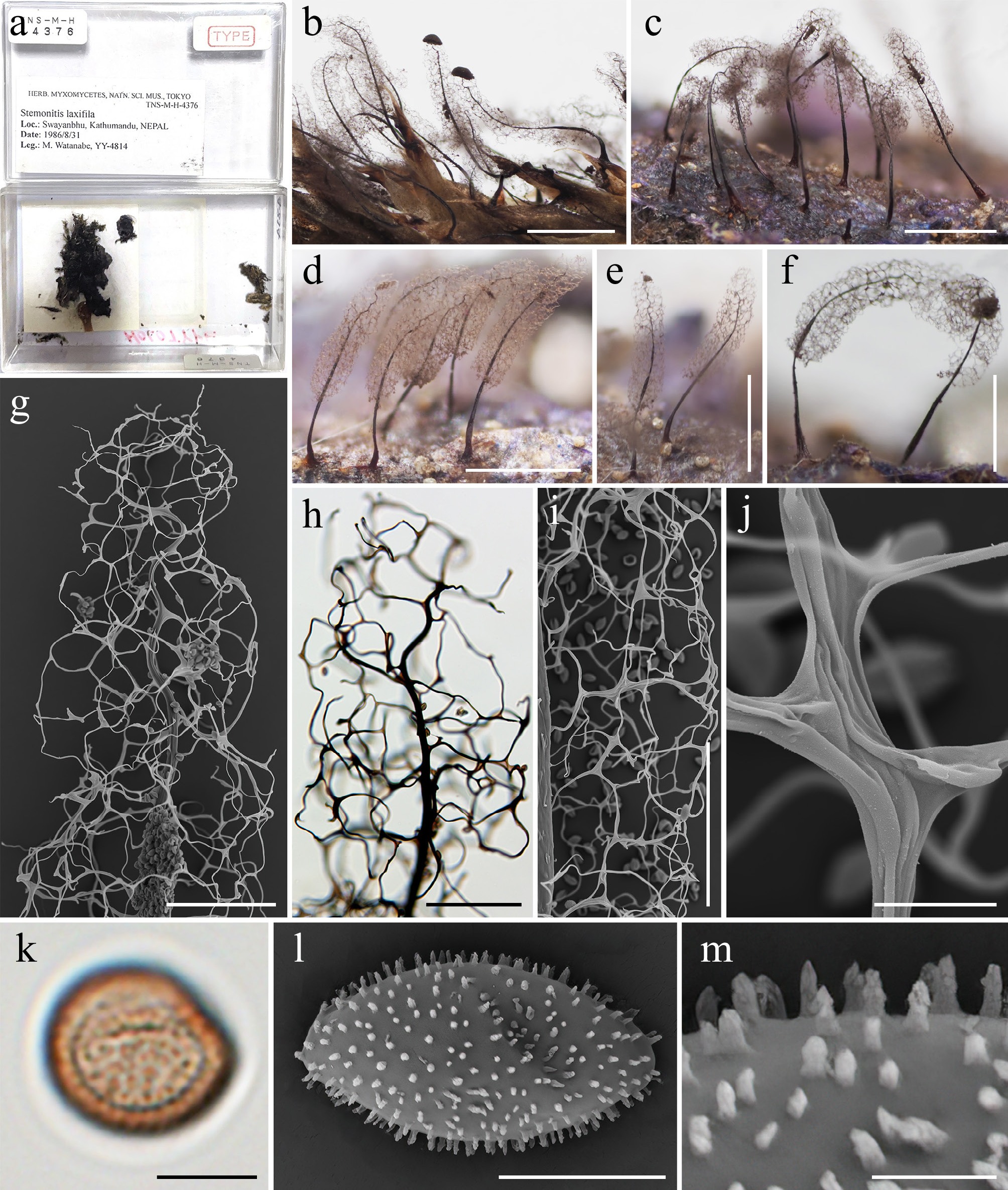
Figure 5.
Morphological characteristics of the holotype (TNS-M-H-4376 = YY-4814) of Corallosporopsis laxifila (≡ Stemonitis laxifila). (a) Herbarium labels and interior views of the herbarium box containing the holotype. (b)–(f) Mature sporocarps (DM). (g) Apical view of the sporotheca (SEM). (h) Apical view of the sporotheca (LM). (i) Capillitium in the middle part of the sporotheca (SEM). (j) Detail of the capillitium threads (SEM). (k) Spore (LM). (l) Spore (SEM). (m) Detail of the spore ornamentation (SEM). Scale bars: (b)–(f) = 1 mm; (g)–(i) = 100 μm; (j) = 10 μm; (k), (l) = 5 μm; (m) = 1 μm.
-
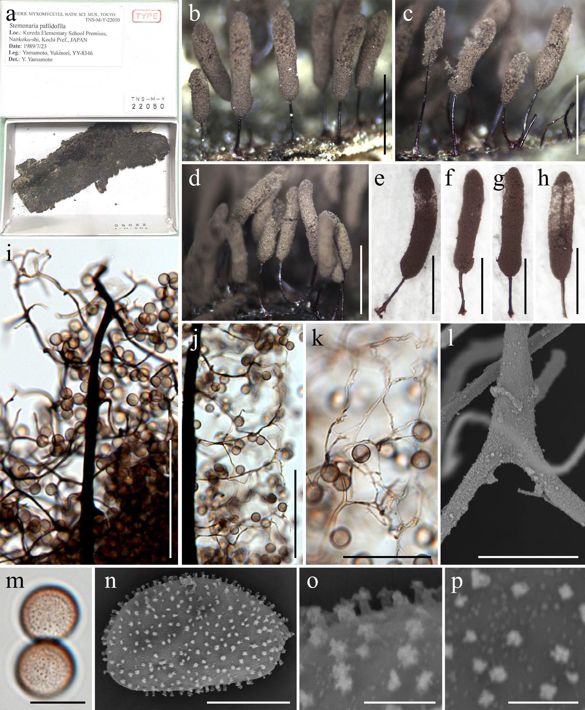
Figure 6.
Morphological characteristics of the holotype (TNS-M-Y-22050 = YY-8346) of Corallosporopsis pallidofila (≡ Stemonaria pallidofila). (a) Herbarium labels and interior views of the herbarium box containing the holotype. (b)–(h) Mature sporocarps (DM). (i) Apical view of the sporotheca (LM). (j) Capillitium in the middle part of the sporotheca (LM). (k) Surface net (LM). (l) Detail of the capillitium threads (SEM). (m) Spores (LM). (n) Spore (SEM). (o), (p) Detail of the spore ornamentation (SEM). Scale bars: (b)–(h) = 1 mm; (i), (j) = 100 μm; (k) = 50 μm; (l), (m) = 10 μm; (n) = 5 μm; (o), (p) = 1 μm.
-
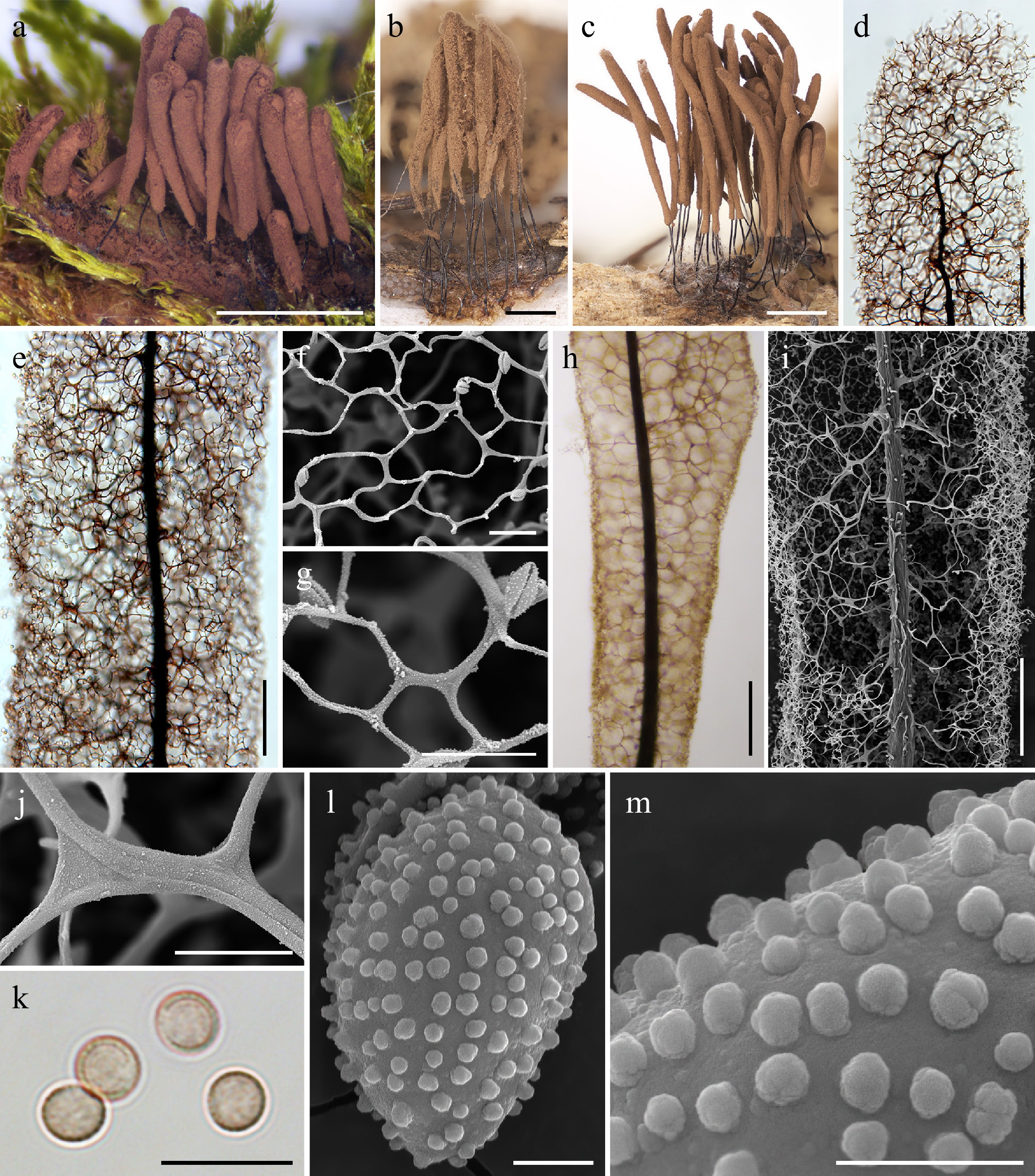
Figure 7.
Morphological characteristics of Grandiverruca cf. smithii (a), (h) MYX 20613; (b), (d)–(g), (i)–(m) LE327934; (c) LE347064. (a)–(c) Mature sporocarps (DM). (d) Apical view of the sporotheca (LM). (e) Surface net (LM). (f), (g) Detail of the surface net (SEM). (h) Capillitium in the middle part of the sporotheca (LM). (i) Capillitium in the middle part of the sporotheca (SEM). (j) Detail of the capillitium threads (SEM). (k) Spores (LM). (l) Spore (SEM). (m) Detail of the spore ornamentation (SEM). Scale bars: (a) = 2 mm; (b), (c) = 1 mm; (d), (e), (h), (i) = 100 μm; (f), (g), (j), (k) = 10 μm; (l), (m) = 1 μm.
-
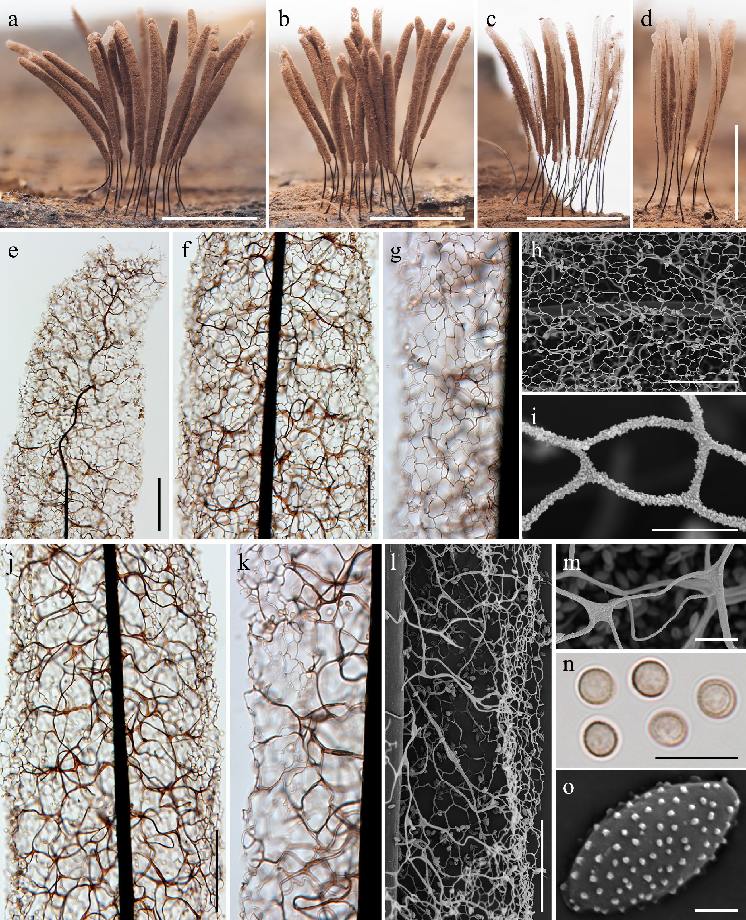
Figure 8.
Morphological characteristics of Grandiverruca verrucosifila (a), (b), (e), (m), (o) HFNNU 9746; (c), (d), (f), (j), (n) HFNNU 4477; (g)–(i), (k), (l) HFNNU 10346. (a)–(d) Mature sporocarps (DM). (e) Apical region of the sporocarp (LM). (f), (g) Surface net (LM). (h), (i) Detail of the surface net (SEM). (j), (k) Capillitium in the middle part of the sporotheca (LM). (l) Capillitium in the middle part of the sporotheca (SEM). (m) Detail of the capillitium threads (SEM). (n) Spores (LM). (o) Spore (SEM). Scale bars: (a)–(d) = 2 mm; (e), (f), (j) = 100 μm; (g), (h), (k), (l) = 50 μm; (m), (n) = 10 μm; (i), (o) = 5 μm.
-
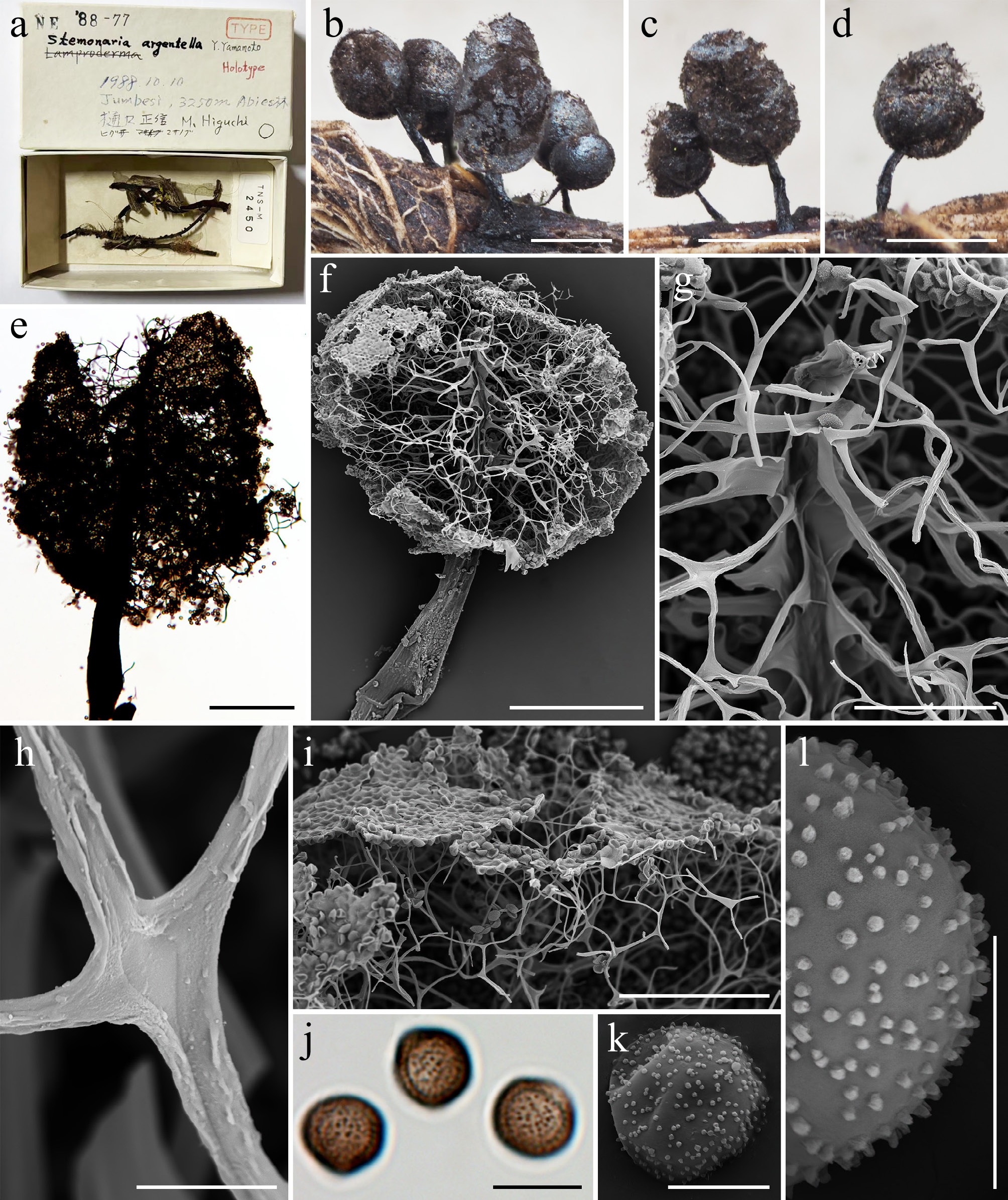
Figure 9.
Morphological characteristics of the holotype (TNS-M-Y-2450 = TNE-88-77) of Stemonaria argentella. (a) Herbarium labels and interior views of the herbarium box containing the holotype. (b)–(d) Mature sporocarps (DM). (e) Apical view of the sporotheca (LM). (f) Apical view of a sporotheca (SEM). (g) Apex of the columella (SEM). (h) Detail of the capillitium threads (SEM). (i) Connection between the capillitium and the peridium (SEM). (j) Spores (LM). (k) Spore (SEM). (l) Detail of the spore ornamentation (SEM). Scale bars: (b)–(d) = 1 mm; (e), (f) = 200 μm; (i) = 100 μm; (g) = 50 μm; (h), (j) = 10 μm; (k), (l) = 5 μm.
-
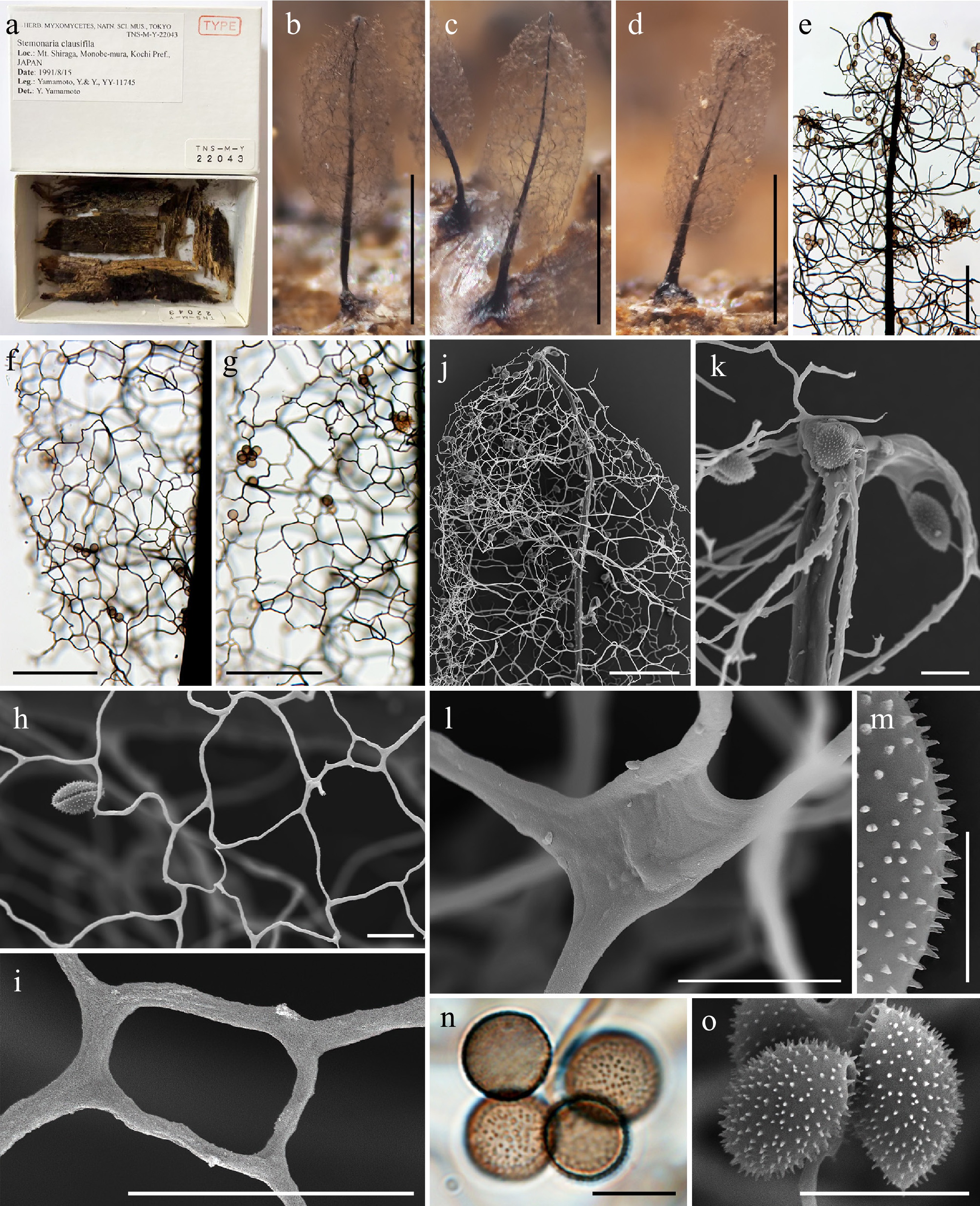
Figure 10.
Morphological characteristics of the holotype (TNS-M-Y-22043 = YY-11745) of Stemonaria clausifila. (a) Herbarium labels and interior views of the herbarium box containing the holotype. (b)–(d) Mature sporocarps (DM). (e) Apical view of the sporotheca (LM). (f), (g) Surface net (LM). (h), (i) Detail of the surface net (SEM). (j) Apical view of the sporotheca (SEM). (k) Apical view of the columella (SEM). (l) Detail of the capillitium threads (SEM). (m) Detail of the spore ornamentation (SEM). (n) Spores (LM). (o) Spores (SEM). Scale bars: (b)–(d) = 1 mm; (e)–(g), (j) = 100 μm; (h), (i), (k), (l), (n), (o) = 10 μm; (m) = 5 μm.
-
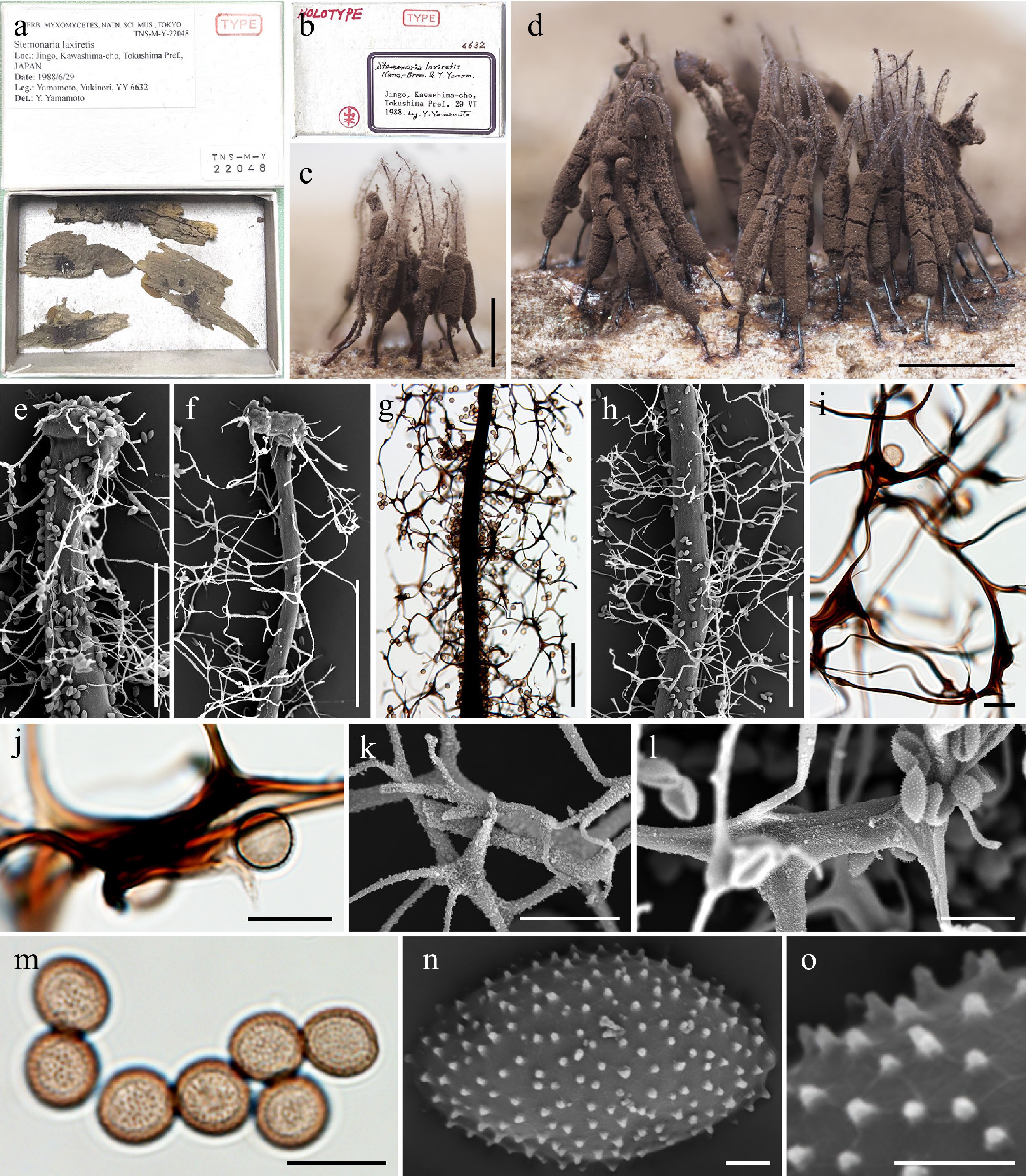
Figure 11.
Morphological characteristics of the holotype (TNS-M-Y-22048 = YY-6632) of Stemonaria laxiretis. (a) Herbarium labels and interior views of the herbarium box containing the holotype. (b) Herbarium labels of the herbarium box containing the holotype. (c), (d) Mature sporocarps (DM). (e), (f) Apical view of the columella (SEM). (g) Capillitium in the middle part of the sporotheca (LM). (h) Capillitium in the middle part of the sporotheca (SEM). (i), (j) Detail of the capillitium threads (LM). (k), (l) Detail of the capillitium threads (SEM). (m) Spores (LM). (n) Spore (SEM). (o) Detail of the spore ornamentation (SEM). Scale bars: (c), (d) = 2 mm; (e)–(h) = 100 μm; (i)–(m) = 10 μm; (n), (o) = 1 μm.
-
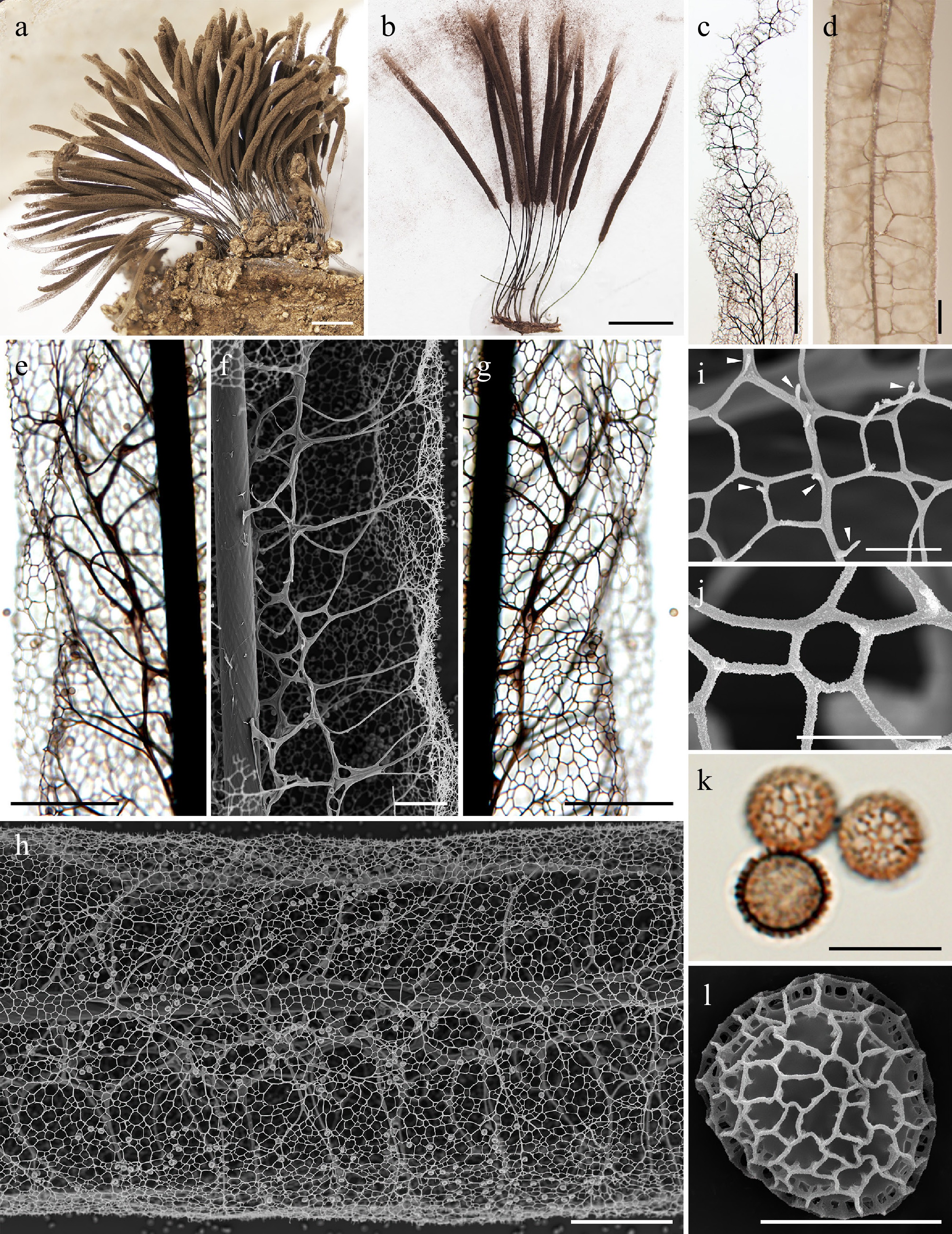
Figure 12.
Morphological characteristics of Stemonitis castillensis (a), (d), (j), (l) LE286562; (b), (c), (e)–(i), (k) LE286564. (a), (b) Mature sporocarps (DM). (c) Apical view of the sporotheca (LM). (d), (e) Capillitium in the middle part of the sporotheca (LM). (f) Capillitium in the middle part of the sporotheca (SEM). (g) Surface net (LM). (h) Surface net (SEM). (i) Spines on the surface net (SEM, arrows). (j) Detail of the surface net (SEM). (k) Spores (LM). (l) Spore (SEM). Scale bars: (a), (b) = 2 mm; (c) = 200 μm; (d), (e), (g), (h) = 100 μm; (f) = 50 μm; (i)–(k) = 10 μm; (l) = 5 μm.
-
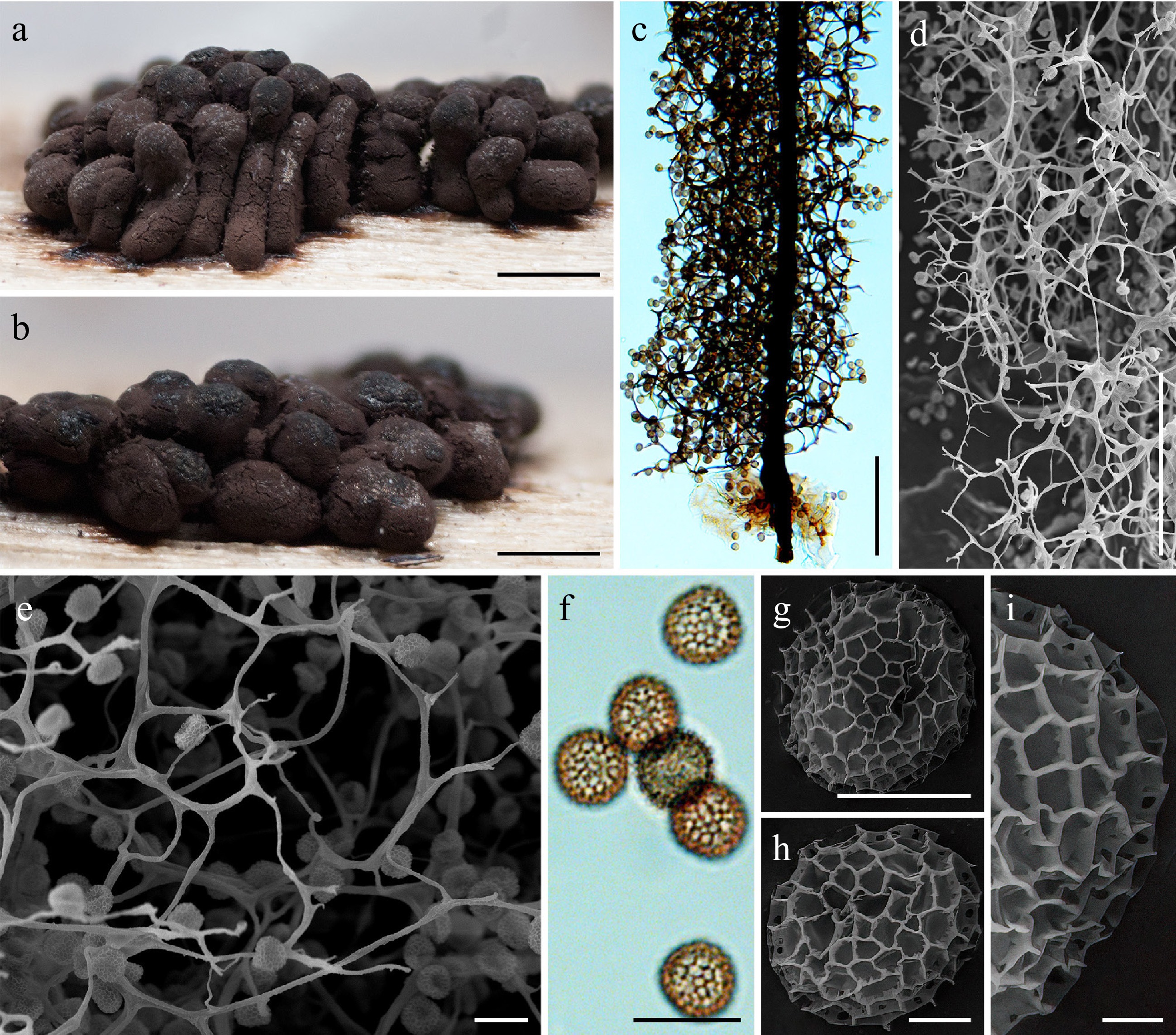
Figure 13.
Morphological characteristics of Stemonitis conferta (HFNNU 10867). (a), (b) Mature sporocarps (DM). (c) Capillitium and stalk of the sporocarp (LM). (d), (e) Edge of the sporotheca (SEM). (f) Spores (LM). (g), (h) Spore (SEM). (i) Detail of the spore ornamentation (SEM). Scale bars: (a), (b) = 1 mm; (c)–(e) = 100 μm; (f) = 10 μm; (g) = 5 μm; (h) = 2 μm; (i) = 1 μm.
-
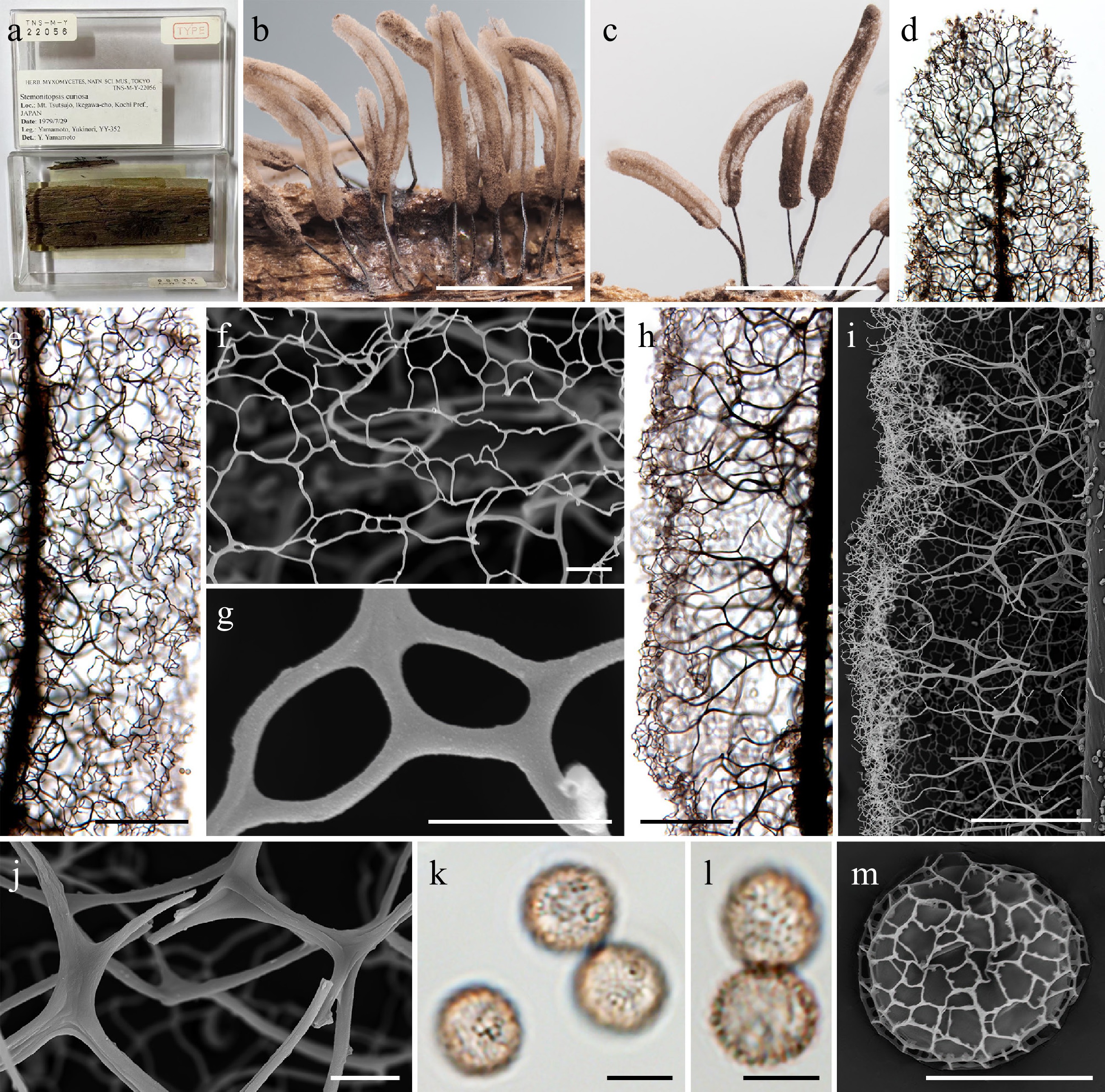
Figure 14.
Morphological characteristics of the holotype (TNS-M-Y-22056 = YY-352) of Stemonitis curiosa (≡ Stemonitopsis curiosa). (a) Herbarium labels and interior views of the herbarium box containing the holotype. (b), (c) Mature sporocarps (DM). (d) Apical view of the sporotheca (LM). (e) Surface net (LM). (f), (g) Detail of the surface net (SEM). (h) Capillitium in the middle part of the sporotheca (LM). (i) Capillitium in the middle part of the sporotheca (SEM). (j) Detail of the capillitium threads (SEM). (k), (l) Spores (LM). (m) Spore (SEM). Scale bars: (b), (c) = 2 mm; (d), (e), (h), (i) = 100 μm; (f), (j), (k) = 10 μm; (g), (k)–(m) = 5 μm.
-
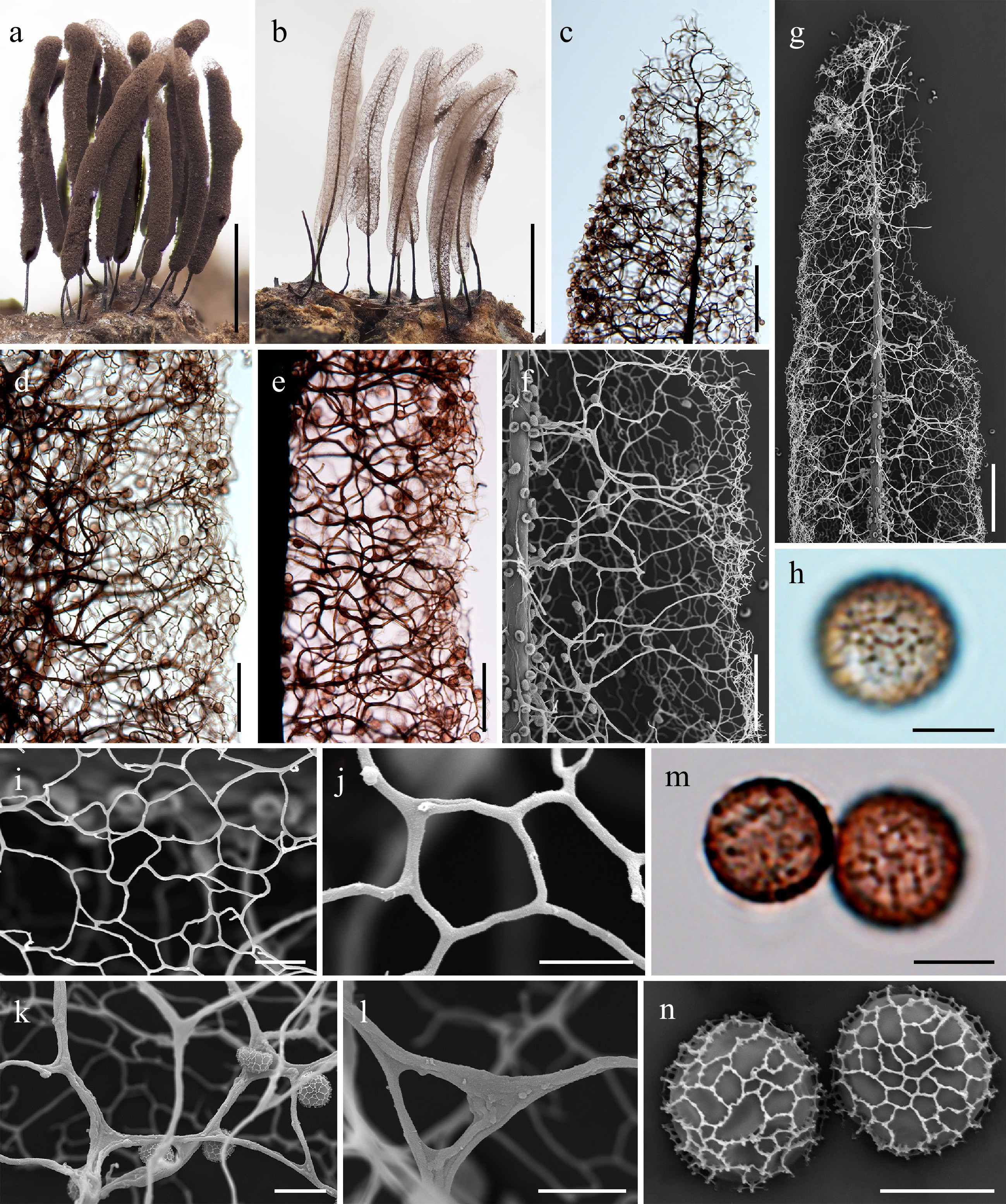
Figure 15.
Morphological characteristics of Stemonitis curvicornis (a)–(e), (h), (m) HFNNU 10970; (g), (f), (i)–(l), (n) HFNNU 10969. (a), (b) Mature sporocarps (DM). (c) Apical view of the sporotheca (LM). (d) Surface net (LM). (e) Capillitium in the middle part of the sporotheca (LM). (f) Capillitium in the middle part of the sporotheca (SEM). (g) Apical view of a sporotheca (SEM). (h) Spore (LM). (i), (j) Detail of the surface net (SEM). (k), (l) Detail of the capillitium threads (SEM). (m) Spores (LM). (n) Spore (SEM). Scale bars: (a), (b) = 2 mm; (c), (g) = 100 μm; (d)–(f) = 50 μm; (i), (k), (l) = 10 μm; (h), (j), (m), (n) = 5 μm.
-
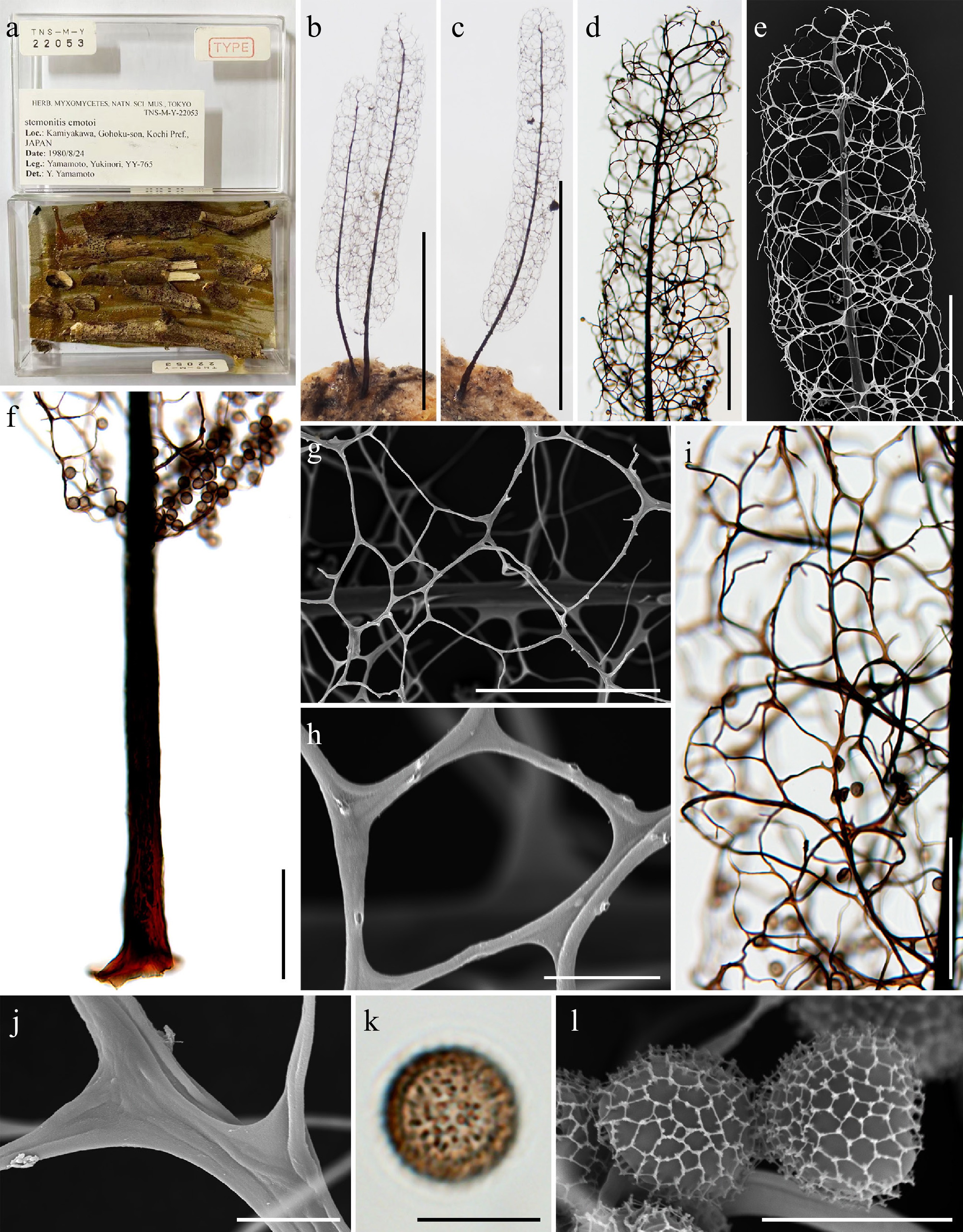
Figure 16.
Morphological characteristics of the holotype (TNS-M-Y-22053 = YY-765) of Stemonitis emotoi. (a) Herbarium label and interior views of the herbarium box containing the holotype. (b), (c) Mature sporocarps (DM). (d) Apical view of the sporotheca (LM). (e) Apical view of the sporotheca (SEM). (f) Stalk (LM). (g), (h) Detail of the surface net (SEM). (i) Capillitium and surface net in the middle part of the sporotheca (LM). (j) Detail of the capillitium threads (SEM). (k) Spore (LM). (l) Spores (SEM). Scale bars: (b), (c) = 2 mm; (d)–(f) = 200 μm; (g), (i) = 100 μm; (h), (j)–(l) = 10 μm.
-
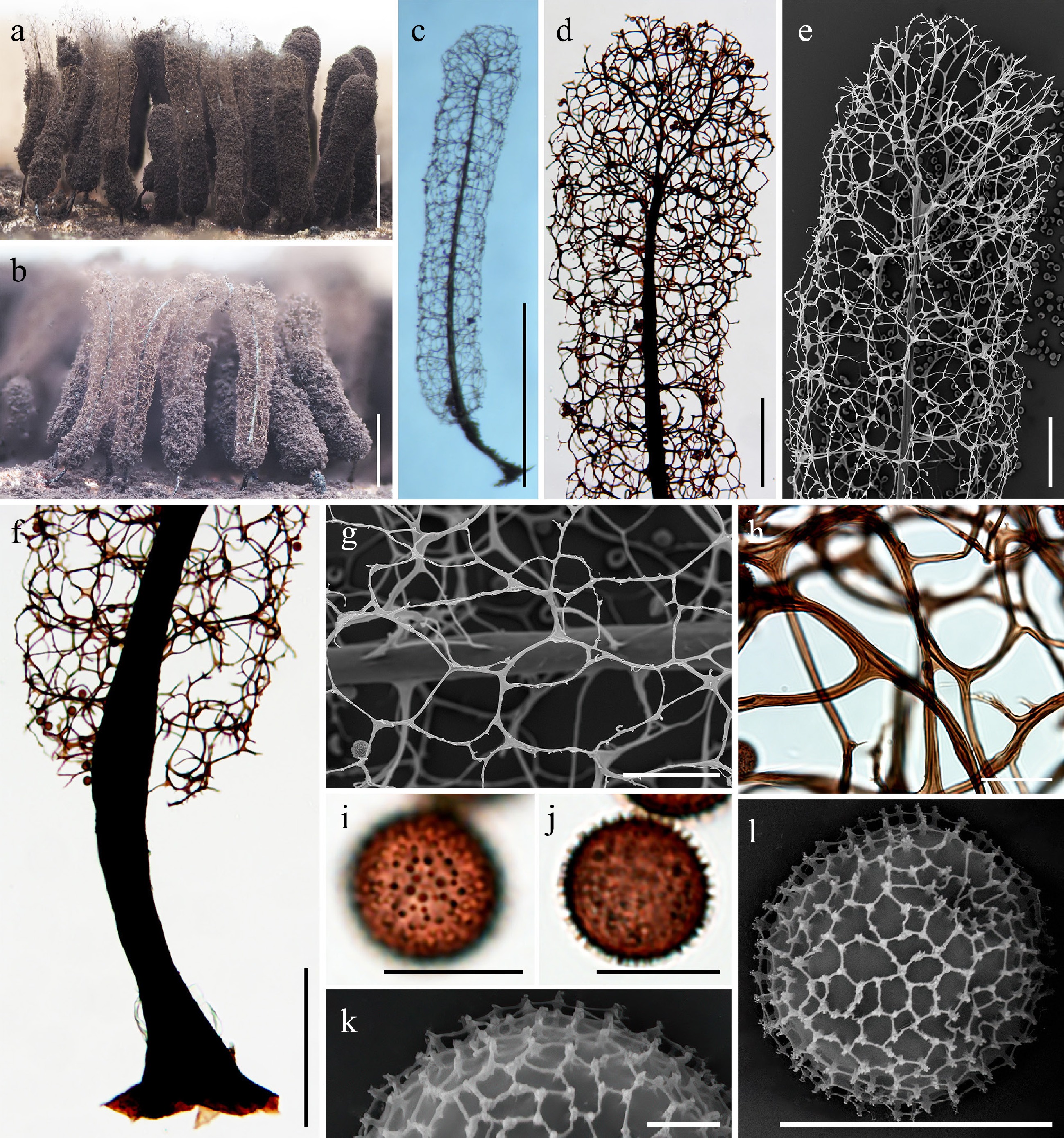
Figure 17.
Morphological characteristics of Stemonitis emotoi (a)–(d), (f), (h), (i), (j) HFNNU 10801; (e), (g), (k), (l) HFNNU 10802. (a)–(c) Mature sporocarps (DM). (d) Apical region of the sporocarp (LM). (e) Apical region of the sporocarp (SEM). (f) Stalk (LM). (g) Detail of the surface net (SEM). (h) Detail of the capillitium threads (LM). (i), (j) Spore (LM). (k) Detail of the spore ornamentation (SEM). (l) Spore (SEM). Scale bars: (a)–(c) = 1 mm; (d), (f) = 200 μm; (e) = 100 μm; (g) = 50 μm; (h) = 20 μm; (i), (j), (l) = 10 μm; (k) = 2 μm.
-
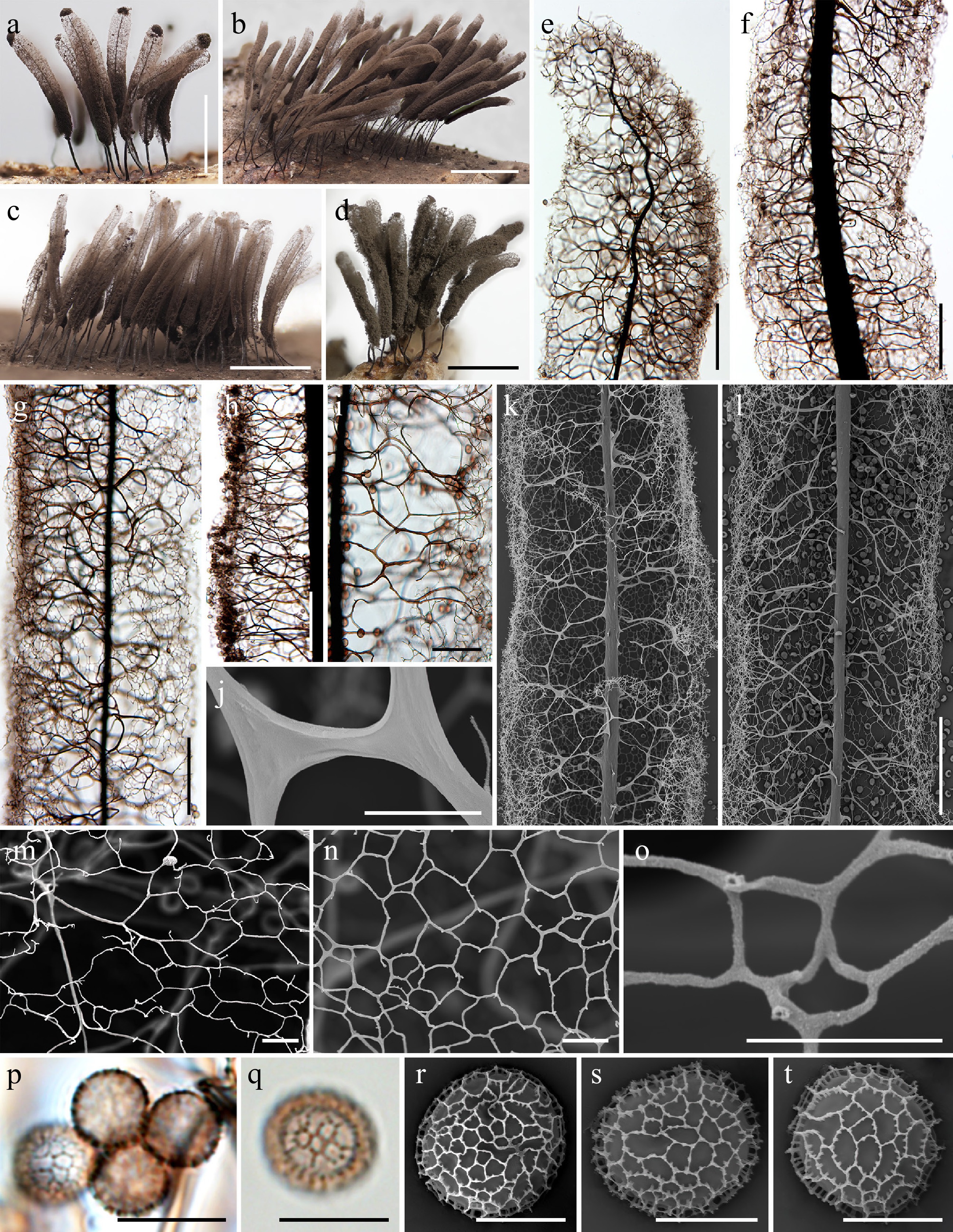
Figure 18.
Morphological characteristics of Stemonitis cf. foliicola (a), (i), (m), (r) HFNNU 10964; (b), (h) HFNNU 12189; (c), (n), (q), (p) MSK-F 43589; (d) LE348878; (e), (g), (l), (o), (s) HFNNU 13264; (f), (j), (k), (t) MSK-F 43592. (a)–(d) Mature sporocarps (DM). (e) Apical view of the sporotheca (LM). (f) Capillitium in the middle part of the sporotheca (LM). (g) Surface net (LM). (h), (i) Capillitium in the middle part of the sporotheca (LM). (j) Detail of the capillitium threads (SEM). (k), (l) Capillitium in the middle part of the sporotheca (SEM). (m)–(o) Detail of the surface net (SEM). (p), (q) Spores (LM). (r)–(t) Spore (SEM). Scale bars: (a)–(d) = 1 mm; (e)–(h), (k), (l) = 100 μm; (i) = 50 μm; (j), (m), (n), (p), (q) = 10 μm; (o), (r)–(t) = 5 μm.
-
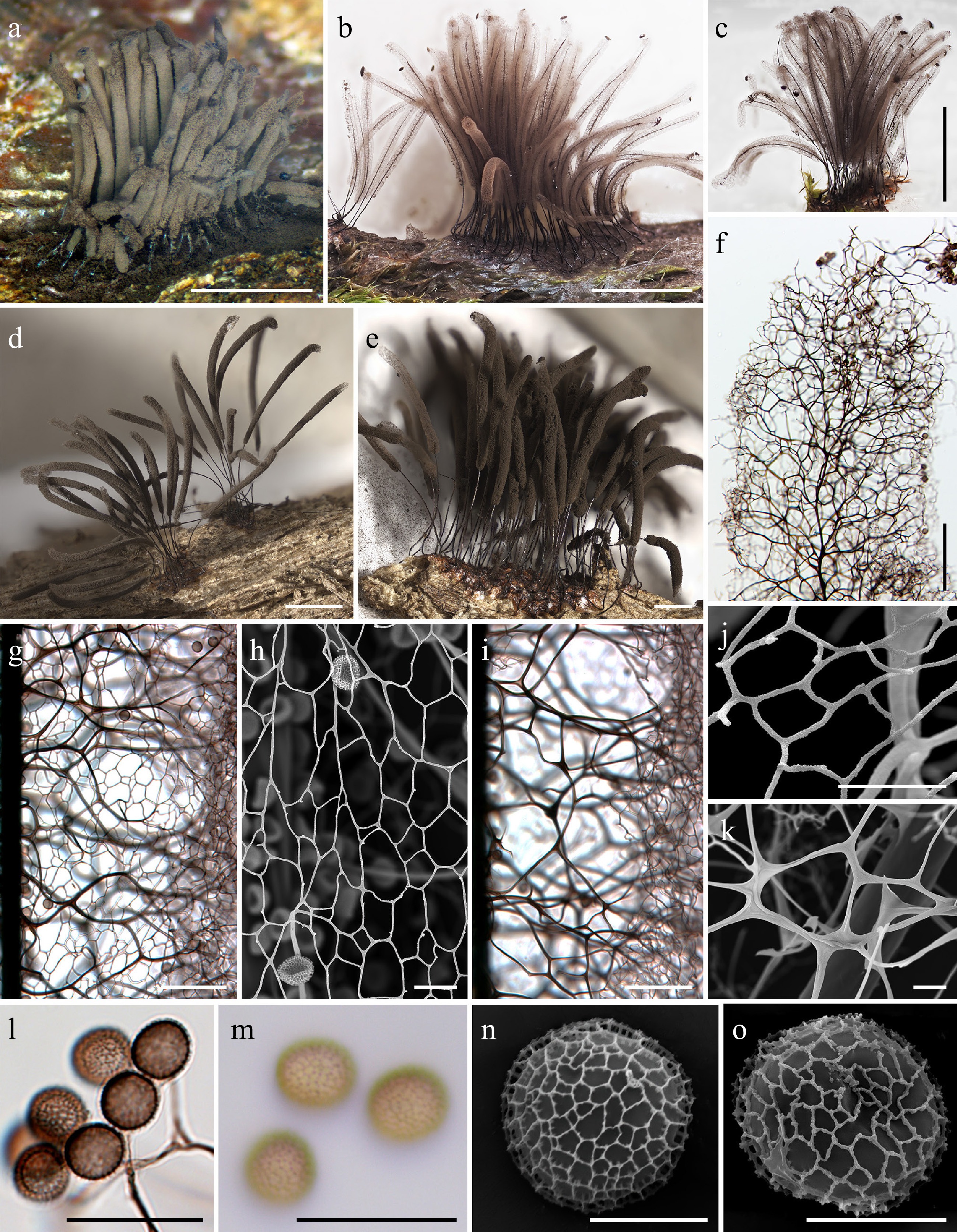
Figure 19.
Morphological characteristics of Stemonitis aff. fusca (a), (j), (m), (o) MYX 17763; (b), (f)–(i), (l), (n) MSK-F 43584; (c) HFNNU 12439; (d) LE348930; (e) LE348891; (k) MSK-F 43606. (a)–(e) Mature sporocarps (DM). (f) Apical region of the sporocarp (LM). (g) Surface net (LM). (h) Detail of the surface net (SEM). (i) Capillitium in the middle part of the sporotheca (LM). (j) Detail of the surface net (SEM). (k) Detail of the capillitium threads (SEM). (l), (m) Spores (LM). (n), (o) Spore (SEM). Scale bars: (a)–(d) = 2 mm; (e) = 1 mm; (f) = 100 μm; (g), (i) = 50 μm; (h,) (j)–(m) = 10 μm; (n), (o) = 5 μm.
-
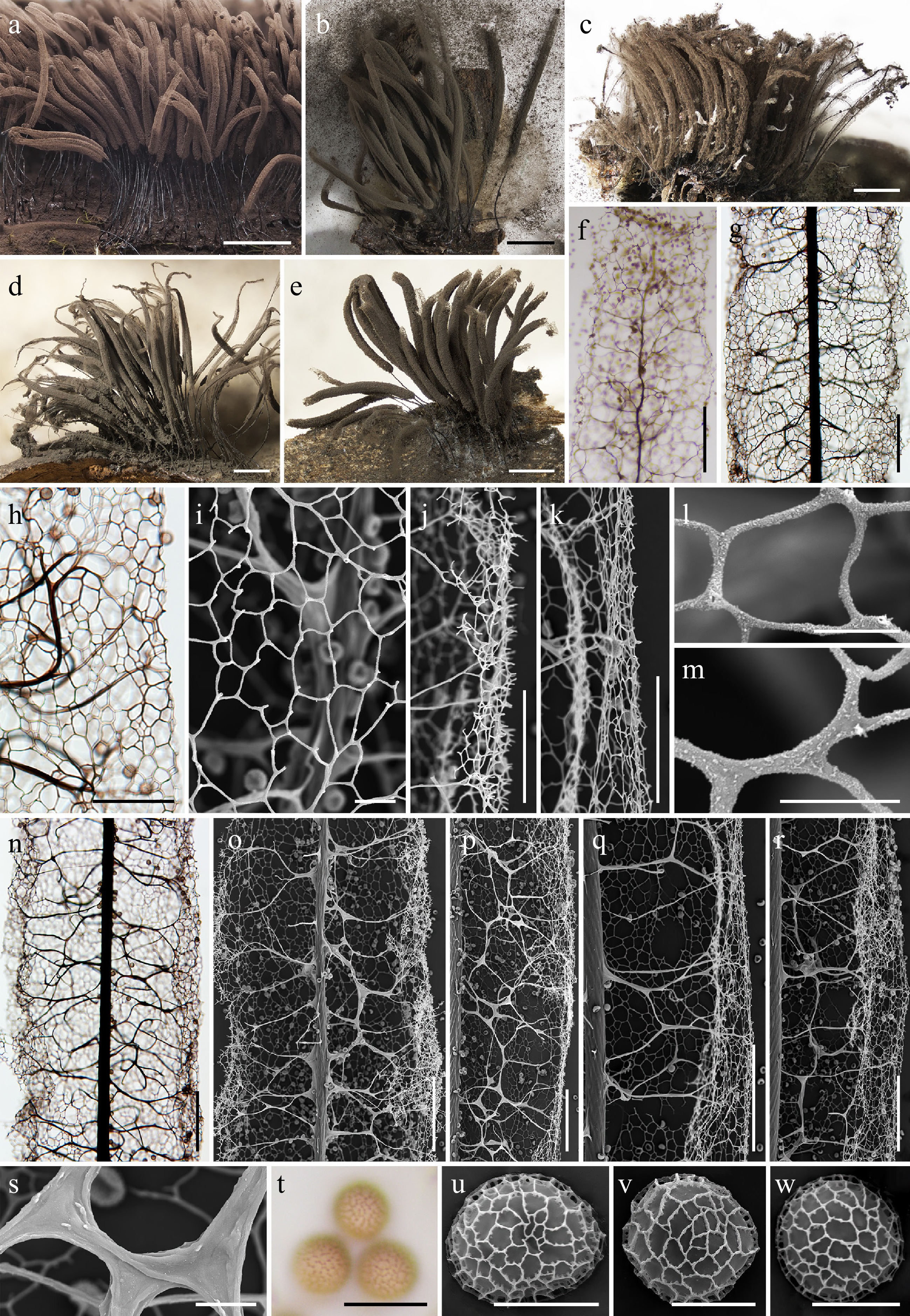
Figure 20.
Morphological characteristics of Stemonitis fusca agg. (a), (i), (j), (l), (o), (t) HFNNU 4212; (b) LE291133; (c) LE290927; (d) LE325855; (e) LE325706; (f), (t) MYX 19501; (g), (h), (k), (n), (q), (r), (w) LE328015; (m), (p), (s) LE346775; (v) MYX 19459. (a)–(e) Mature sporocarps (DM). (f) Apical region of the sporocarp (LM). (g), (h) Surface net (LM). (i) Detail of the surface net (SEM). (j), (k) Edge of the surface net (SEM). (l), (m) Detail of the surface net (SEM). (n) Capillitium in the middle part of the sporotheca (LM). (o)–(r) Capillitium in the middle part of the sporotheca (SEM). (s) Detail of the capillitium threads (SEM). (t) Spores (LM). (u)–(w) Spore (SEM). Scale bars: (a)–(e) = 2 mm; (f), (g), (n), (o)–(r) = 100 μm; (h), (j), (k) = 50 μm; (i), (s), (t) = 10 μm; (l), (m), (u)–(w) = 5 μm.
-

Figure 21.
Morphological characteristics of the holotype (TNS-M-Y-22046 = YY-1127) of Stemonitis gracilis (≡ Stemonaria gracilis). (a) Herbarium labels and interior views of the herbarium box containing the holotype. (b) Herbarium labels of the herbarium box containing the holotype. (c)–(f) Mature sporocarps (DM). (g) Apical view of the sporotheca (LM). (h), (i) Surface net in the middle part of the sporotheca (LM). (j) Capillitium and surface net in the middle part of the sporotheca (SEM). (k) Capillitium and surface net in the edge of the sporotheca (SEM). (l) Surface net (SEM). (m), (n) Capillitium in the middle part of the sporotheca (LM). (o), (p) Detail of the capillitium threads (SEM). (q), (r) Spores (LM). (s), (t) Spore (SEM). Scale bars: (c)–(f) = 1 mm; (g), (j), (l) = 100 μm; (h), (i), (m), (n) = 50 μm; (k), (o)–(r) = 10 μm; (s), (t) = 5 μm.
-
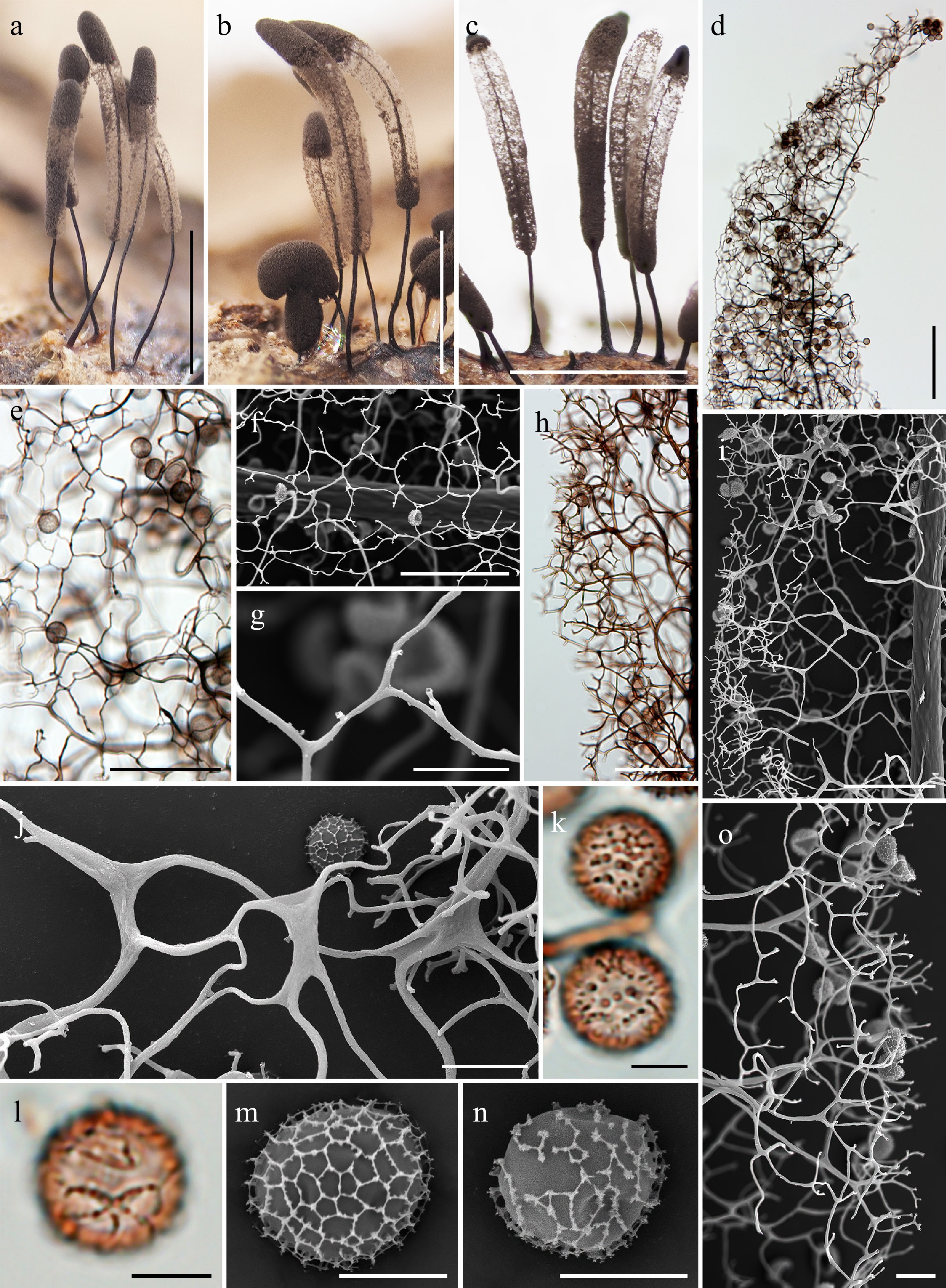
Figure 22.
Morphological characteristics of Stemonitis imperfecta (a), (b), (f)–(o) HFNNU 8808; (c)–(e) HFNNU 12493. (a)–(c) Mature sporocarps (DM). (d) Apical region of the sporocarp (LM). (e) Surface net (LM). (f), (g) Detail of the surface net (SEM). (h) Capillitium in the middle part of the sporotheca (LM). (i) Capillitium in the middle part of the sporotheca (SEM). (j) Detail of the capillitium threads (SEM). (k), (l) Spores (LM). (m), (n) Spore (SEM). (o) Edge of a sporotheca (SEM). Scale bars: (a)–(c) = 1 mm; (d) = 200 μm; (e), (f), (h), (i) = 50 μm; (g), (j), (o) = 10 μm; (k)–(n) = 5 μm.
-
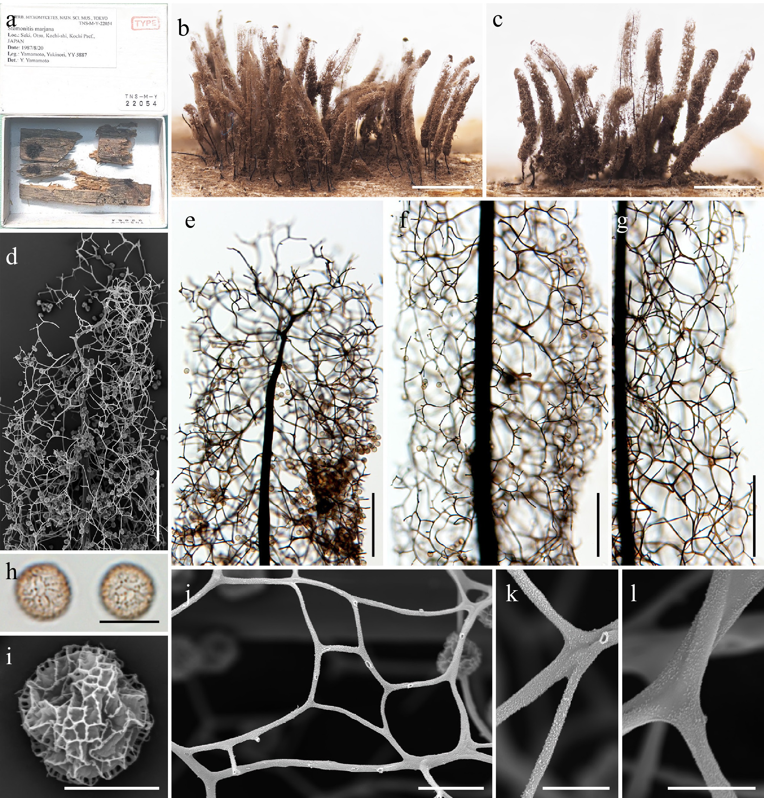
Figure 23.
Morphological characteristics of the holotype (TNS-M-Y-22054 = YY-5887) of Stemonitis marjana. (a) Herbarium label and interior view of the herbarium box containing the holotype. (b), (c) Mature sporocarps (DM). (d) Apical view of the sporotheca (SEM). (e) Apical view of the sporotheca (LM). (f), (g) Surface net (LM). (h) Spores (LM). (i) Spore (SEM). (j), (k) Detail of the surface net (SEM). (l) Detail of the capillitium threads (SEM). Scale bars: (b), (c) = 1 mm; (d)–(g) = 100 μm; (h), (j) = 10 μm; (i), (k), (l) = 5 μm.
-
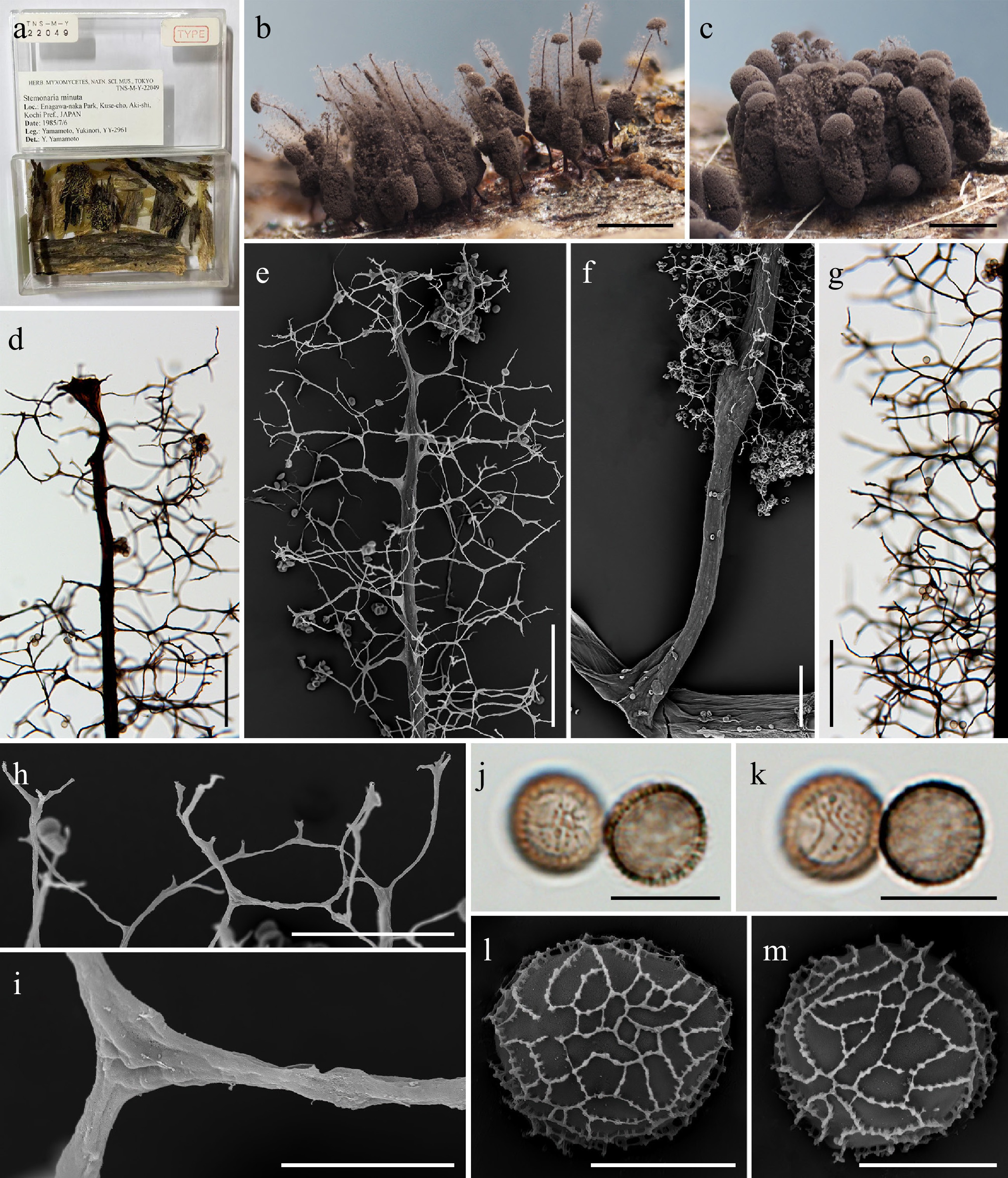
Figure 24.
Morphological characteristics of the isotype (TNS-M-Y-22049 = YY-2961) of Stemonitis minuta (≡ Stemonaria minuta). (a) Herbarium label and interior view of the herbarium box containing the holotype. (b), (c) Mature sporocarps (DM). (d) Apical view of the sporotheca (LM). (e) Apical view of the sporotheca (SEM). (f) Stalk (SEM). (g) Capillitium in the middle part of the sporotheca (LM). (h) Edge of the sporotheca (SEM). (i) Detail of the capillitium threads (SEM). (j), (k) Spores (LM). (l), (m) Spore (SEM). Scale bars: (b), (c) = 0.5 mm; (d)–(g) = 100 μm; (h) = 50 μm; (i)–(k) = 10 μm; (l), (m) = 5 μm.
-
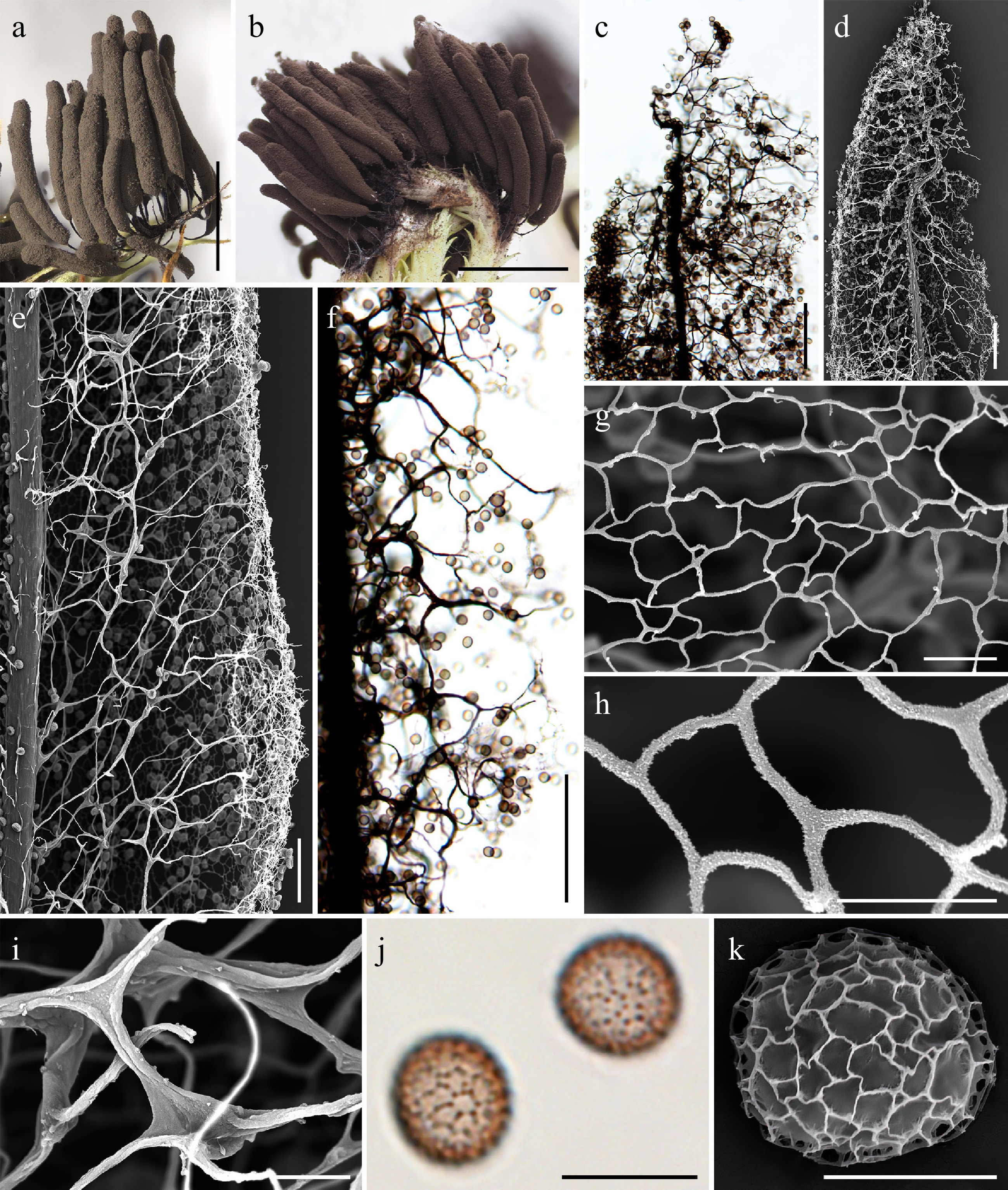
Figure 25.
Morphological characteristics of Stemonitis palustris (LE346780). (a), (b) Mature sporocarps (DM). (c) Apical region of the sporocarp (LM). (d) Apical region of the sporocarp (SEM). (e) Capillitium in the middle part of the sporotheca (SEM). (f) Capillitium in the middle part of the sporotheca (LM). (g), (h) Detail of the surface net (SEM). (i) Detail of the capillitium threads (SEM). (j) Spores (LM). (k) Spore (SEM). Scale bars: (a), (b) = 2 mm; (c), (d), (f) = 100 μm; (e) = 50 μm; (g), (i), (j) = 10 μm; (h), (k) = 5 μm.
-
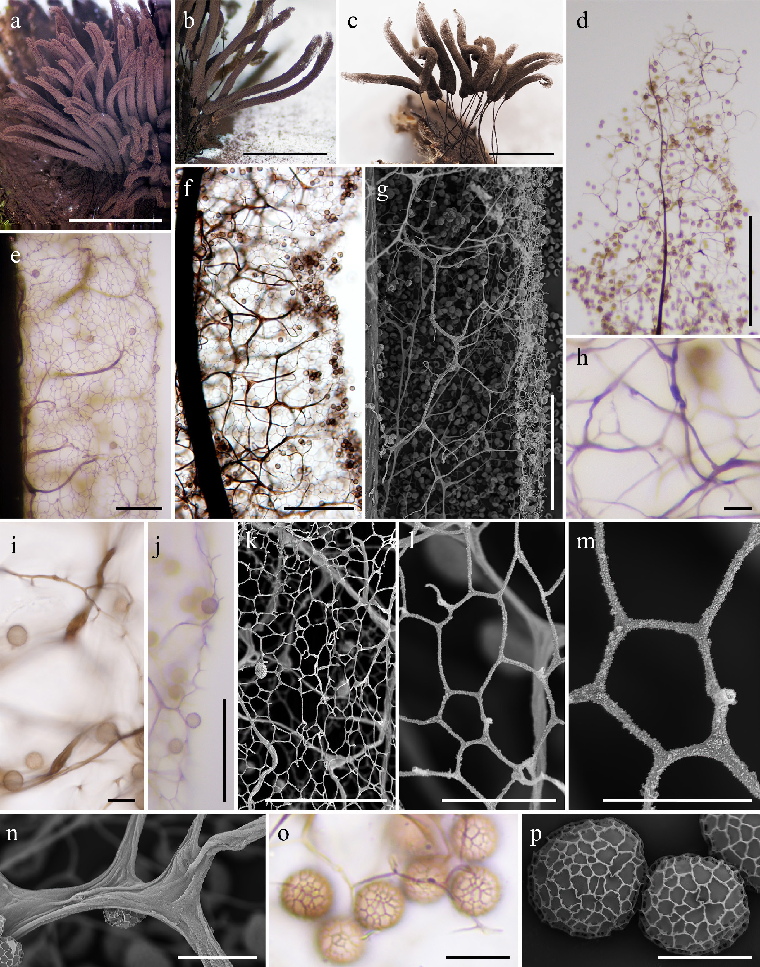
Figure 26.
Morphological characteristics of Stemonitis cf. pilosa (a), (d), (h), (o) MYX 24292; (b) MYX 20696; (c), (f), (g), (i), (k)–(n), (p) LE325048; (e), (j) MYX 24418. (a)–(c) Mature sporocarps (DM). (d) Apical view of the sporotheca (LM). (e) Surface net (LM). (f) Capillitium in the middle part of the sporotheca (LM). (g) Capillitium in the middle part of the sporotheca (SEM). (h), (i) Rounded thickenings in the capillitium (LM). (j) Edge of the sporotheca (LM). (k)–(m) Detail of the surface net (SEM). (n) Detail of the capillitium threads (SEM). (o) Spores (LM). (p) Spores (SEM). Scale bars: (a)–(c) = 5 mm; (d)–(g), (j) = 100 μm; (k) = 50 μm; (h), (i), (l), (n), (o) = 10 μm; (m), (p) = 5 μm.
-
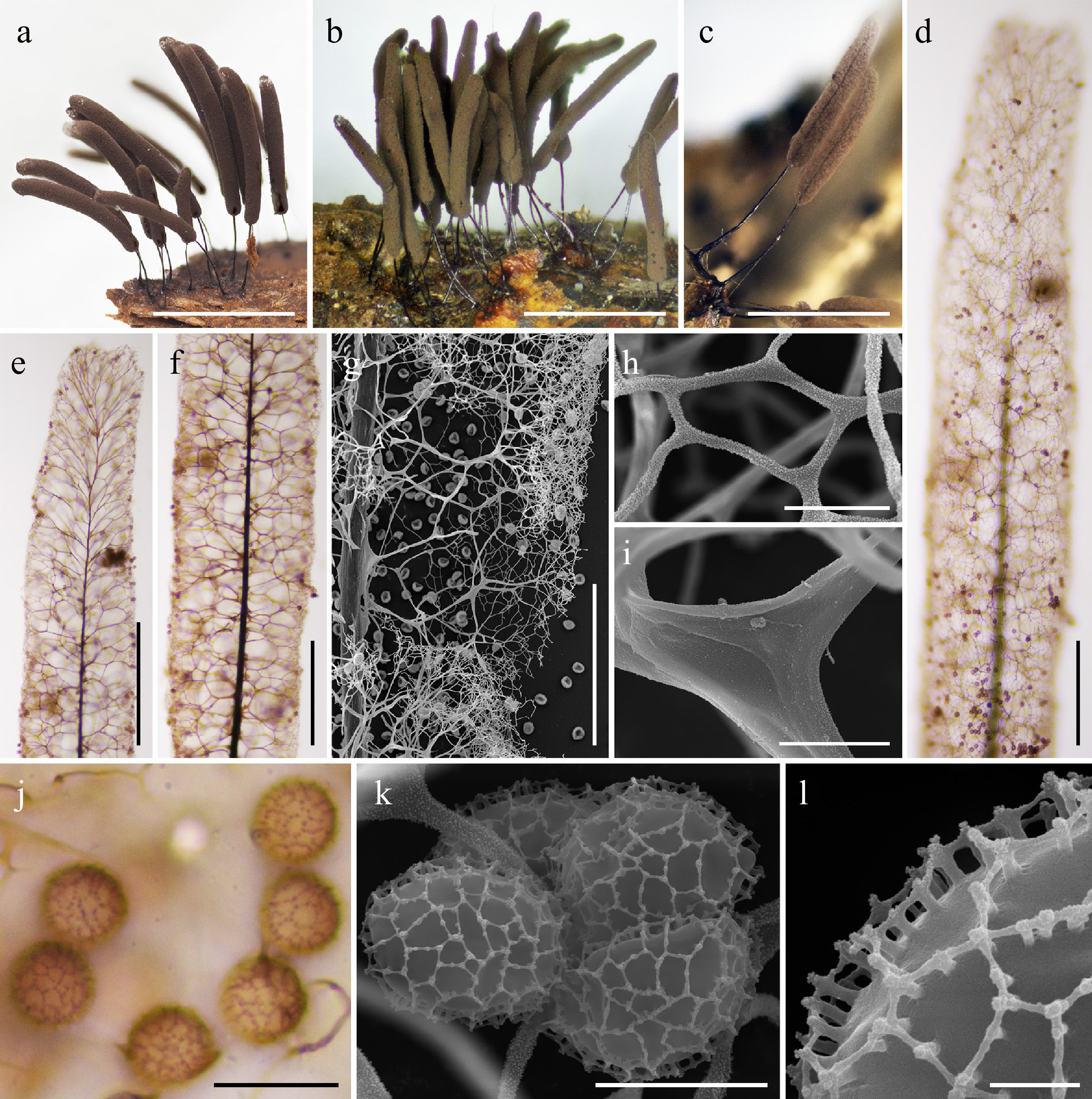
Figure 27.
Morphological characteristics of Stemonitis pinnatiapicalis (a) LE325372; (b), (d)–(g), (j), (l) MYX 24965; (c), (h), (i), (k) MYX 22874. (a)–(c) Mature sporocarps (DM). (d), (e) Apical region of the sporocarp (LM). (f) Capillitium in the middle part of the sporotheca (LM). (g) Capillitium in the middle part of the sporotheca (SEM). (h) Detail of the surface net (SEM). (i) Detail of the capillitium threads (SEM). (j) Spores (LM). (k) Spores (SEM). (l) Detail of the spore ornamentation (SEM). Scale bars: (a)–(c) = 2 mm; (d)–(g), (j) = 10 μm; (h), (i), (k) = 5 μm; (l) = 1 μm.
-
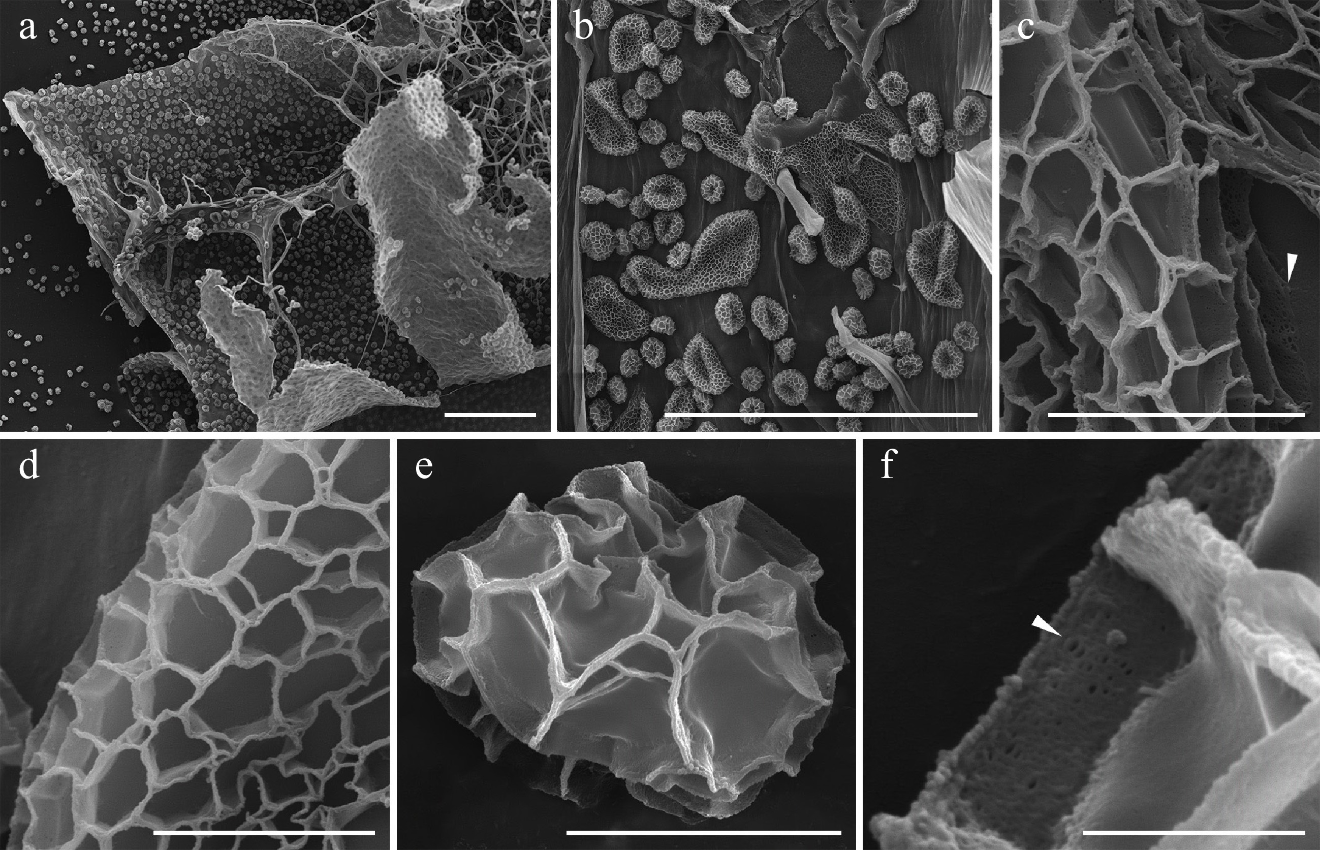
Figure 28.
Morphological characteristics of Stemonitis rispaudii (LE303142) under SEM. (a) Capillitium and peridium. (b)–(d) Flattened spore-like bodies and spores on the inner peridium surface. (e), (f) Spores. The punctiform perforations in the ridges of the spore ornamentation are shown by arrows (c, f). Scale bars: (a), (b) = 100 μm, (c), (d) = 5 μm, (f) = 1 μm.
-
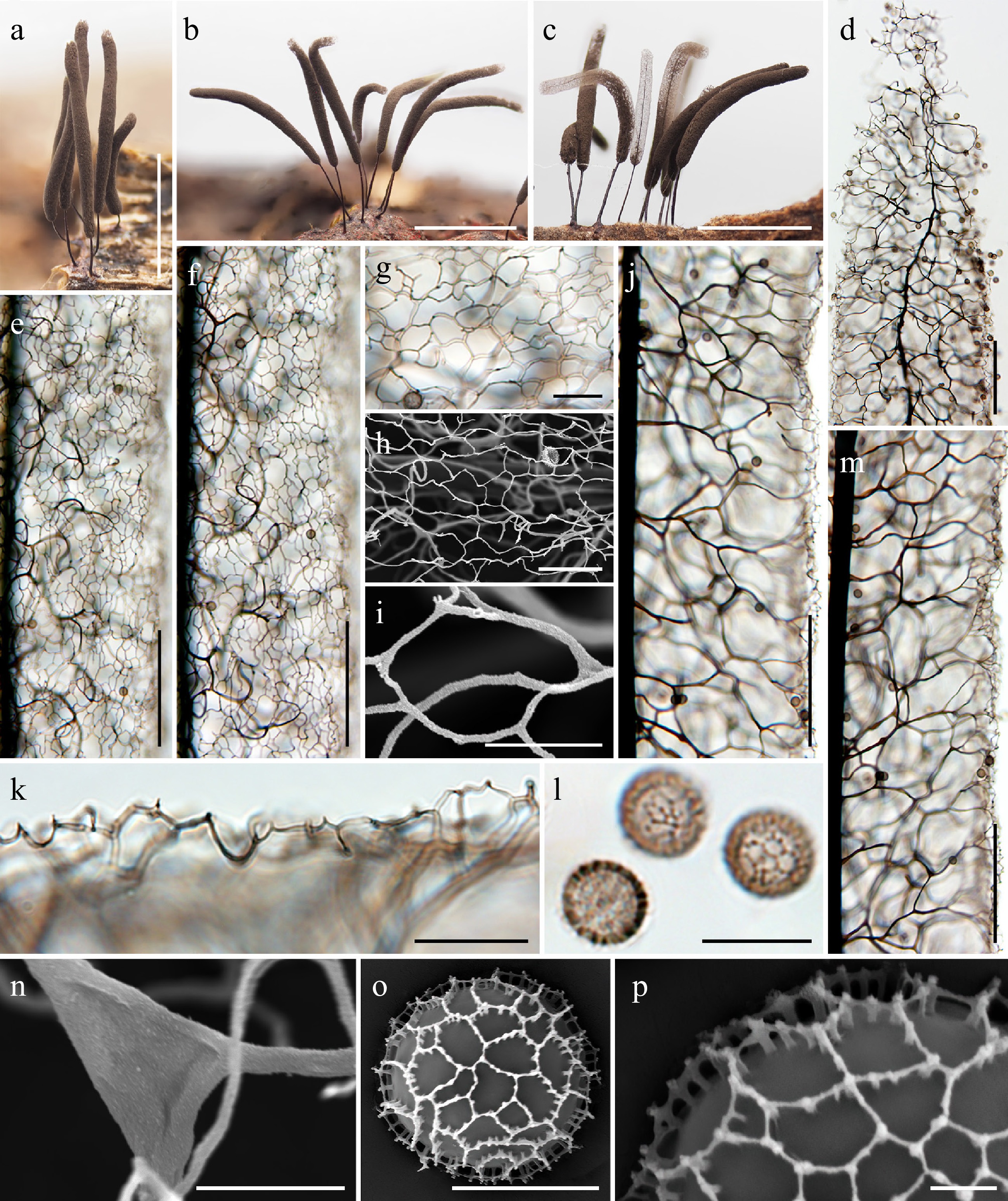
Figure 29.
Morphological characteristics of Stemonitis spinimuralis (a)–(m) HFNNU 2815; (n), (o) HMAS 0257700; (p) HFNNU 12498. (a)–(c) Mature sporocarps (DM). (d) Apical view of the sporotheca (LM). (e)–(g) Surface net (LM). (h), (i) Detail of the surface net (SEM). (j) Capillitium in the middle part of the sporotheca (LM). (k) Edge of the sporotheca (LM). (l) Spores (LM). (m) Capillitium in the middle part of the sporotheca (LM). (n) Detail of the capillitium threads (SEM). (o) Spore (SEM). (p) Detail of the spore ornamentation (SEM). Scale bars: (a)–(c) = 1 mm; (d)–(f), (j), (m) = 100 μm; (g), (h), (k) = 20 μm; (l) = 10 μm; (i), (n), (o) = 5 μm; (p) = 1 μm.
-
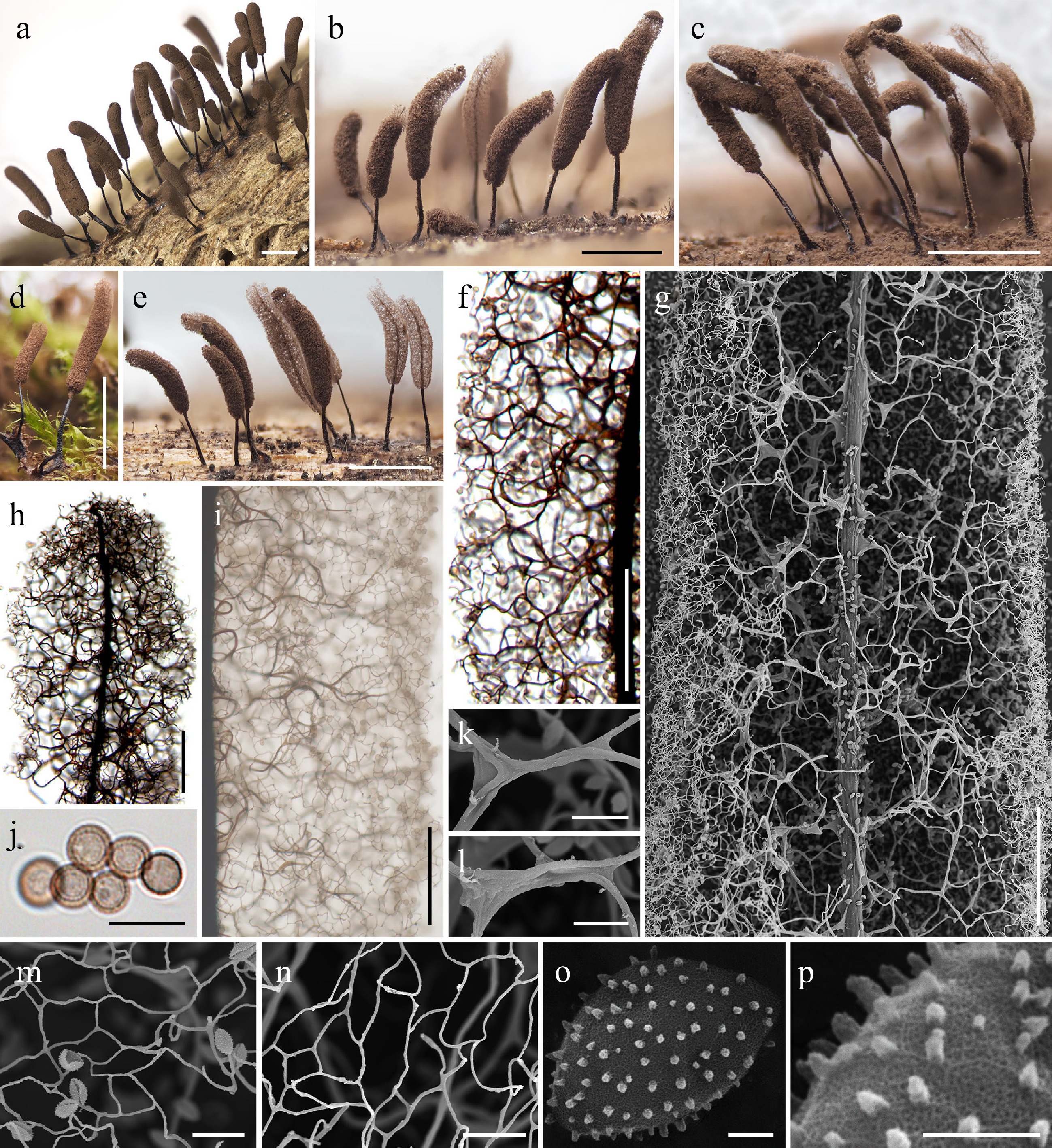
Figure 30.
Morphological characteristics of Symphytocarpus dispersus (a) LE327886; (b), (e), (f) HMAS 0258209; (c) HMAS 0258208; (g), (k)–(n) LE348844; (h), (j) HMAS 0258217; (d), (i), (o), (p) LE352558. (a)–(e) Mature sporocarps (DM). (f) Capillitium in the middle part of the sporotheca (LM). (g) Capillitium in the middle part of the sporotheca (SEM). (h) Apical region of the sporotheca (LM). (i) Surface net (LM). (j) Spores (LM). (k), (l) Detail of the capillitium threads (SEM). (m), (n) Detail of the surface net (SEM). (o) Spore (SEM). (p) Detail of spore ornamentation (SEM). Scale bars: (a)–(e) = 1 mm; (f)–(i) = 100 μm; (j)–(n) = 10 μm; (o), (p) = 1 μm.
-
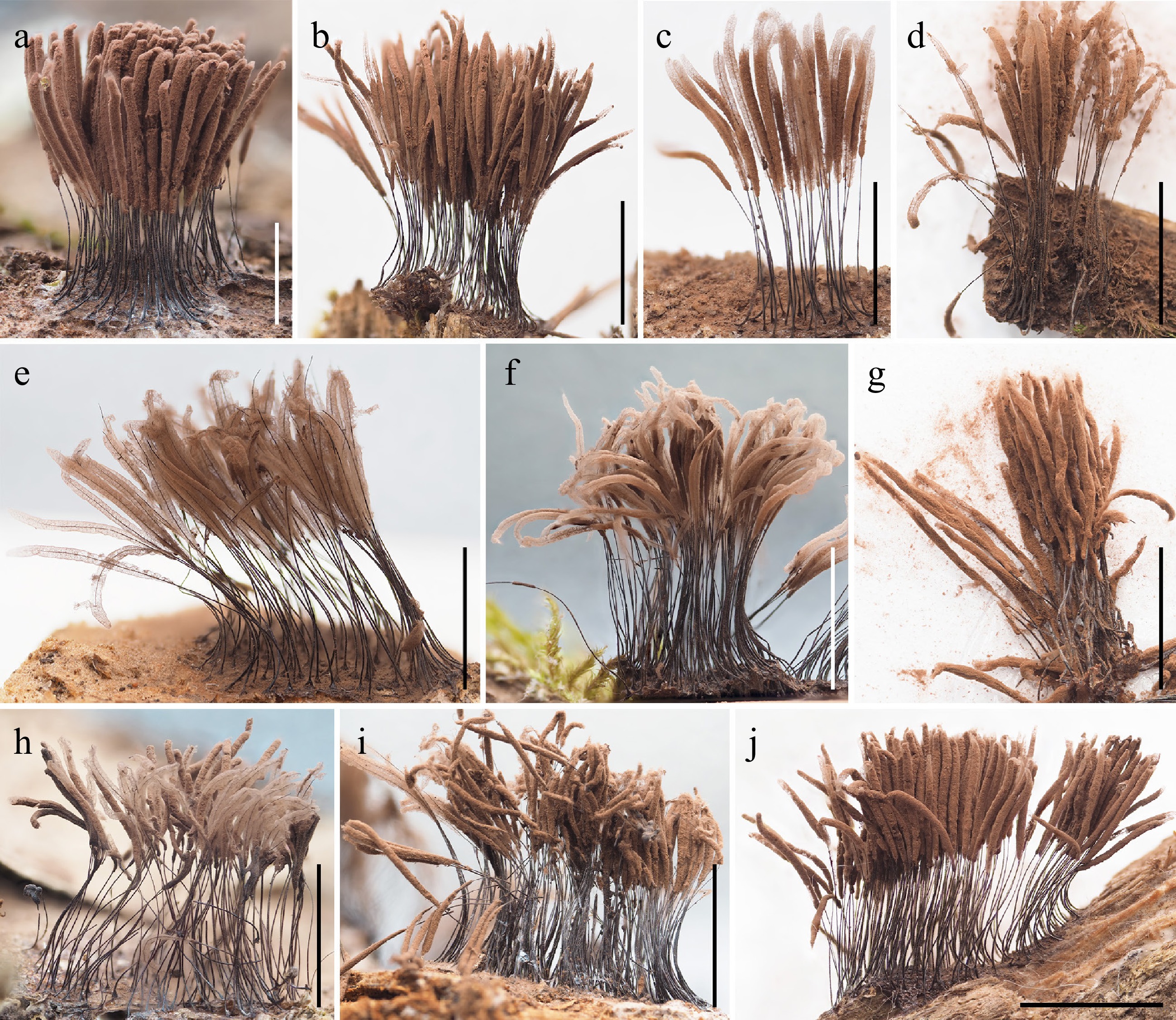
Figure 31.
Mature sporocarps of Symphytocarpus ferrugineus (DM). (a) Specimen HFNNU 11590. (b) Specimen HFNNU 11586. (c) Specimen MSK-F 43579. (d) Specimen LE328196. (e) Specimen MSK-F 41151-1. (f) Specimen MSK-F 43508. (g) Specimen LE259805. (h) Specimen HFNNU 12499. (i) Specimen HMAS 75392. (j) Specimen HFNNU 11577. Scale bars: 2 mm.
-
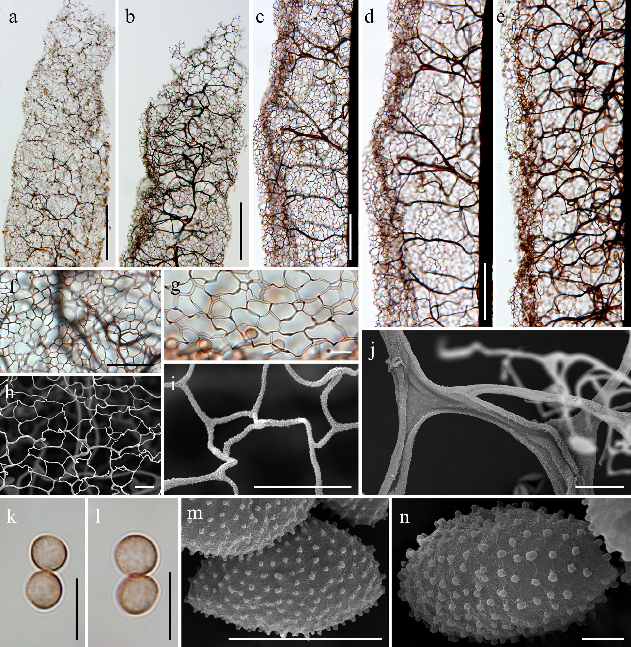
Figure 32.
Morphological characteristics of Symphytocarpus ferrugineus (a) HFNNU 11590; (b) HFNNU 11586; (c), (k), (l) LE320987; (d) LE328196; (e) MSK-F 41151-1; (f) MSK-F 43508; (g) LE259805; (h) HFNNU 12499; (i) HMAS 75392; (j) HFNNU 11577; (m) MYX 19217; (n) MYX 24329. (a), (b) Apical region of the sporotheca (LM). (c) Surface net (LM). (d), (e) Capillitium in the middle part of the sporotheca (LM). (f), (g) Detail of the surface net (LM). (h), (i) Detail of the surface net (SEM). (j) Detail of the capillitium threads (SEM). (k), (l) Spores (LM). (m), (n) Spore (SEM). Scale bars: (a), (b) = 200 μm; (c)–(e) = 100 μm; (f) = 50 μm; (g)–(l) = 10 μm; (m) = 5 μm; (n) = 1 μm.
-
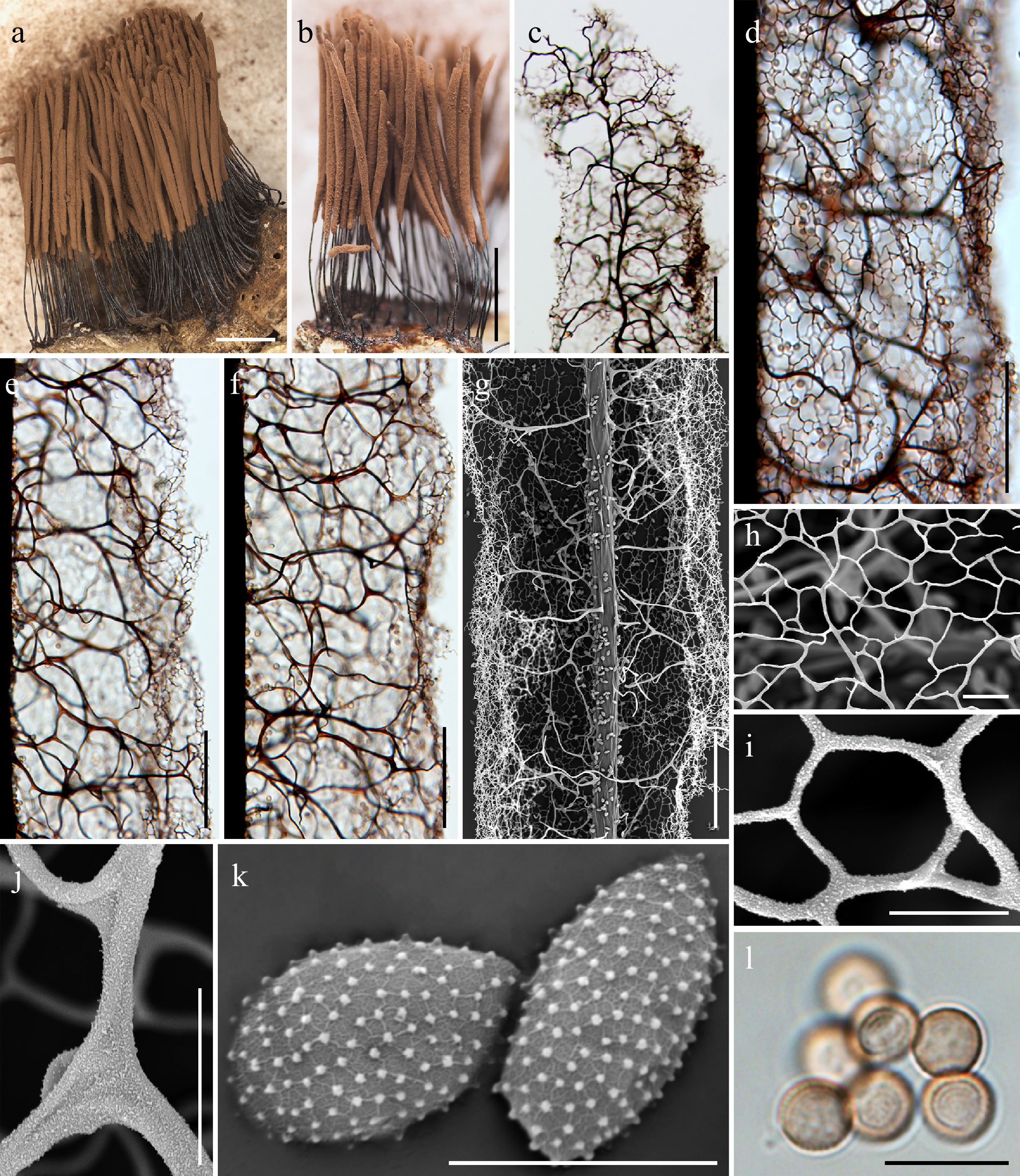
Figure 33.
Morphological characteristics of Symphytocarpus ferrugineus (LE245001, USA). (a), (b) Mature sporocarps (DM). (c) Apical region of the sporotheca (LM). (d) Surface net (LM). (e), (f) Capillitium in the middle part of the sporotheca (LM). (g) Capillitium in the middle part of the sporotheca (SEM). (h), (i) Detail of the surface net (SEM). (j) Detail of the capillitium threads (SEM). (k) Spores (SEM). (l) Spores (LM). Scale bars: (a), (b) = 2 mm; (c)–(g) = 100 μm; (h), (l) = 10 μm; (i)–(k) = 5 μm.
-
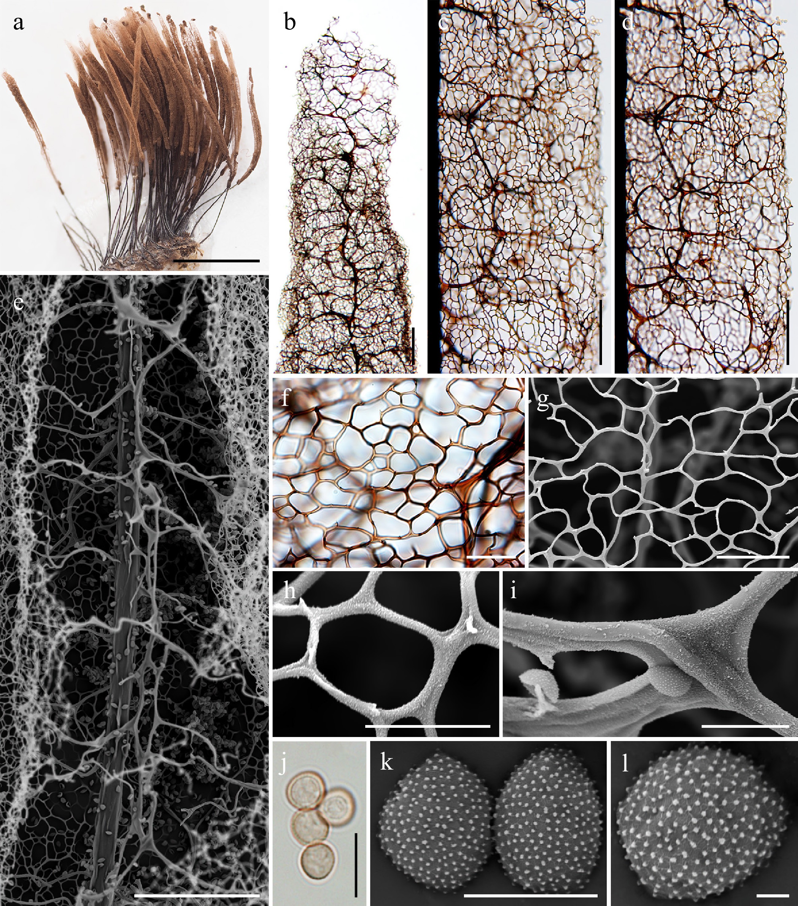
Figure 34.
Morphological characteristics of Symphytocarpus ferrugineus (LE254641). (a) Mature sporocarps (DM). (b) Apical region of the sporotheca (LM). (c) Surface net (LM). (d) Capillitium in the middle part of the sporotheca (LM). (e) Capillitium in the middle part of the sporotheca (SEM). (f) Detail of the surface net (LM). (g), (h) Detail of the surface net (SEM). (i) Detail of the capillitium threads (SEM). (j) Spores (LM). (k), (l) Spores (SEM). Scale bars: (a) = 1.5 mm; (b)–(e) = 100 μm; (f), (g) = 20 μm; (h)–(j) = 10 μm; (k)= 5 μm; (l)= 1 μm.
-
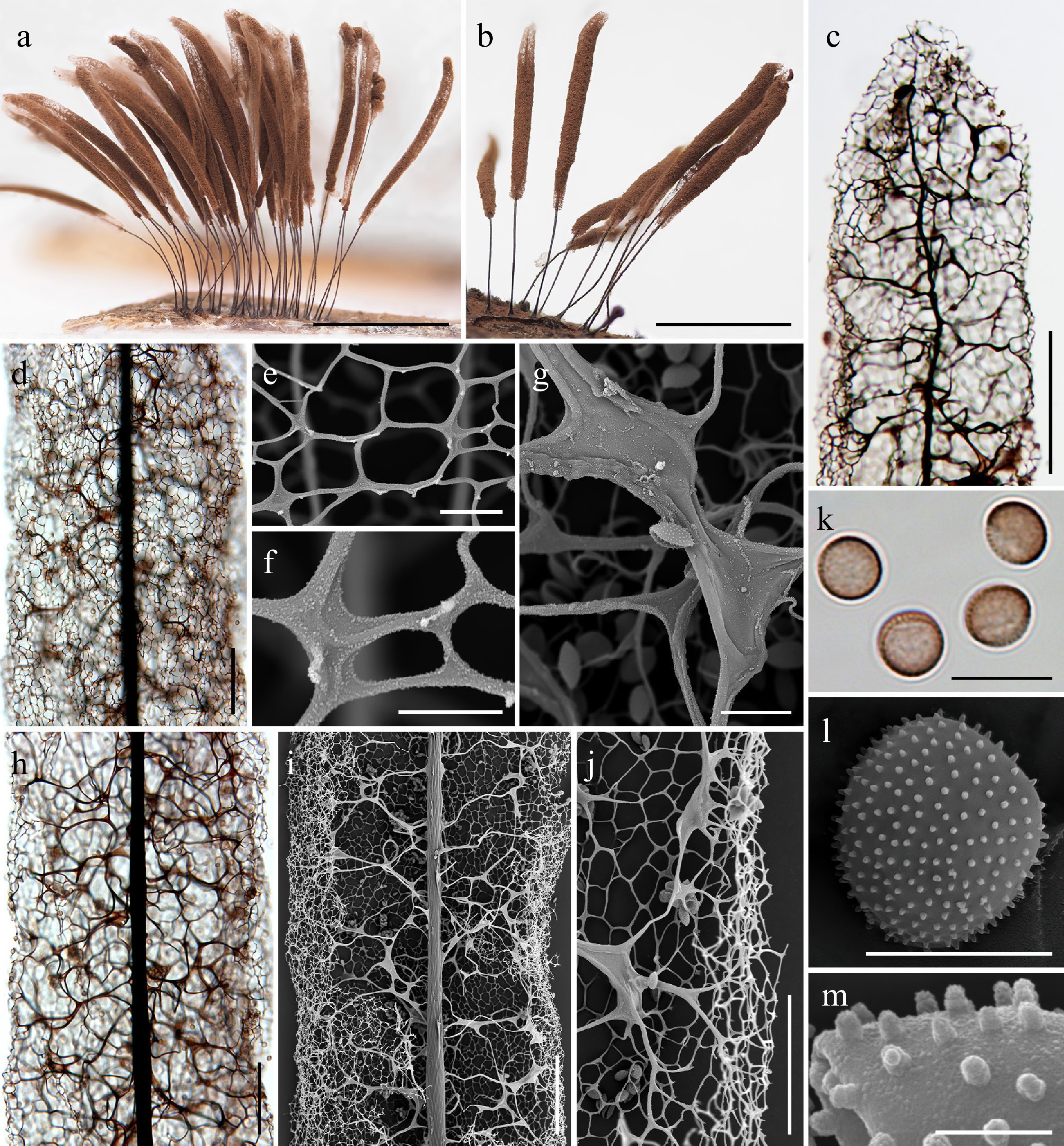
Figure 35.
Morphological characteristics of Symphytocarpus laxicarpicus (a), (b), (g), (l) LE348807; (c)–(f), (h)–(j) HFNNU 12497; (m) MYX 23608). (a), (b) Mature sporocarps (DM). (c) Apical part of the sporocarp (LM). (d) Surface net (LM). (e), (f) Detail of the surface net (SEM). (g) Detail of the capillitium threads (SEM). (h) Capillitium in the middle part of the sporotheca (LM). (i) Capillitium in the middle part of the sporotheca (SEM). (j) Edge of the sporotheca (SEM). (k) Spores (LM). (l) Spore (SEM). (m) Detail of the spore ornamentation (SEM) Scale bars: (a), (b) = 3 mm; (c) = 200 μm; (d), (h), (i) = 100 μm; (j) = 50 μm; (e), (g), (k) = 10 μm; (f), (l) = 5 μm; (m) = 1 μm.
-
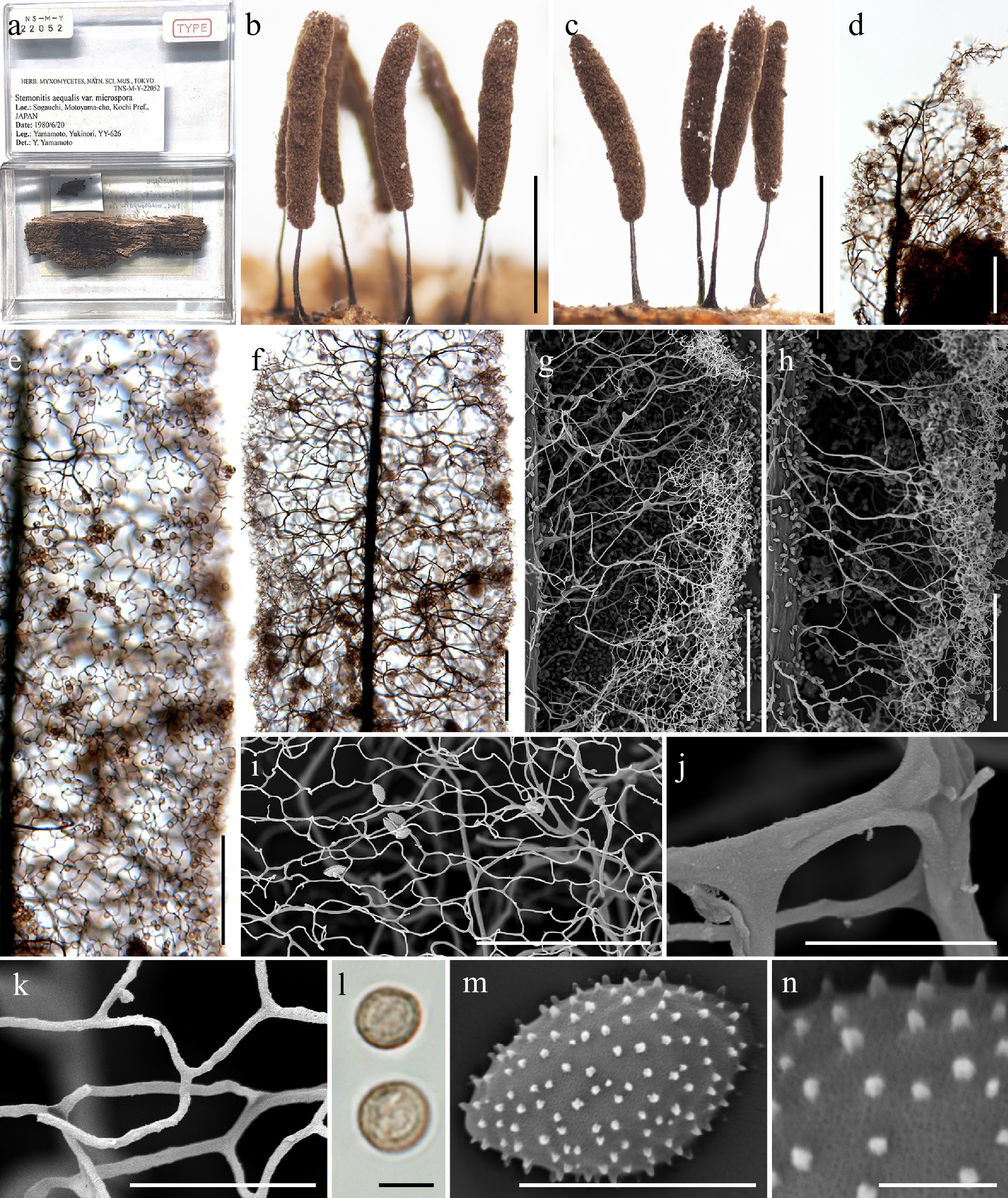
Figure 36.
Morphological characteristics of the holotype (TNS-M-Y 22052 = YY-626) of Symphytocarpus microsporus (≡ Stemonitis aequalis var. microspora). (a) Herbarium labels and interior views of the herbarium box containing the holotype. (b), (c) Mature sporocarps (DM). (d) Apical view of the sporotheca (LM). (e) Surface net (LM). (f) Capillitium in the middle part of the sporotheca (LM). (g), (h) Capillitium in the middle part of the sporotheca (SEM). (i) Detail of the surface net (SEM). (j) Detail of the capillitium threads (SEM). (k) Detail of the surface net (SEM). (l) Spores (LM). (m) Spore (SEM). (n) Detail of the spore ornamentation (SEM). Scale bars: (b), (c) = 2 mm; (d)–(h) = 100 μm; (i) = 50 μm; (j), (k) = 10 μm; (l)–(n) = 5 μm.
-
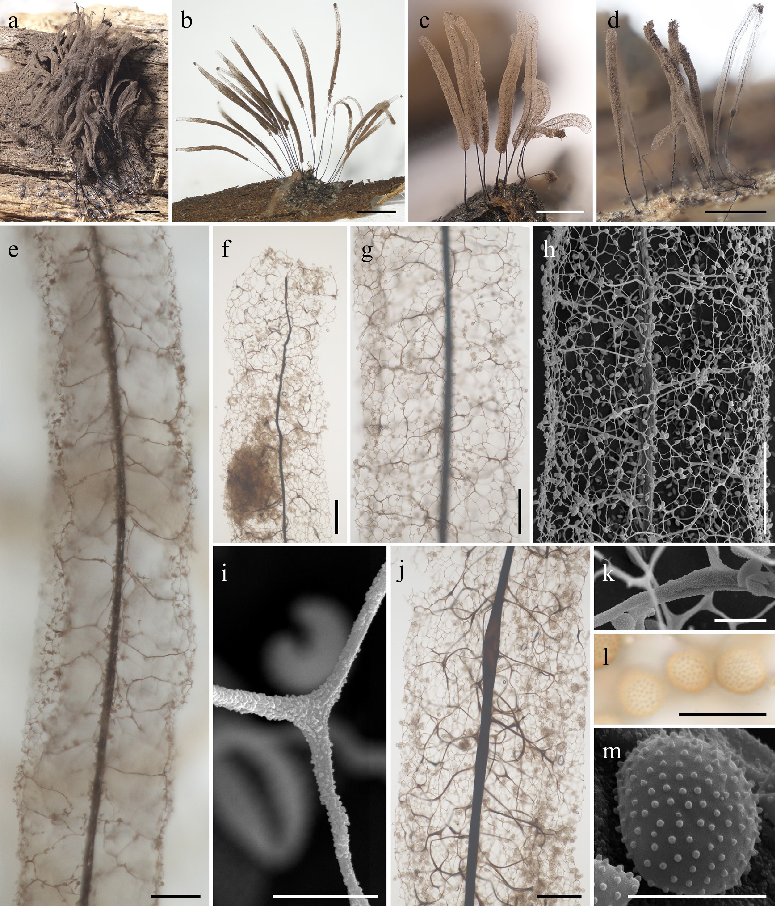
Figure 37.
Morphological characteristics of Symphytocarpus minor (a), (j) LE306599; (b), (l) LE348772; (c), (e), (g)–(i), (k), (m) LE348776; (d), (f) LE307469. (a)–(d) Mature sporocarps (DM). (e) Capillitium in the middle part of the sporotheca (LM). (f) Apical region of the sporocarp (LM). (g) Surface net (LM). (h) Surface net (SEM). (i) Detail of the surface net (SEM). (j) Capillitium in the middle part of the sporotheca (LM). (k) Detail of the capillitium threads (SEM). (l) Spores (LM). (m) Spores (SEM). Scale bars: (a)–(d) = 2 mm; (e), (f), (g), (h) = 100 μm; (j) = 50 μm; (k), (l) = 10 μm; (i), (m) = 5 μm.
-
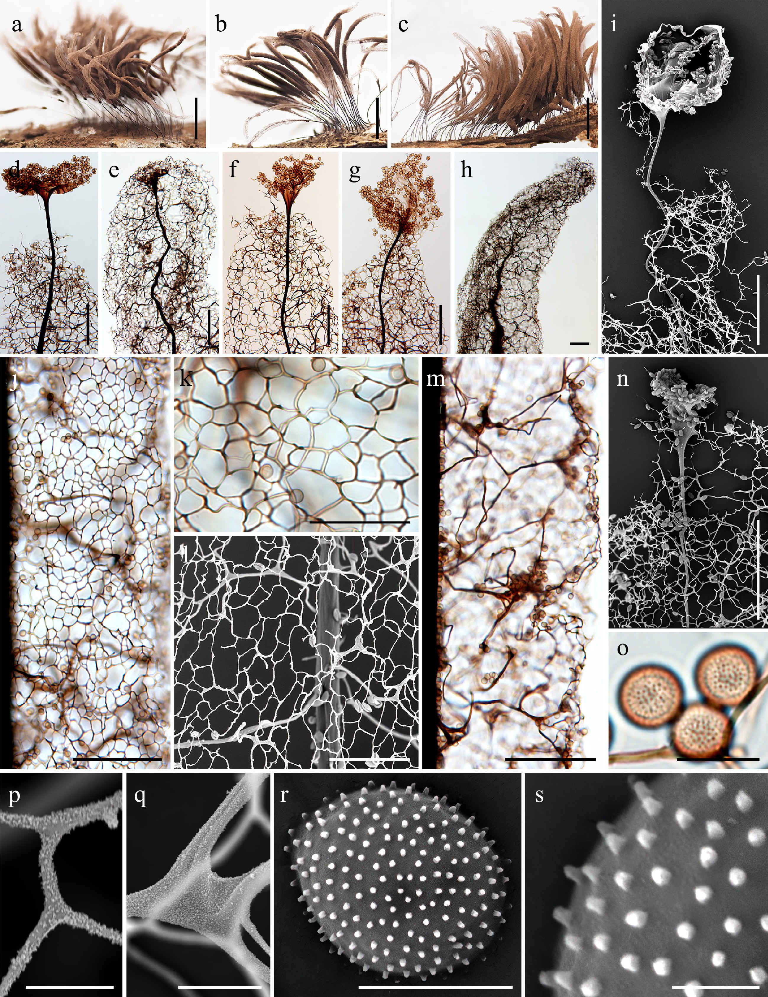
Figure 38.
Morphological characteristics of Symphytocarpus pseudoflavogenitus (a), (d), (f), (g), (i), (o)–(s) HFNNU 11433; (b), (l), (n) HFNNU 11574; (c), (e), (h), (j), (k), (m) HMAS 75422. (a)–(c) Mature sporocarps (DM). (d)–(h) Apical view of the sporotheca (LM). (i) Apical view of the sporotheca (SEM). (j) Surface net (LM). (k) Detail of the surface net (LM). (l) Detail of the surface net (SEM). (m) Capillitium in the middle part of the sporotheca (LM). (n) Apical view of the columella (SEM). (o) Spores (LM). (p) Detail of the surface net (SEM). (q) Detail of the capillitium threads (SEM). (r) Spore (SEM). (s) Detail of the spore ornamentation (SEM). Scale bars: (a)–(c) = 2 mm; (d)–(j), (m), (n) = 100 μm; (k), (l) = 50 μm; (q) = 10 μm; (p), (r) = 5 μm; (s) = 1 μm.
-
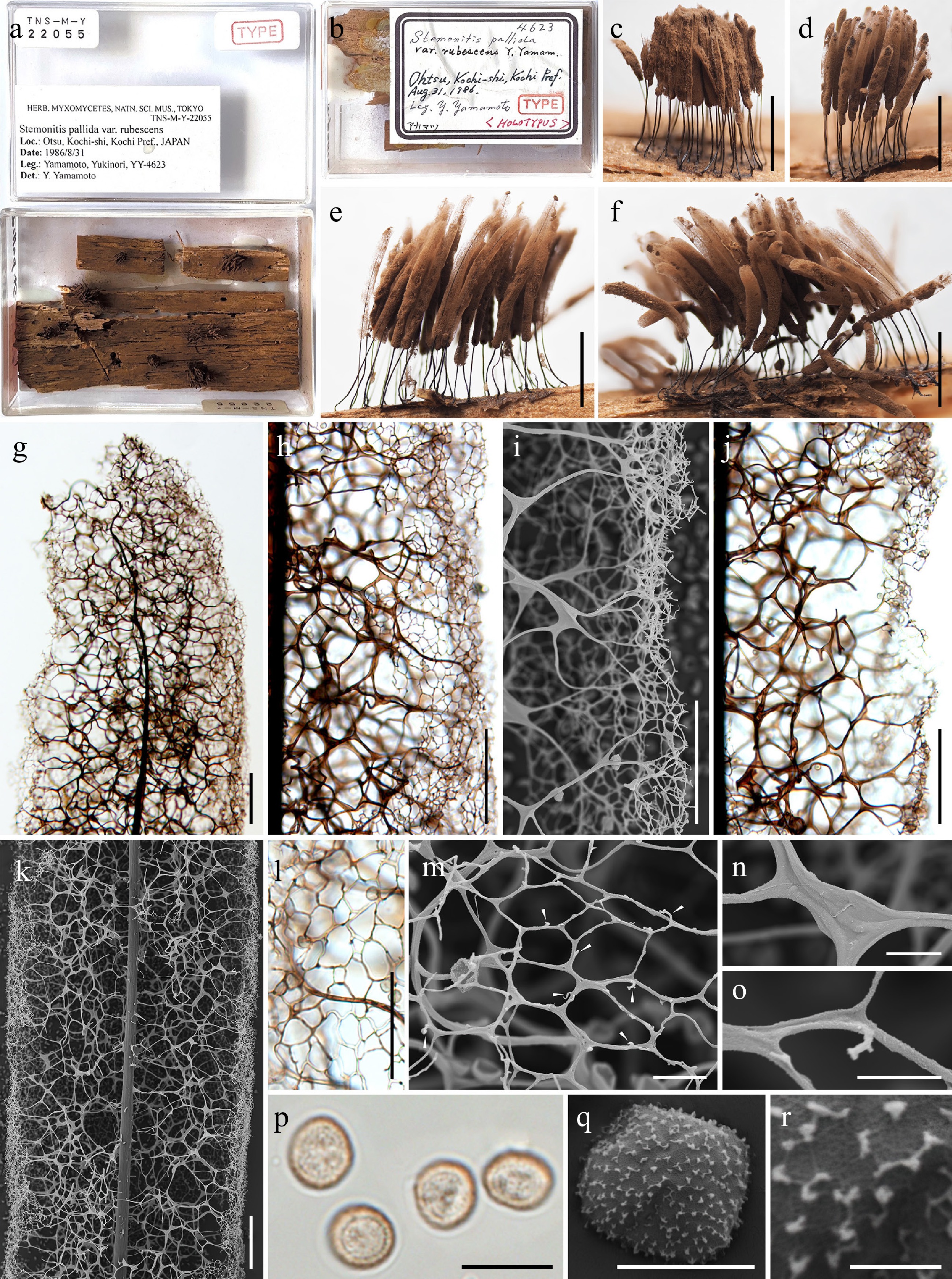
Figure 39.
Morphological characteristics of the holotype (TNS-M-Y-22055 = YY-4623) of Symphytocarpus rubescens (≡ Stemonitis pallida var. rubescens). (a) Herbarium labels and interior views of the herbarium box containing the holotypes. (b) Herbarium labels of the herbarium box containing the holotypes. (c)–(f) Mature sporocarps (DM). (g) Apical view of the sporotheca (LM). (h) Capillitium and surface net in the middle part of the sporotheca (LM). (i) Capillitium and surface net in the edge of the sporotheca (SEM). (j) Capillitium in the middle part of the sporotheca (LM). (k) Capillitium in the middle part of the sporotheca (SEM). (l) Surface net (LM). (m) Spines on the surface net (SEM, arrow). (n) Detail of the capillitium threads (SEM). (o) Detail of the surface net (SEM). (p) Spore (LM). (q) Spores (SEM). (r) Detail on the spore ornamentation (SEM). Scale bars: (c)–(f) = 3 mm; (g), (h), (j), (k) = 100 μm; (i), (l) = 50 μm; (m), (n), (p) = 10 μm; (o), (q) = 5 μm; (r) = 1 μm.
-
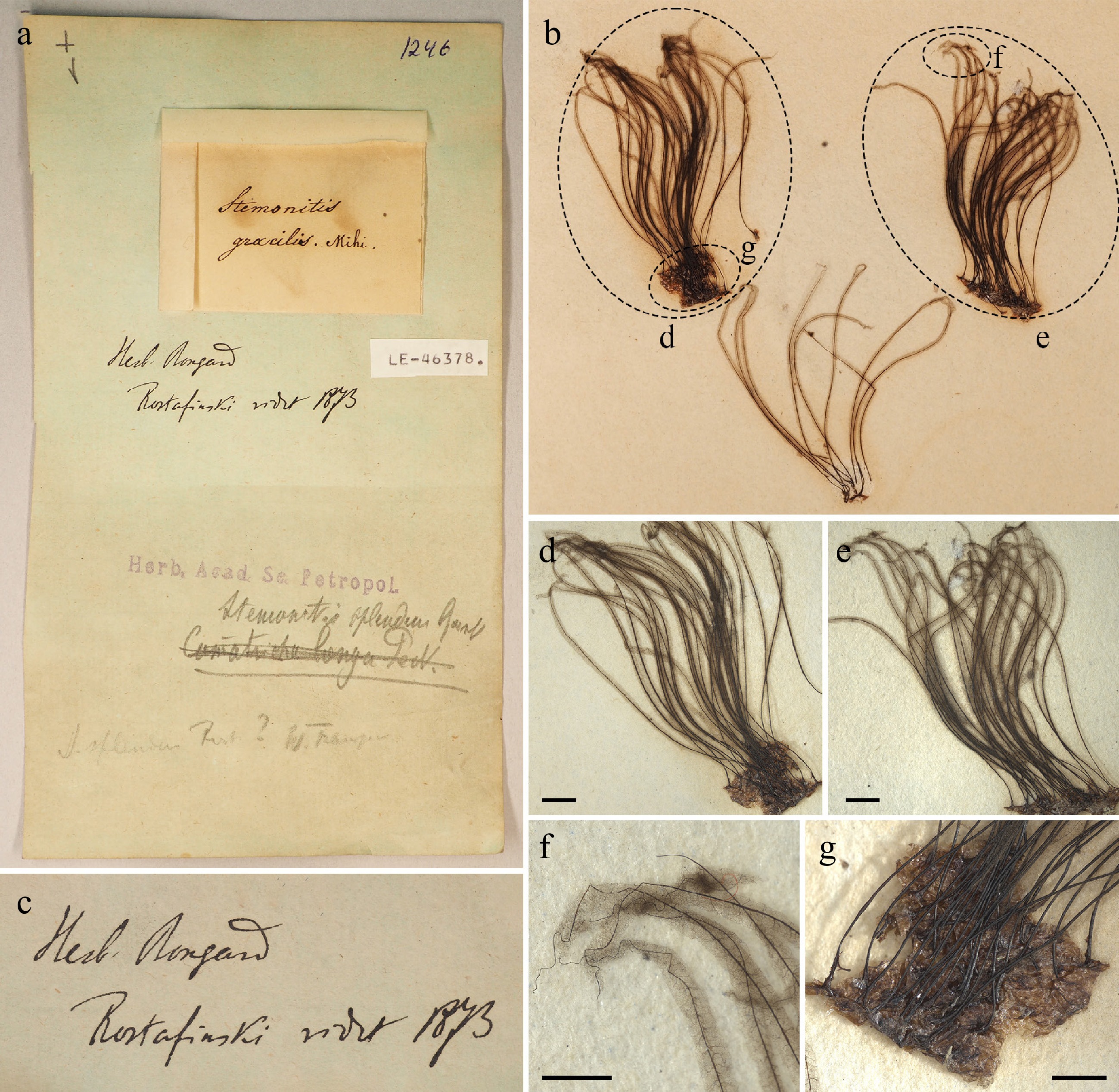
Figure 40.
Herbarium envelope and mature sporocarps of the lectotype (LE46378) of Symphytocarpus splendens. (a) Herbarium labels and interior views of the herbarium box containing the lectotype. (b) The interior views of the herbarium box. (c) The notes 'Herb. Bongard' and 'Rostafinski vidit 1873'. (d), (e) Mature sporocarps (DM). (f) Apical view of the sporotheca (DM). (g) Hypothallus (DM). Scale bars: (d), (e) = 2 mm; (f), (g) = 1 mm.
-
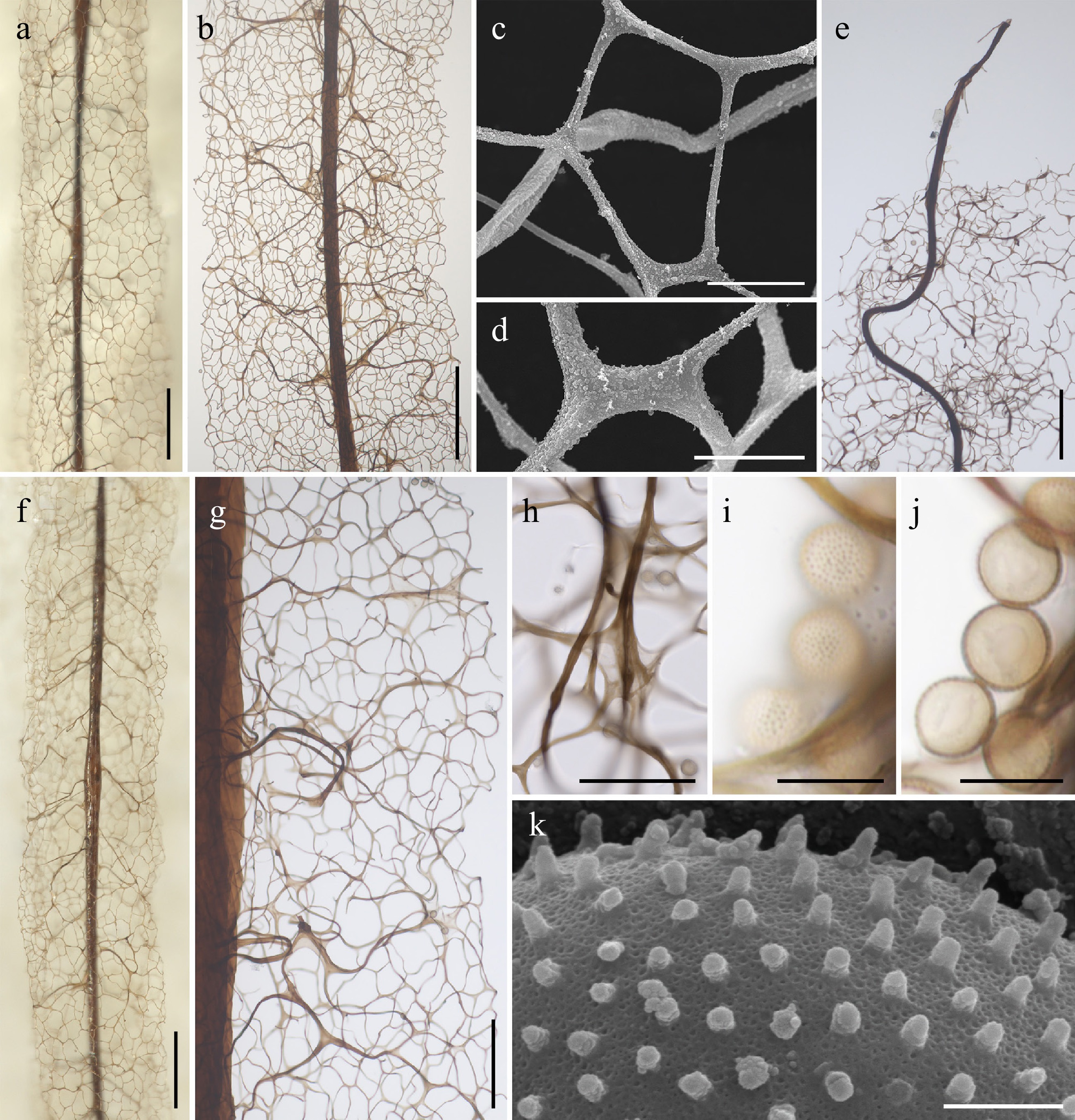
Figure 41.
Morphological characteristics of the lectotype (LE46378) of Symphytocarpus splendens. (a) Surface net (DM). (b) Surface net (LM). (c), (d) Detail of the surface net (SEM). (e) Apical view of the sporotheca (LM). (f) Capillitium and surface net in the middle part of the sporotheca (DM). (g) Capillitium and surface net in the middle part of the sporotheca (LM). (h) Extensions of the internal capillitium (LM). (i), (j) Spores (LM). (k) Detail of the spore ornamentation (SEM). Scale bars: (a), (b), (f) = 200 μm; (e), (g) = 100 μm; (c), (d), (h)–(j) = 10 μm; (k) = 1 μm.
-
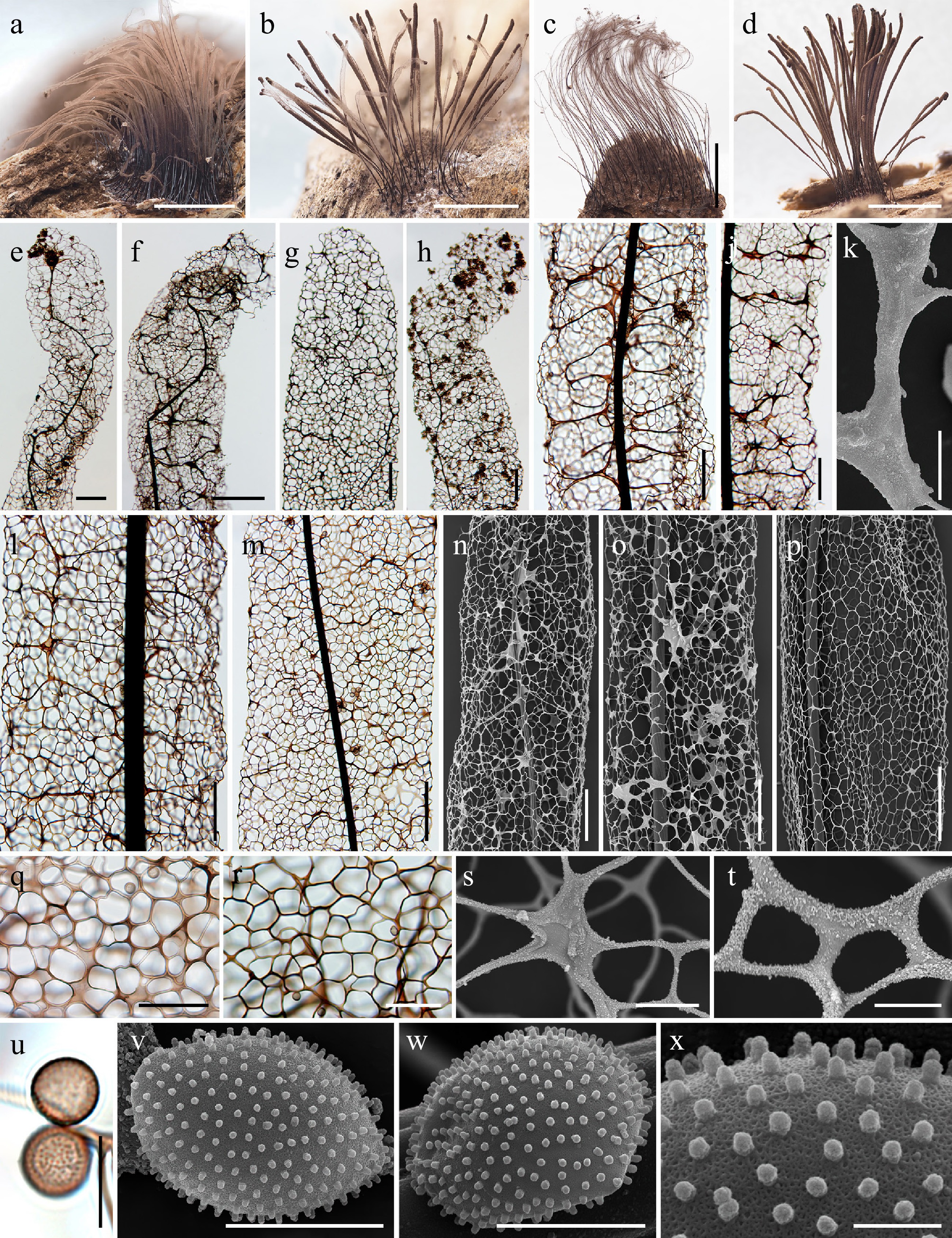
Figure 42.
Morphological characteristics of Symphytocarpus splendens (a), (p) HFNNU 13261; (b) HFNNU 10977; (c) HMAS 74774; (d) HFNNU 10990; (e), (l) HMAS 72488; (f) HMAS 78091; (g) HMAS 0294767; (h) HMAS 72518; (i) HFNNU 12500; (j) HMAS 0295162; (k), (w) MYX 25029; (m) HMAS 0295149; (n)–(o) HMAS 0294776; (q) HMAS 0294767; (r) HMAS 0295150; (s)–(t) HFNNU 10977; (u) HMAS 0258263; (v), (x) MYX 19746. (a)–(d) Mature sporocarps (DM). (e)–(h) Apical region of the sporocarp (LM). (i), (j) Capillitium in the middle part of the sporotheca (LM). (k) Detail of the capillitium threads (SEM). (l), (m) Capillitium in the middle part of the sporotheca (LM). (n)–(p) Surface net (SEM). (q), (r) Detail of the surface net (LM). (s), (t) Detail of the surface net (SEM). (u) Spores (LM). (v), (w) Spore (SEM). (x) Detail of the spore ornamentation (SEM). Scale bars: (a)–(d) = 3 mm; (e)–(j), (l)–(p) = 100 μm; (q), (r) = 10 μm; (k), (s), (u) = 10 μm; (t), (v), (w) = 5 μm; (x) = 1 μm.
-
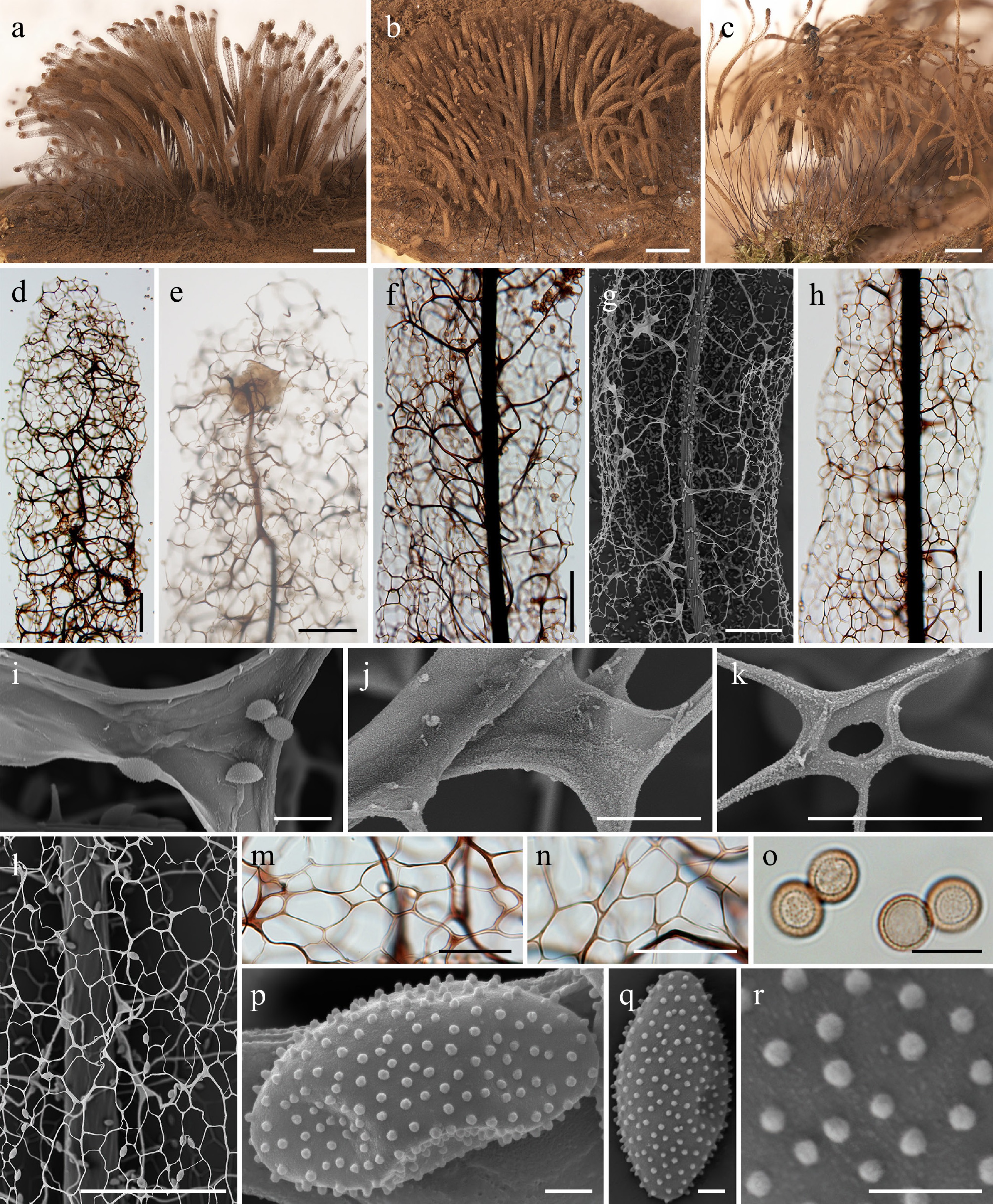
Figure 43.
Morphological characteristics of Symphytocarpus tenuipes (a), (f), (h), (j), (l), (q), (r) LE289704; (b), (e), (p) LE289716; (c) LE289730; (d), (g), (i), (k), (m)–(o). LE285125. (a)–(c) Mature sporocarps (DM). (d), (e) Apical region of the sporotheca (LM). (f) Capillitium in the middle part of the sporotheca (LM). (g) Capillitium in the middle part of the sporotheca (SEM). (h) Surface net (LM). (i), (j) Detail of the capillitium threads (SEM). (k), (l) Detail of the surface net (SEM). (m), (n) Detail of the surface net (LM). (o) Spores (LM). (p), (q) Spore (SEM). (r) Detail of the spore ornamentation (SEM). Scale bars: (a)–(c) = 2 mm; (d)–(h), l = 100 μm; (m), (n) = 10 μm; (i)–(k), (o) = 10 μm; (p)–(r) = 1 μm.
-
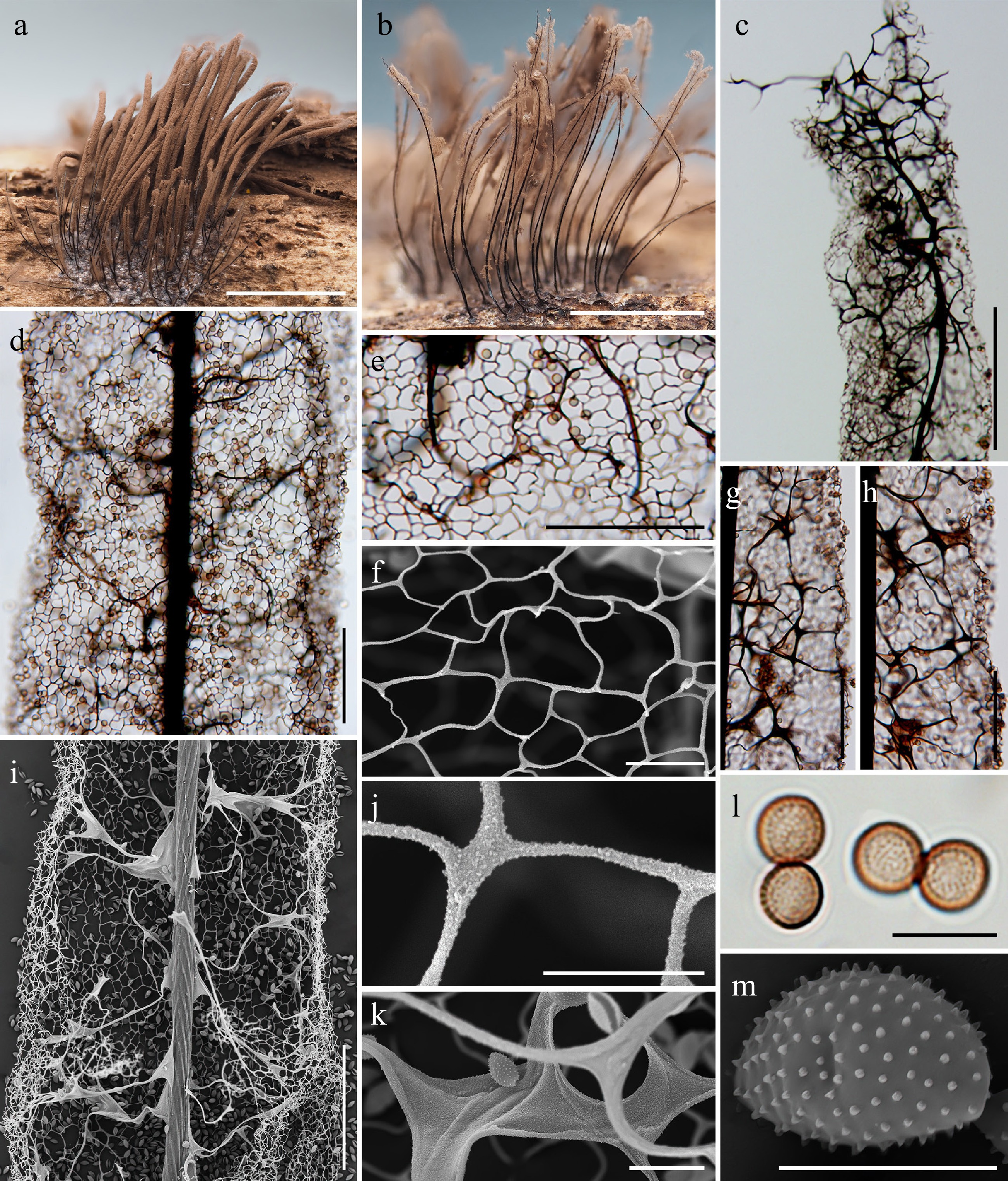
Figure 44.
Morphological characteristics of Symphytocarpus uniforatus (a), (c), (f)–(m) HFNNU 4214; (b), (d), (e) HFNNU 12194. (a), (b) Mature sporocarps (DM). (c) Apical view of the sporotheca (LM). (d) Surface net (LM). (e) Detail of the surface net (LM). (f) Detail of the surface net (SEM). (g), (h) Capillitium in the middle part of the sporotheca (LM). (i) Capillitium in the middle part of the sporotheca (SEM). (j) Detail of the surface net (SEM). (k) Detail of the capillitium threads (SEM). (l) Spores (LM). (m) Spore (SEM). Scale bars: (a), (b) = 3 mm; (c) = 200 μm; (d), (e), (g)–(i) = 100 μm; (f), (k), (l) = 10 μm; (j), (m) = 5 μm.
-
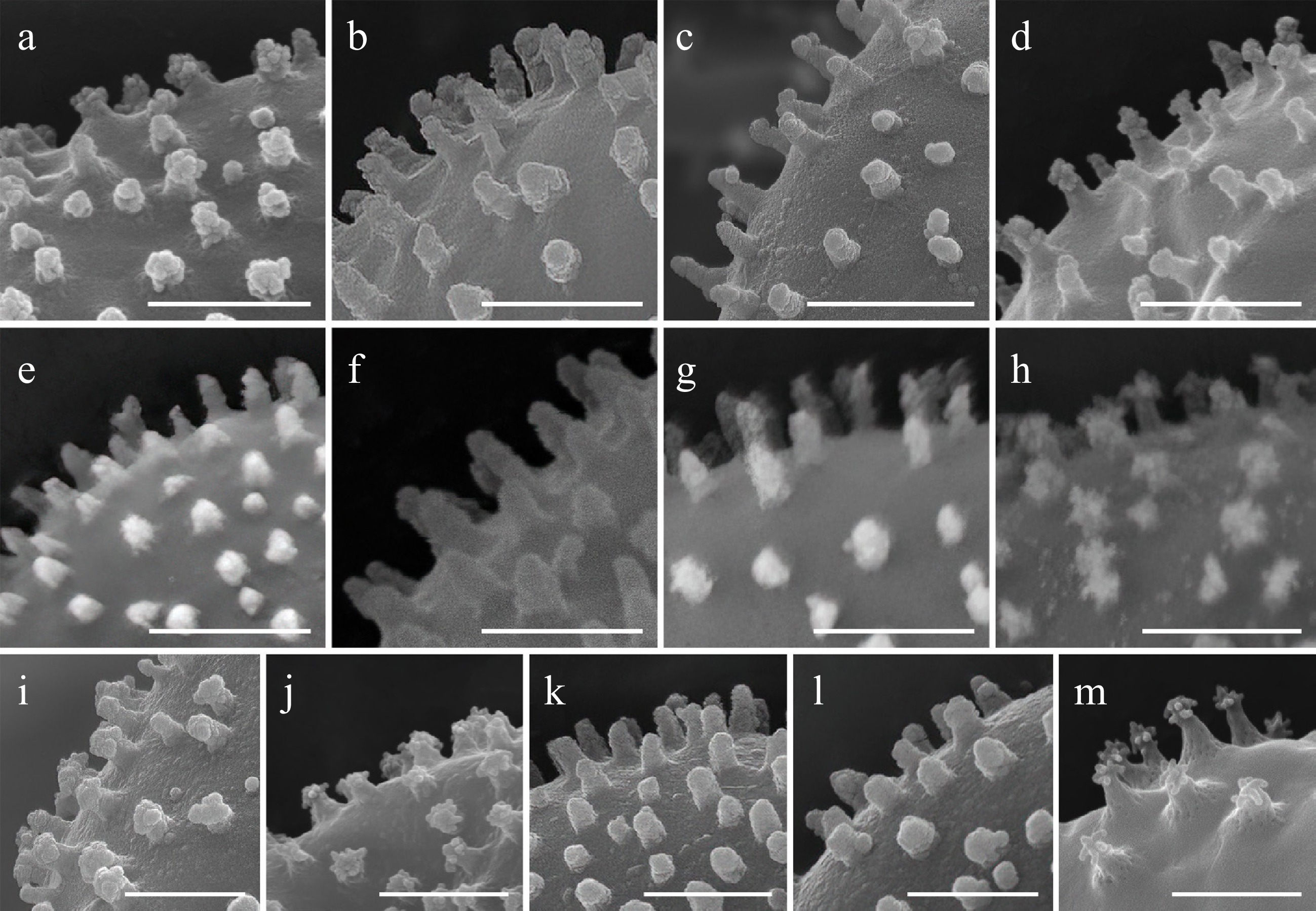
Figure 45.
Comparison of spore ornamentation among Corallosporopsis species involved in this study. (a) Corallosporopsis flavogenita LE325169. (b) Corallosporopsis flavogenita LE325481. (c) Corallosporopsis cf. gracilis MYX 22279. (d) Corallosporopsis cf. gracilis LE348922. (e) Corallosporopsis cf. herbatica HMAS 98461. (f) Corallosporopsis cf. herbatica LE348810. (g) Corallosporopsis laxifila TNS-M-H-4376. (h) Corallosporopsis pallidofila TNS-M-Y-22050. (i) Corallosporopsis gabrieliana LE306548. (j) Corallosporopsis gabrieliana LE306833. (k) Stemonitopsis cf. aequalis MYX 15509. (l) Corallosporopsis sp. LE325737. (m) Corallosporopsis spinispora LE297432. Scale bars: 1 μm.
-
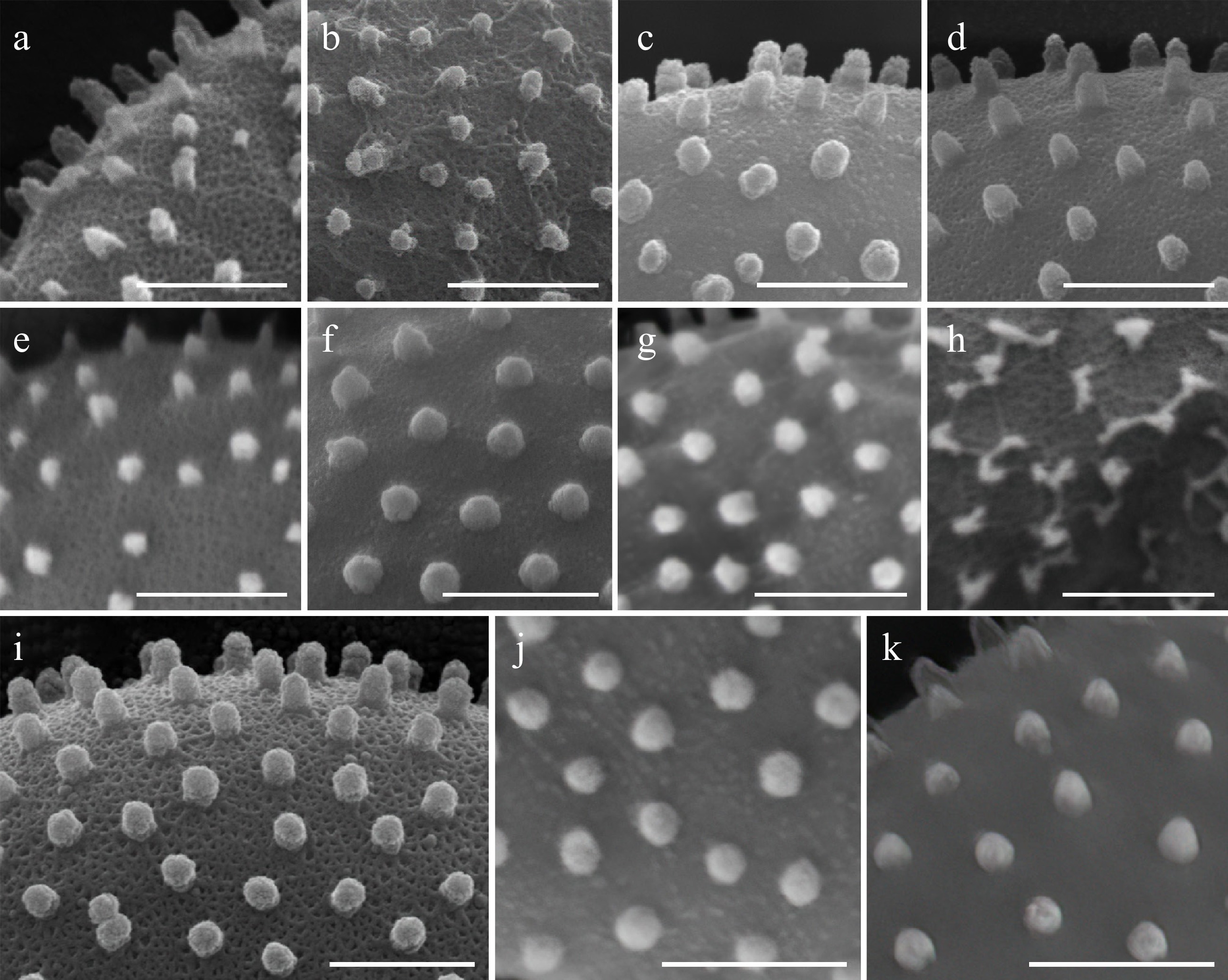
Figure 46.
Comparison of spore ornamentation among Symphytocarpus species involved in this study. (a) Symphytocarpus dispersus LE352558. (b) Symphytocarpus ferrugineus MYX 19217. (c) Symphytocarpus laxicarpicus MYX 23608. (d) Symphytocarpus flaccidus LE47610. (e) Symphytocarpus microsporus TNS-M-Y 22052. (f) Symphytocarpus minor LE348776. (g) Symphytocarpus pseudoflavogenitus HFNNU 11433. (h) Symphytocarpus rubescens TNS-M-Y 22055. (i) Symphytocarpus splendens MYX19796. (j) Symphytocarpus tenuipes LE289704. (k) Symphytocarpus uniforatus HFNNU4214. Scale bars: 1 μm.
-
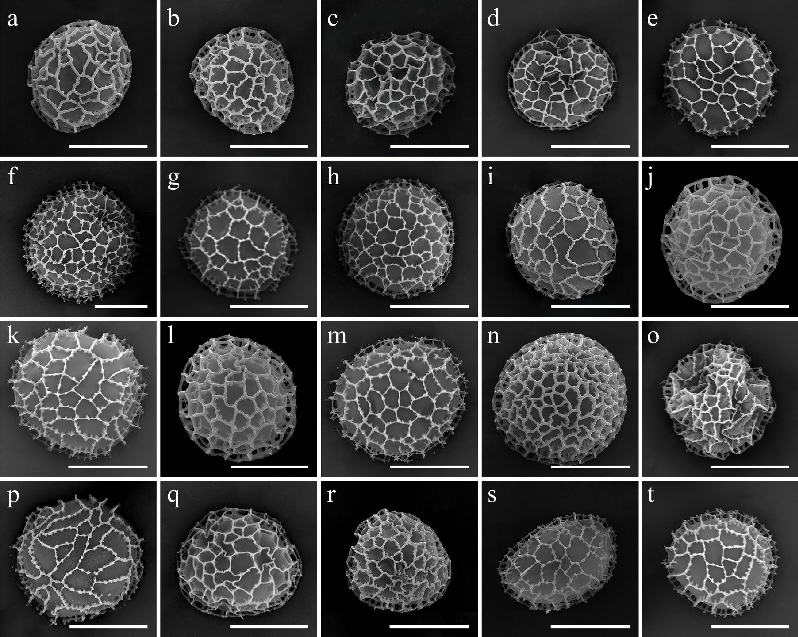
Figure 47.
Comparison of spore ornamentation among Stemonitis s.str. species involved in this study. (a) Stemonitis amaurochaetoides MYX 7126. (b) Stemonitis castillensis LE286562. (c) Stemonitis conferta HFNNU 8578. (d) Stemonitis curiosa TNS-M-Y-22056. (e) Stemonitis curvicornis HFNNU 10969. (f) Stemonitis emotoi HFNNU 10802. (g) Stemonitis cf. foliicola MSK-F 43589. (h) Stemonitis aff. fusca MSK-F 43584. (i) Stemonitis fusca agg. MYX 19459. (j) Stemonitis fuscoides MYX 12516. (k) Stemonitis gracilis TNS-M-Y-22046. (l) Stemonitis cf. gracilis MYX 10257. (m) Stemonitis imperfecta HFNNU 8808. (n) Stemonitis longa MYX 24803. (o) Stemonitis marjana TNS-M-Y-22054. (p) Stemonitis minuta TNS-M-Y-22049. (q) Stemonitis palustris LE346780. (r) Stemonitis cf. pilosa MYX 24418. (s) Stemonitis pinnatiapicalis LE325372. (t) Stemonitis spinimuralis HMAS 0257700. Scale bars: 5 μm.
-
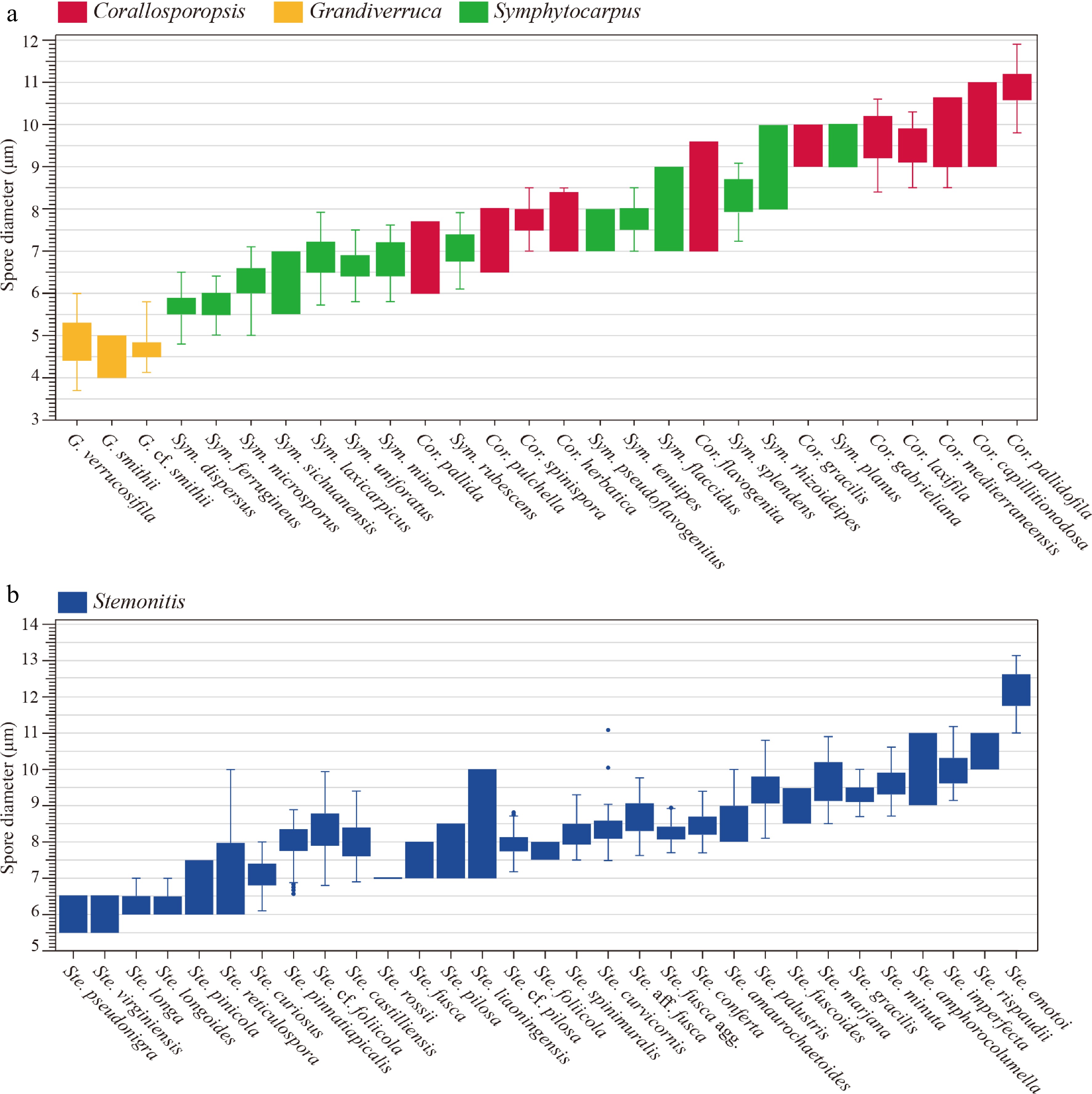
Figure 48.
Comparative analysis of spore diameters. (a) Warted-spore species belonging to Corallosporopsis (red), Grandiverruca (yellow) and Symphytocarpus (green). (b) Reticulate-spore species of Stemonitis (blue). Only species assigned to categories A (Accepted species) and categories B (Morphospecies included but not yet phylogenetically studied) in the taxonomic framework of this study are included; category C species are excluded. Within each panel, species are arranged left-to-right in order of increasing minimum spore diameter; when minima are equal, the order is based on increasing maximum diameter; if both minima and maxima coincide, species are listed alphabetically. Data from specimens examined in this study (including re-examined type material) or original descriptions for species not directly studied.
-
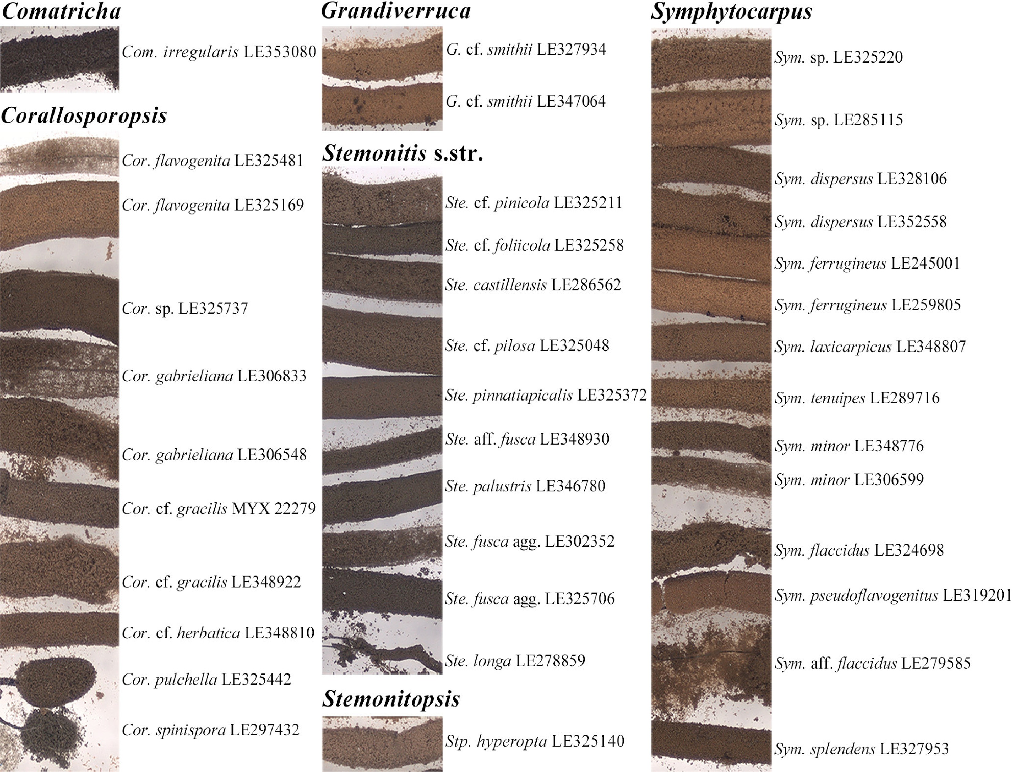
Figure 49.
Comparison of spore mass (sporotheca) coloration under standardized conditions for representative specimens of the genera Comatricha, Corallosporopsis, Grandiverruca, Stemonitis s.str., Stemonitopsis, and Symphytocarpus.
-
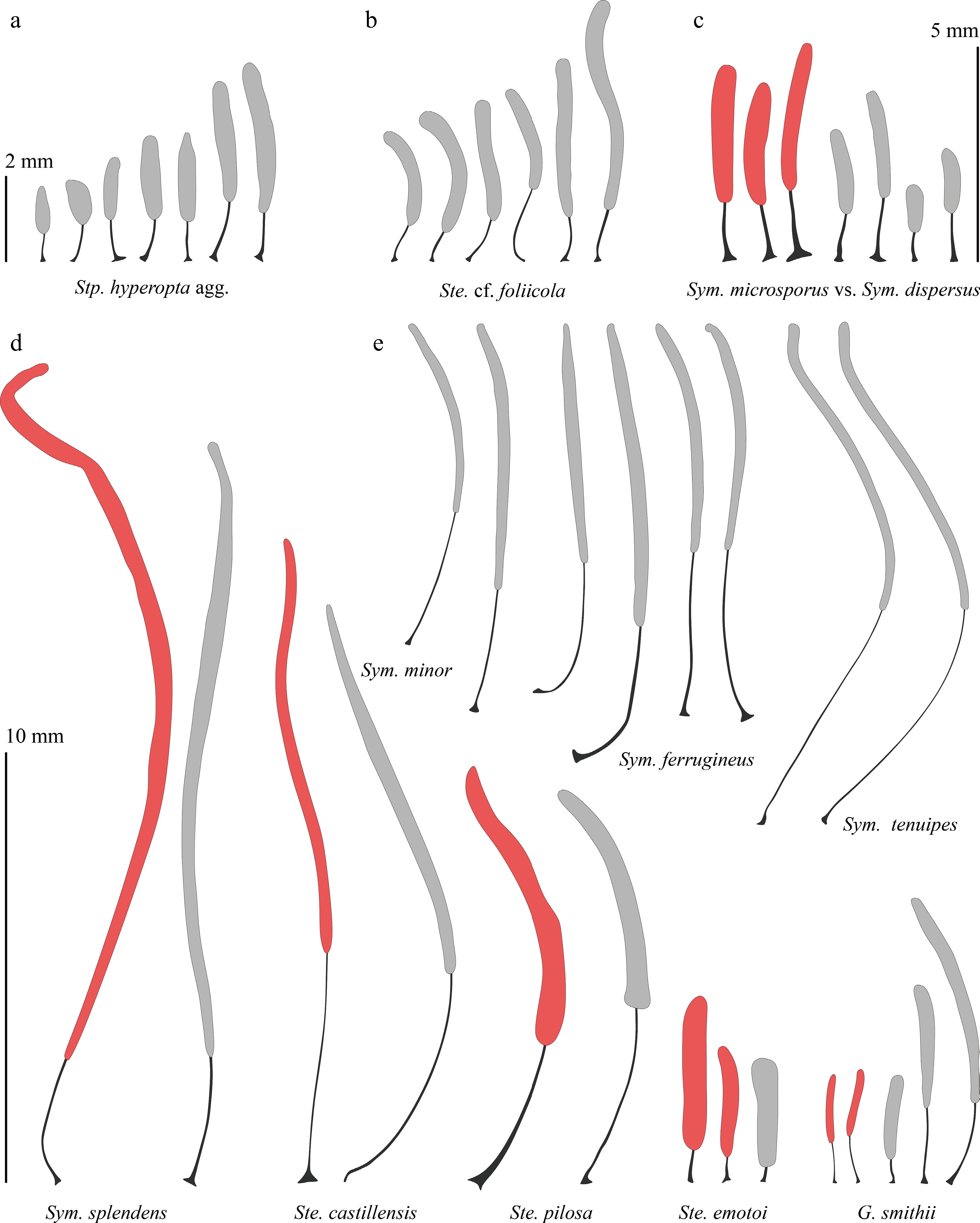
Figure 50.
Silhouettes of some of the discussed species on the same scale. The type specimens are highlighted in red. (a) Variations in the Stemonitopsis hyperopta species complex (LE229360, LE279778, LE325006, LE325108, LE325140, LE353078, mixed). (b) Variations in the species Stemonitis cf. foliicola (LE325205, LE325258, LE348878, mixed). (c) Comparison of related species Symphytocarpus microsporus and Sym. dispersus (TNS-M-Y-22052 [2 pcs.], type from Fig. 20b of Nannenga-Bremekamp & Yamamoto[99], LE348844, LE328106, LE327886, LE352558, from left to right). (d) Comparison of some studied specimens with the types of the corresponding taxa: Sym. splendens (LE46378 and LE327953), Ste. castillensis (type from Macbride [1893], Plate X, Fig. 5, and LE286564), Ste. pilosa (type from Nannenga-Bremekamp et al. [1984], Fig. 2b and LE325048), Ste. emotoi (TNS-M-Y-22053, type from Nannenga-Bremekamp et al., (1984), Fig. 7, HFNNU10801), Grandiverruca smithii (type from Macbride [1893], Plate X, Fig. 4, LE327934 [2 pcs.], LE347064). Everything is from left to right. (e) Comparison of similar species Sym. minor, Sym. ferrugineus and Sym. tenuipes (LE348772 [2 pcs.], LE245001 [2 pcs.], LE328110 [2 pcs.], LE289730 [2 pcs.]). Everything is from left to right.
-
Gene Name F/R Sequence (5′–3′) Amplification protocol nrSSU S1 F AACCTGGTTGATCCTGCC 2 min at 98 °C, 35 cycles (10 s at 95 °C, 10 s at 56 °C, 15 s at 72 °C) and 5 min at 72 °C S2 F TGGTTGATCCTGCCAGTAGTGT SU19R R GACTTGTCCTCTAATTGTTACTCG SSU_rev R AGACTTGTCCTCYAATTGTTAC EF-1α PB1F F ACCCGTGAGCACGCTCTCCT 2 min at 98 °C, 35 cycles (10 s at 98 °C, 10 s at 65.4 °C, 10 s at 72 °C) and 5 min at 72 °C PB1R R CGCACATGGGCTTGGAGGGG EF03 F TGATCTACAAGTGCGGTG 2 min at 98 °C, 35 cycles (10 s at 98 °C, 10 s at 60 °C, 15 s at 72 °C) and 5 min at 72 °C EF04* F TGGGTGTTGGACAAACTC KEF_R3 R CCGTTCTTGATGTTCTTGG STE_EF_F1 F TTCGAGGCTGGTATTGCCAAG 2 min at 98 °C, 35 cycles (10 s at 98 °C, 10 s at 60.4 °C, 15 s at 72 °C) and 5 min at 72 °C STE_EF_R1 R ACGTTGAACCCGACGTTGT mtSSU Kmit_F F AGTGTTATTCGTGATGACTGG 2 min at 98 °C, 35 cycles (10 s at 95 °C, 10 s at 56 °C, 15 s at 72 °C) and 5 min at 72 °C Kmit_Fi* F ATGACTGGGCGTAGGGTA Kmit_R R CGAATTAAACCACATCTCCACC Note: nrSSU, nuclear small subunit ribosomal RNA; EF-1α, elongation factor-1 alpha; mtSSU, mitochondrial small subunit ribosomal RNA. The asterisk (*) designates that the primer was used in a semi-nested polymerase chain reaction. Table 1.
Primer pairs and amplification protocols used in this study.
Figures
(50)
Tables
(1)