-
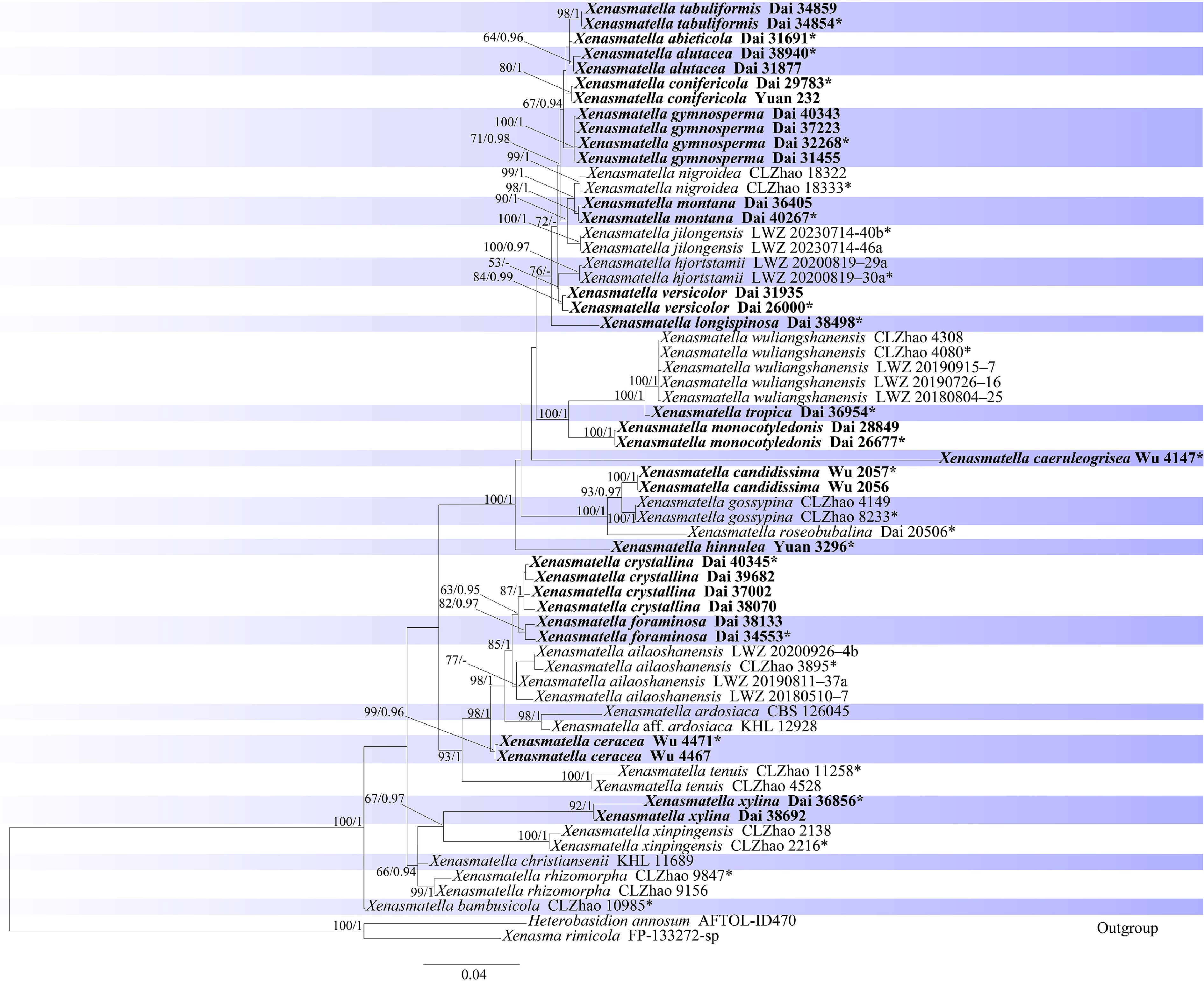
Figure 1.
Maximum likelihood strict consensus tree illustrating the phylogeny of species of order Xenasmatales based on a dataset of ITS + nLSU. ML bootstrap values higher than 60% and Bayesian posterior probability values more than 0.90 are shown. New species are in bold; * indicates type material.
-
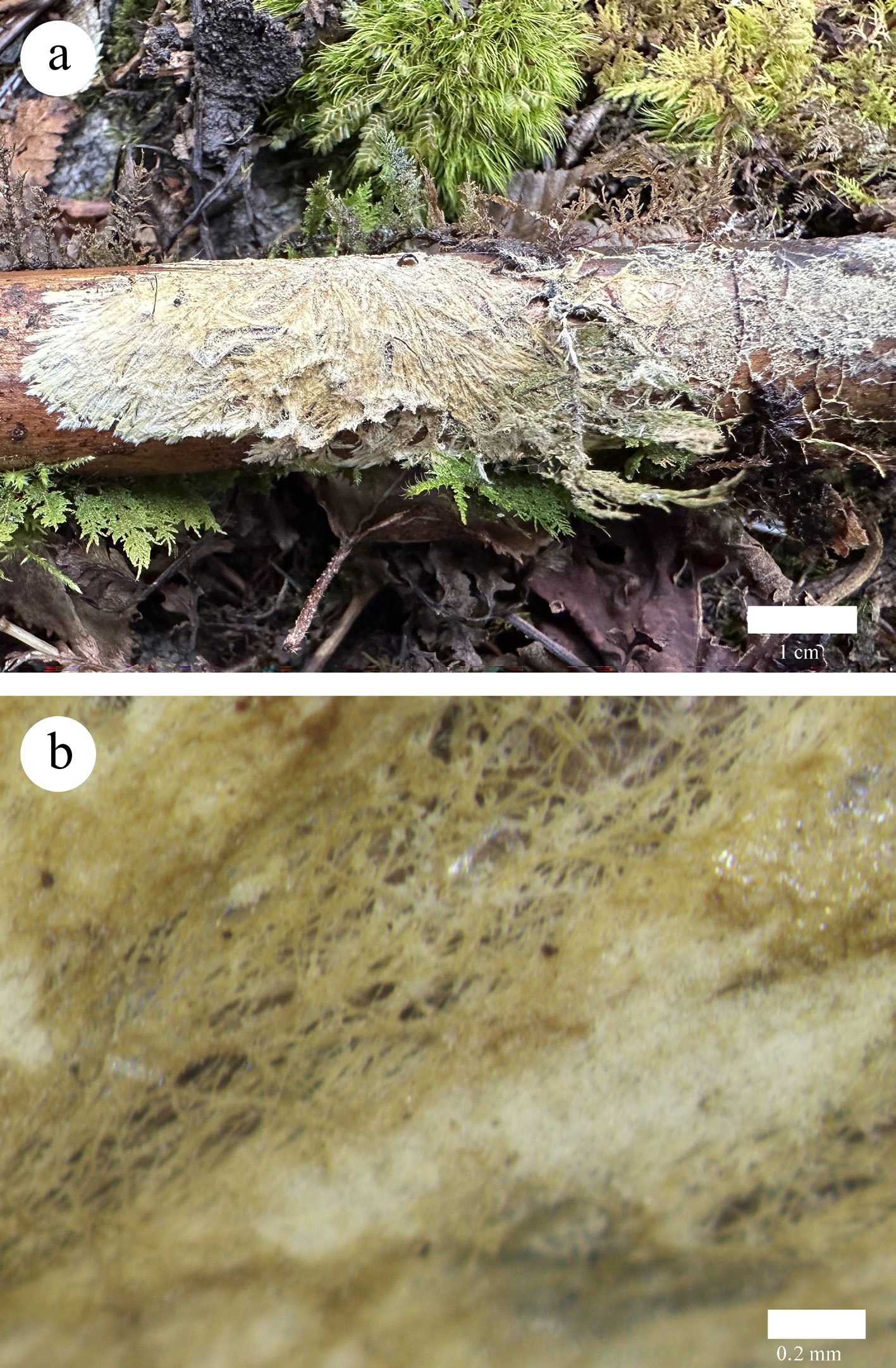
Figure 2.
Basidiomata of Xenasmatella abieticola (Dai 31691, holotype). (a) A fresh basidioma. (b) Dry hymenophore.
-
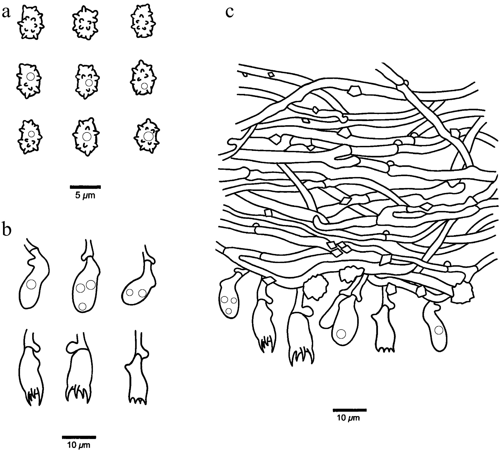
Figure 3.
Microscopic structures of Xenasmatella abieticola (Dai 31691, holotype). (a) Basidiospores. (b) Basidia and basidioles. (c) A section of hymenium.
-
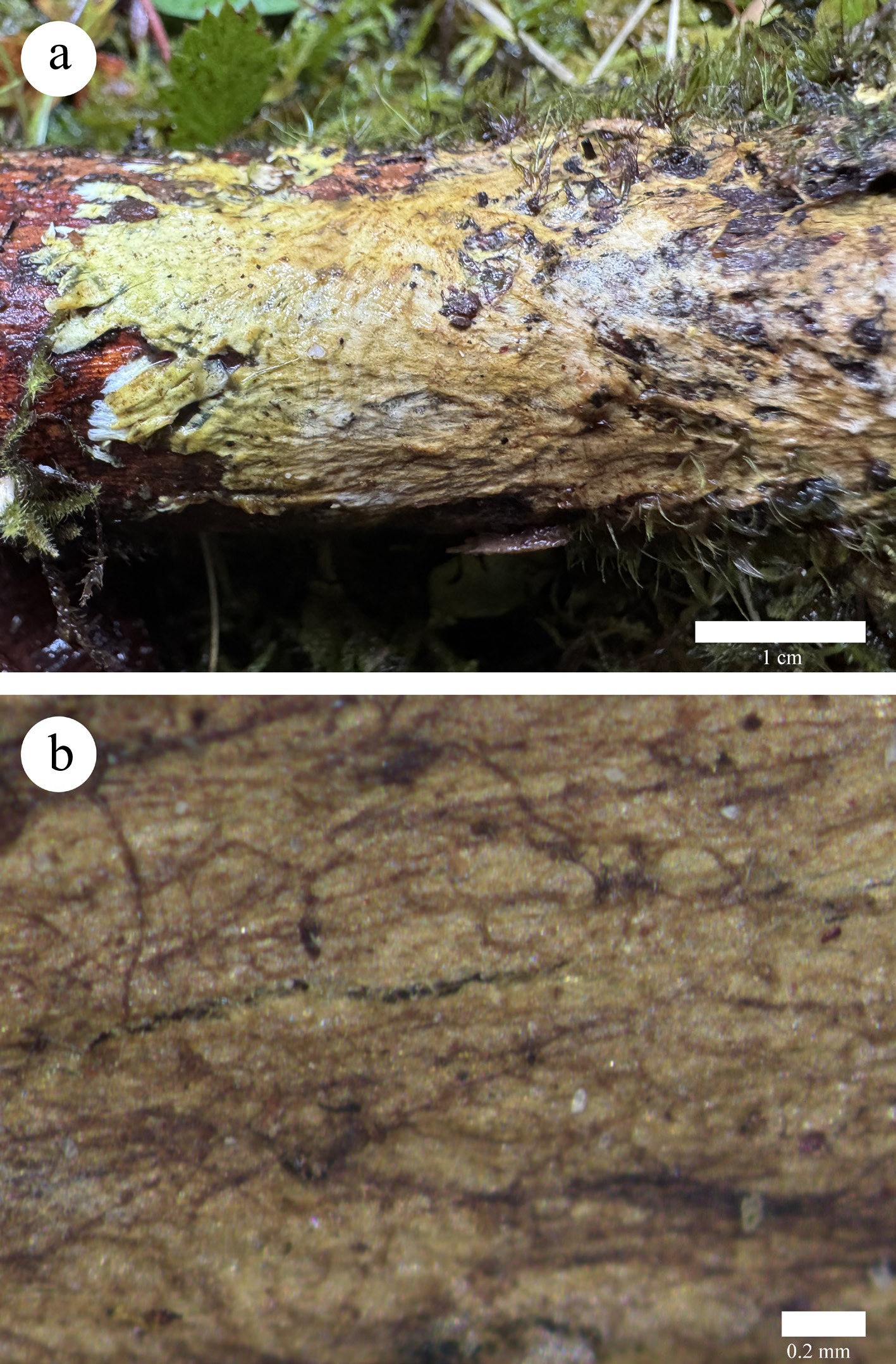
Figure 4.
Basidiomata of Xenasmatella alutacea (Dai 38940, holotype). (a) A fresh basidioma. (b) Dry hymenophore.
-
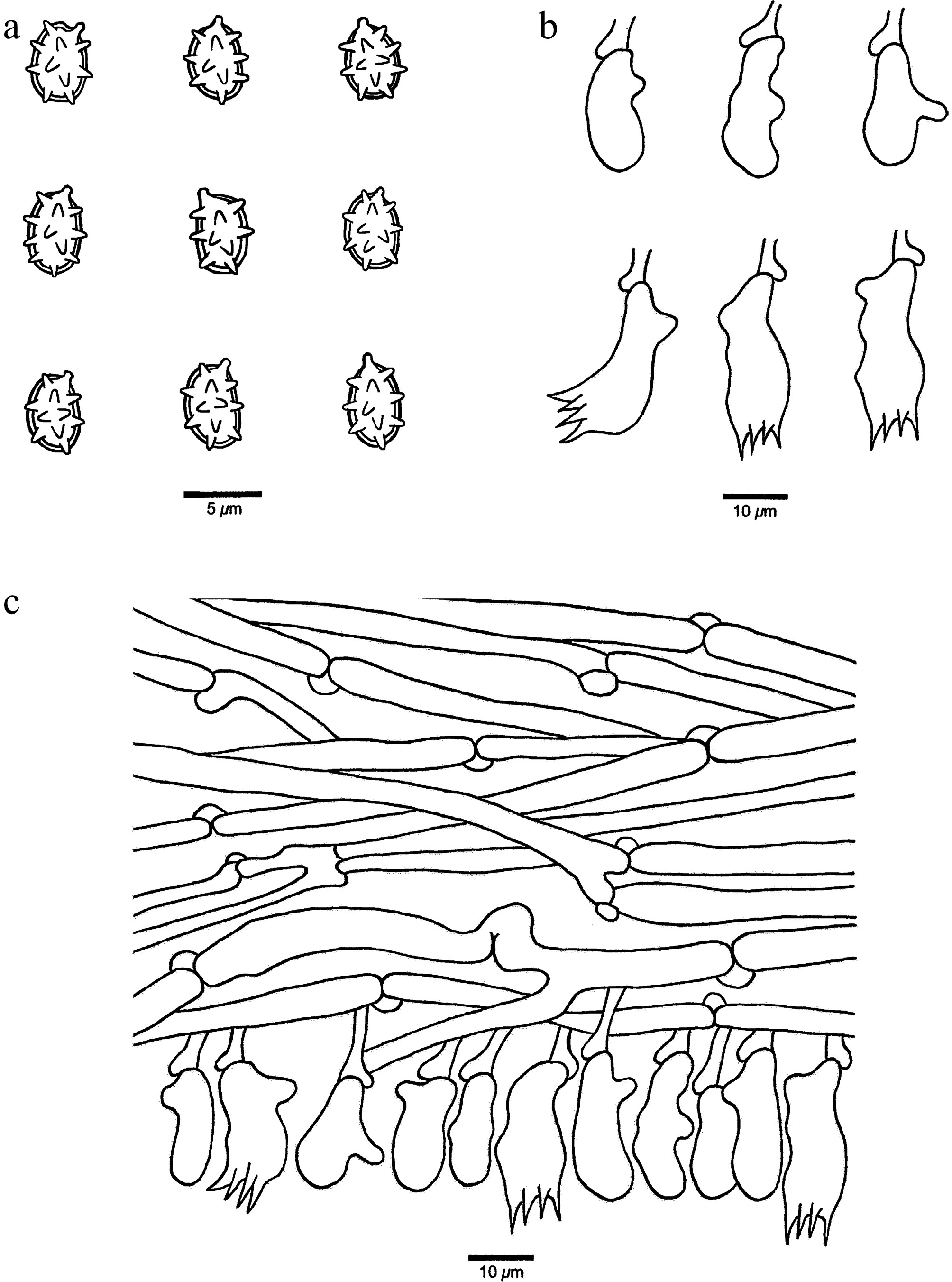
Figure 5.
Microscopic structures of Xenasmatella alutacea (Dai 38940, holotype). (a) Basidiospores. (b) Basidia and basidioles. (c) A section of hymenium.
-
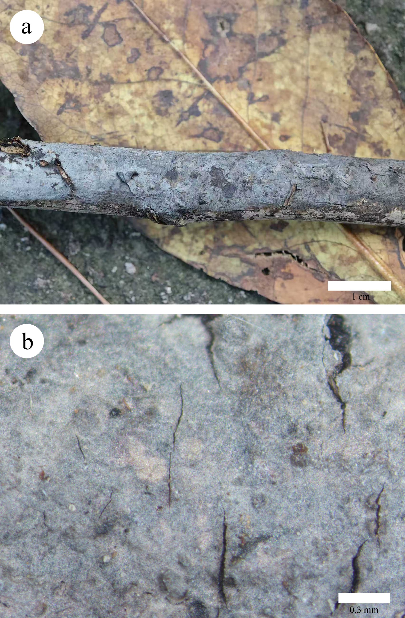
Figure 6.
Basidiomata of Xenasmatella caeruleogrisea (Wu 4147, holotype). (a) A fresh basidioma. (b) Dry hymenophore.
-
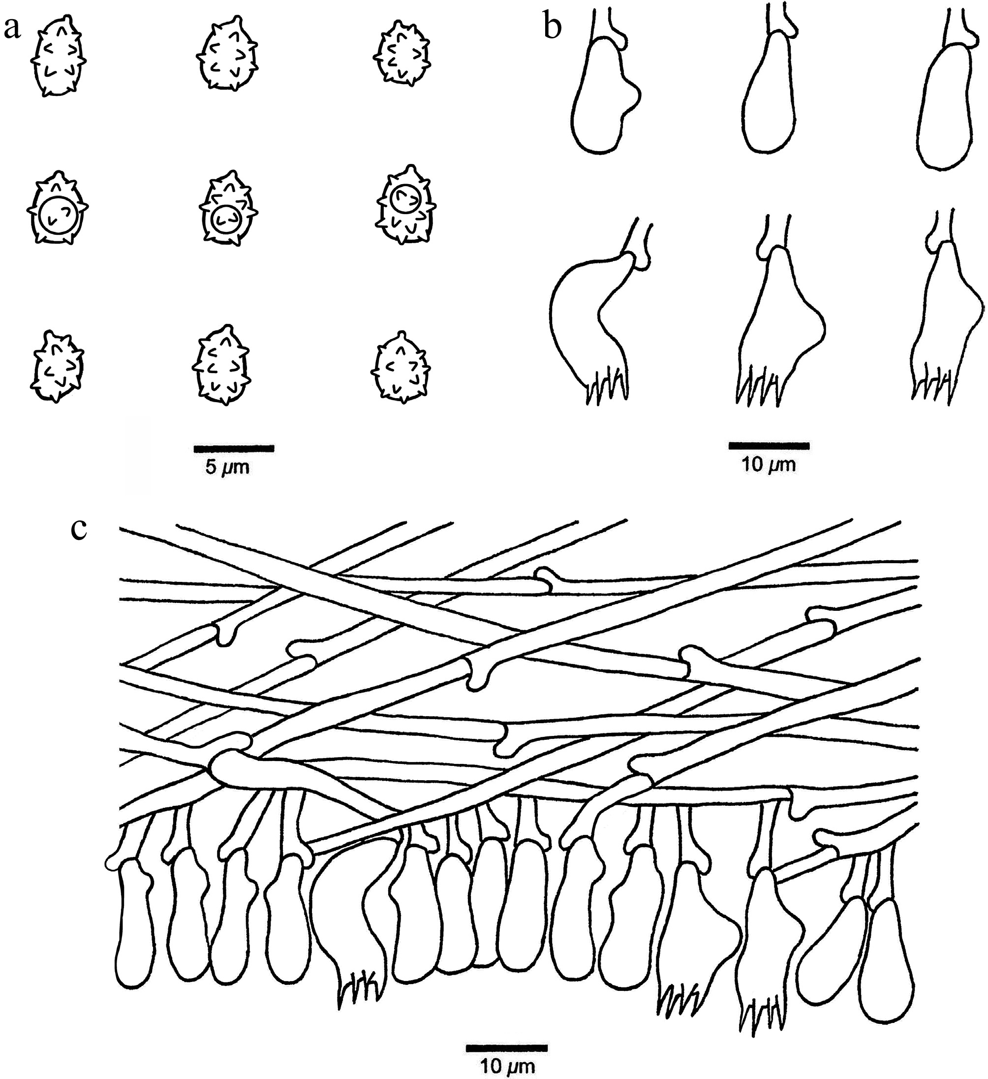
Figure 7.
Microscopic structures of Xenasmatella caeruleogrisea (Wu 4147, holotype). (a) Basidiospores. (b) Basidia and basidioles. (c) A section of hymenium.
-
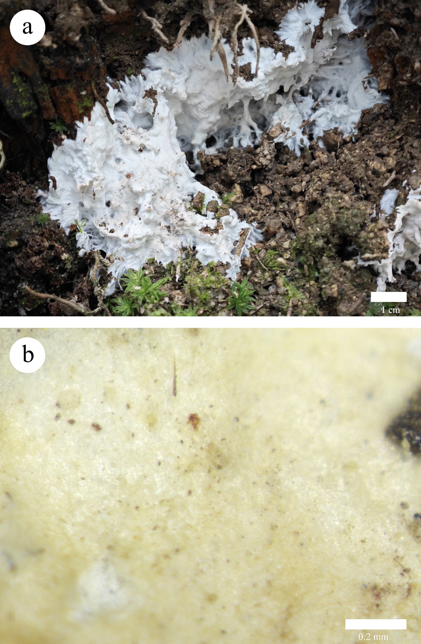
Figure 8.
Basidiomata of Xenasmatella candidissima (Wu 2057, holotype). (a) A fresh basidioma. (b) Dry hymenophore.
-
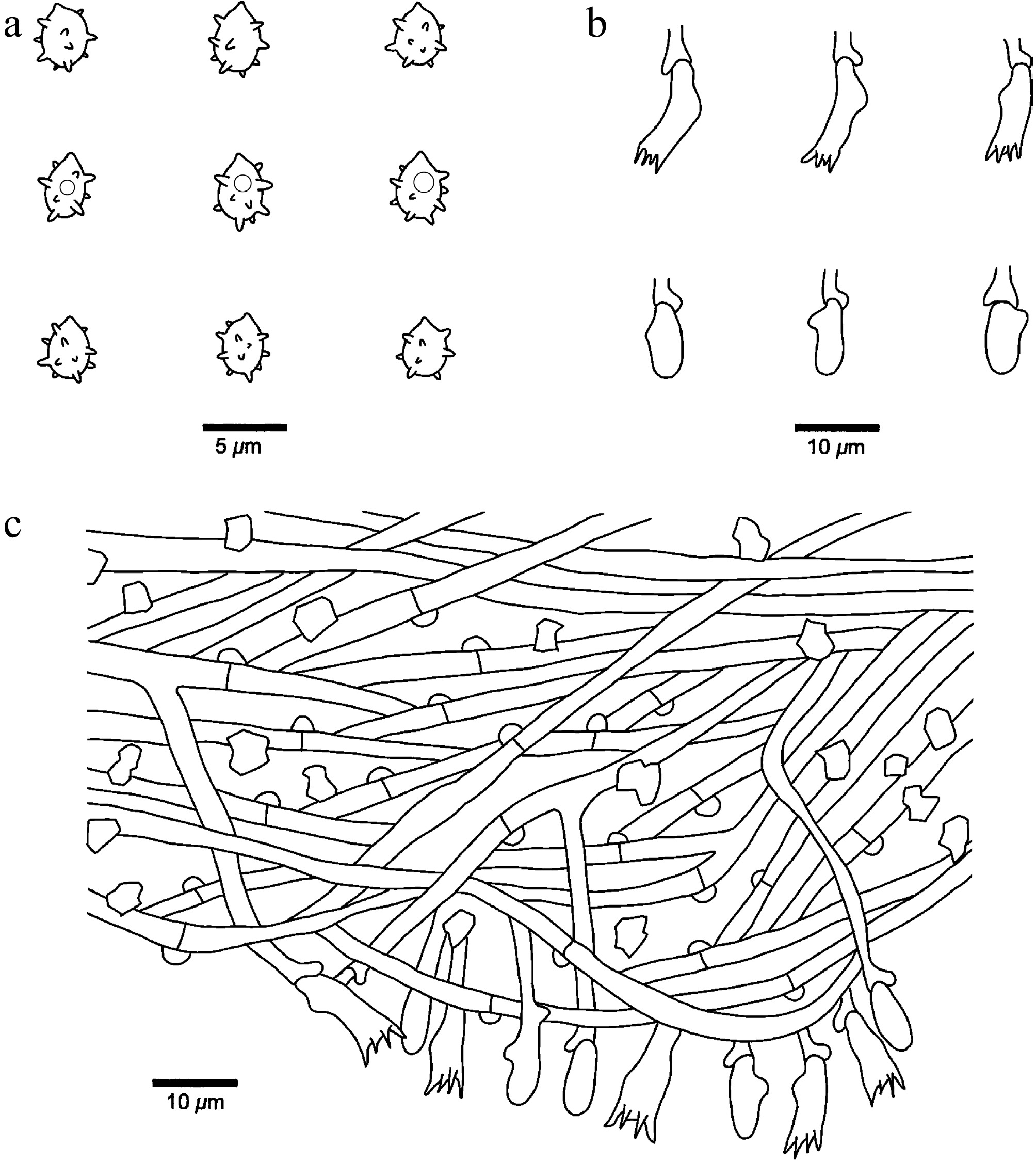
Figure 9.
Microscopic structures of Xenasmatella candidissima (Dai 2057, holotype). (a) Basidiospores. (b) Basidia and basidioles. (c) A section of hymenium.
-
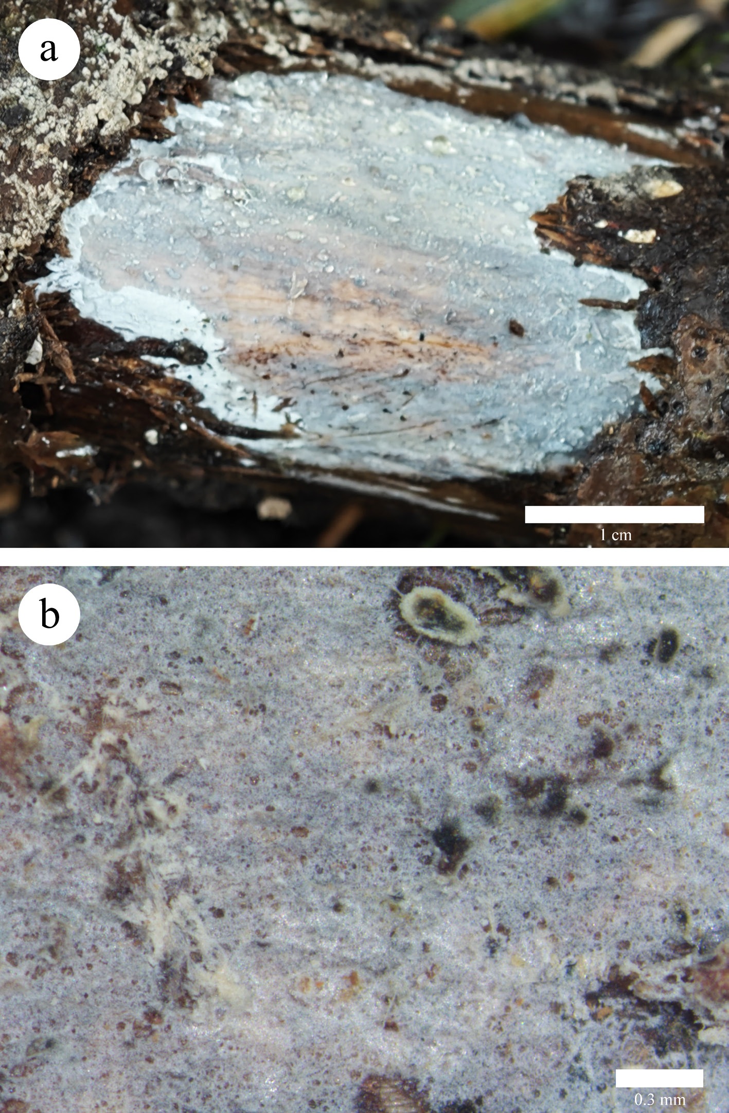
Figure 10.
Basidiomata of Xenasmatella ceracea (Wu 4471, holotype). (a) A fresh basidioma. (b) Dry hymenophore.
-
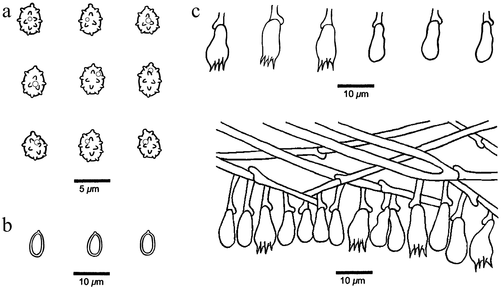
Figure 11.
Microscopic structures of Xenasmatella ceracea (Wu 4471, holotype). (a) Basidiospores. (b) Chlamydospores. (c) Basidia and basidioles. (d) A section of hymenium.
-
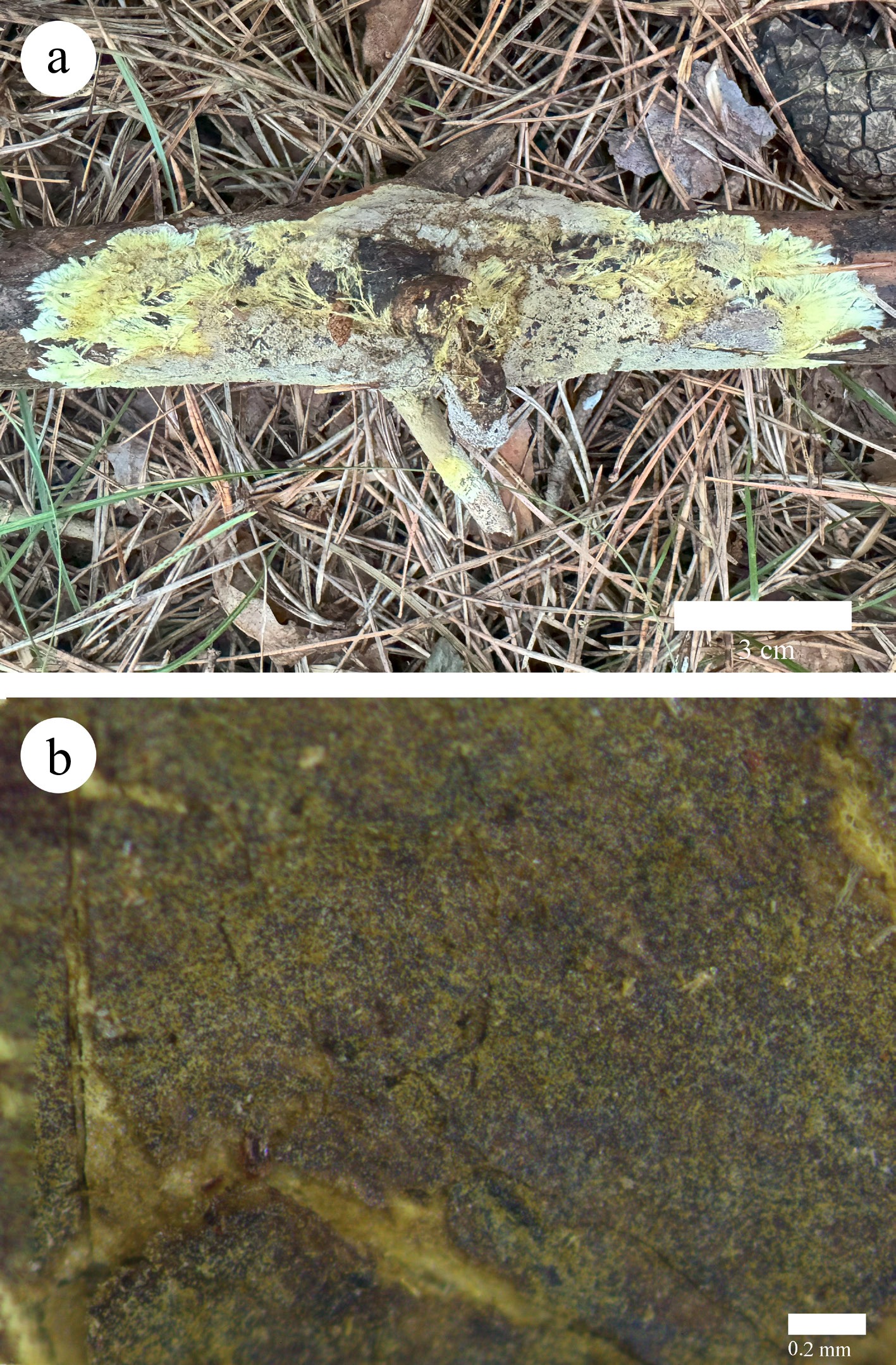
Figure 12.
Basidiomata of Xenasmatella conifericola (Dai 29783, holotype). (a) A fresh basidioma. (b) Dry hymenophore.
-
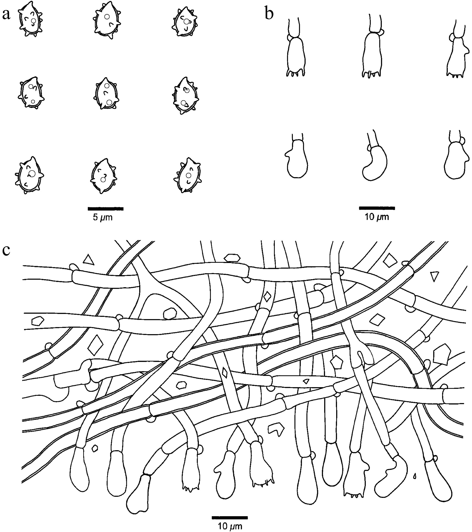
Figure 13.
Microscopic structures of Xenasmatella conifericola (Dai 29783, holotype). (a) Basidiospores. (b) Basidia and basidioles. (c) A section of hymenium.
-
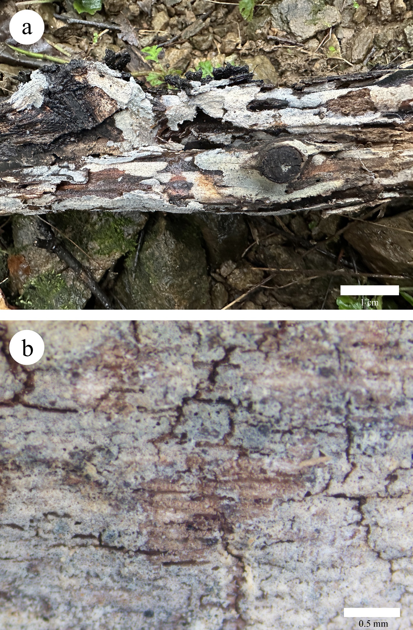
Figure 14.
Basidiomata of Xenasmatella crystallina (Dai 40345, holotype). (a) A fresh basidioma. (b) Dry hymenophore.
-
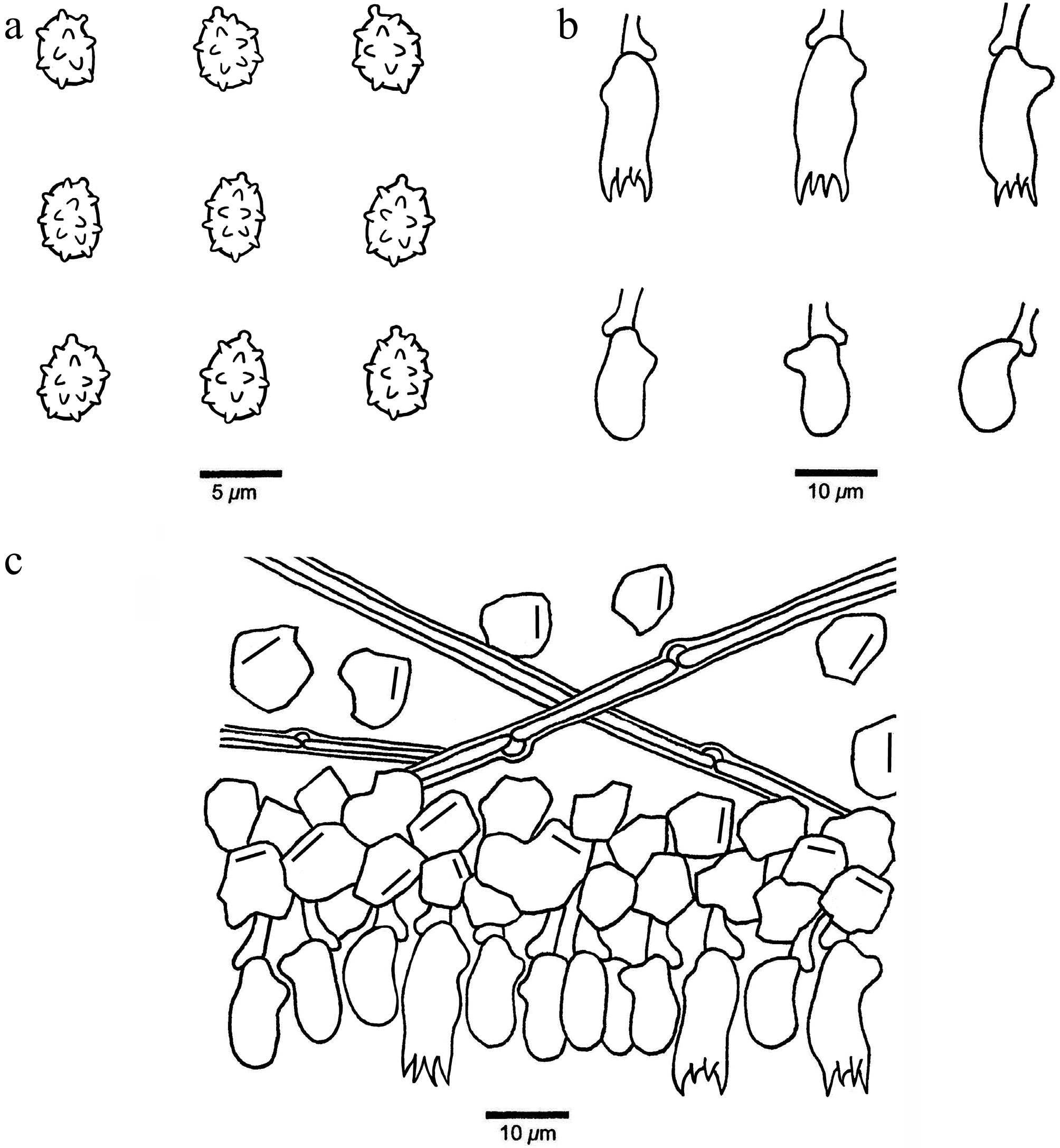
Figure 15.
Microscopic structures of Xenasmatella crystallina (Dai 40345, holotype). (a) Basidiospores. (b) Basidia and basidioles. (c) A section of hymenium.
-
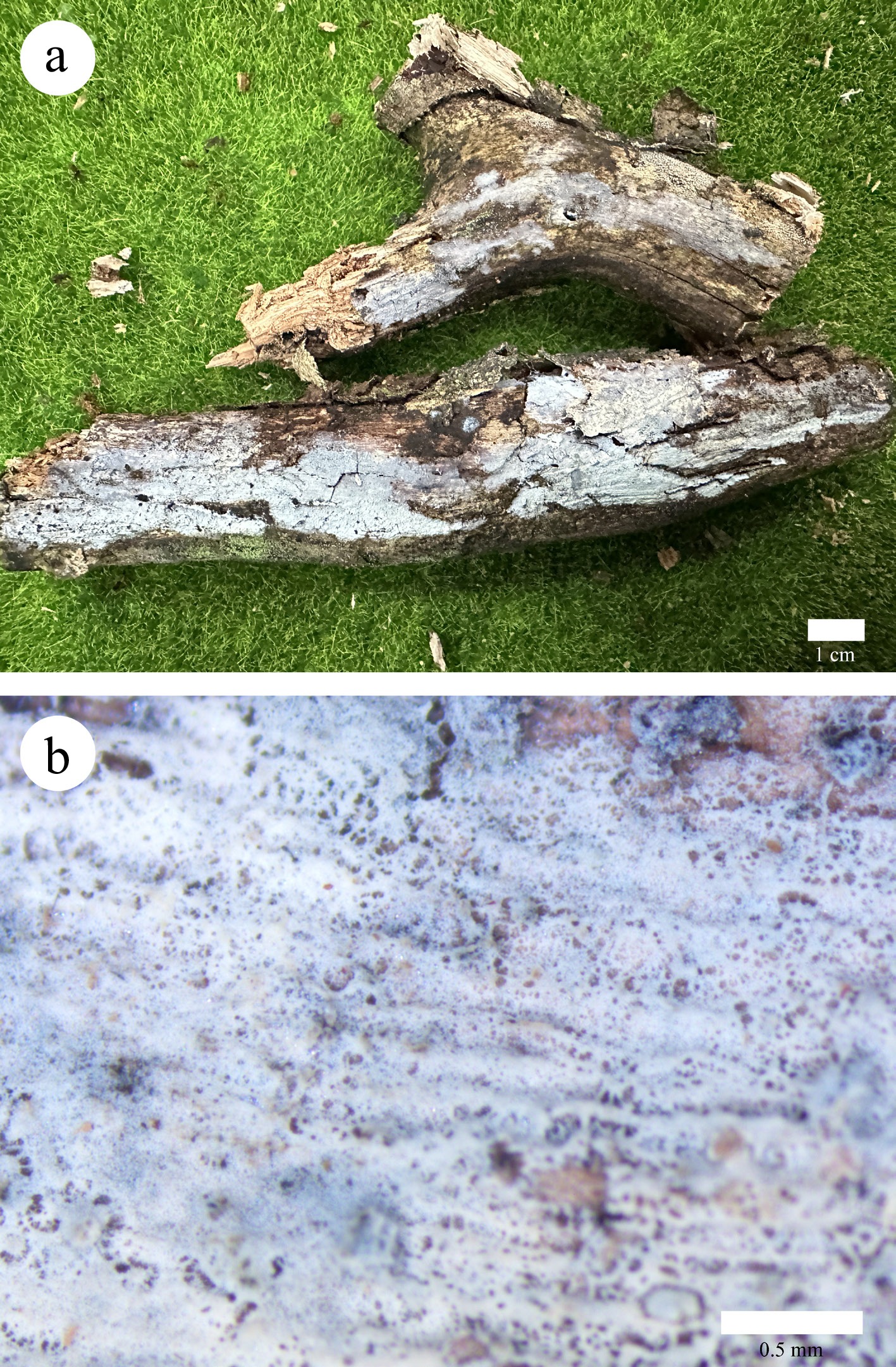
Figure 16.
Basidiomata of Xenasmatella foraminosa (Dai 34553, holotype). (a) A fresh basidioma. (b) Dry hymenophore.
-
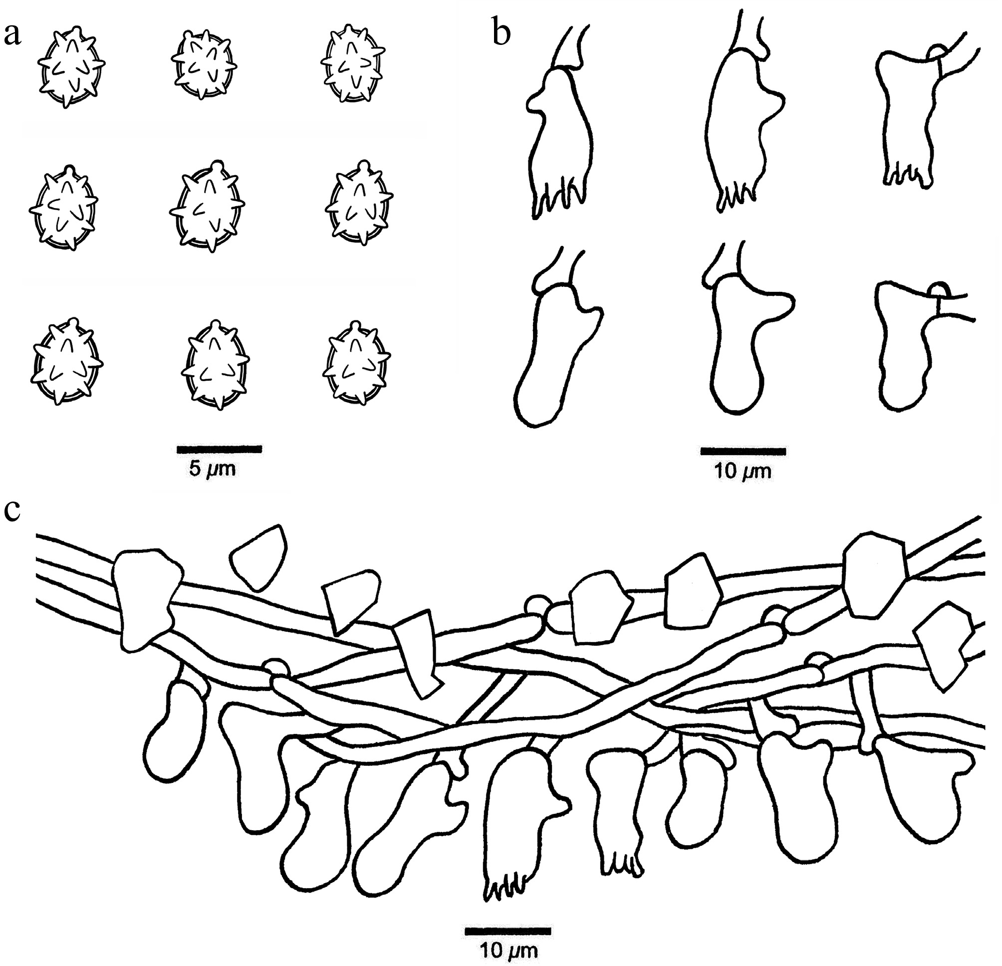
Figure 17.
Microscopic structures of Xenasmatella foraminosa (Dai 34553, holotype). (a) Basidiospores. (b) Basidia and basidioles. (c) A section of hymenium.
-
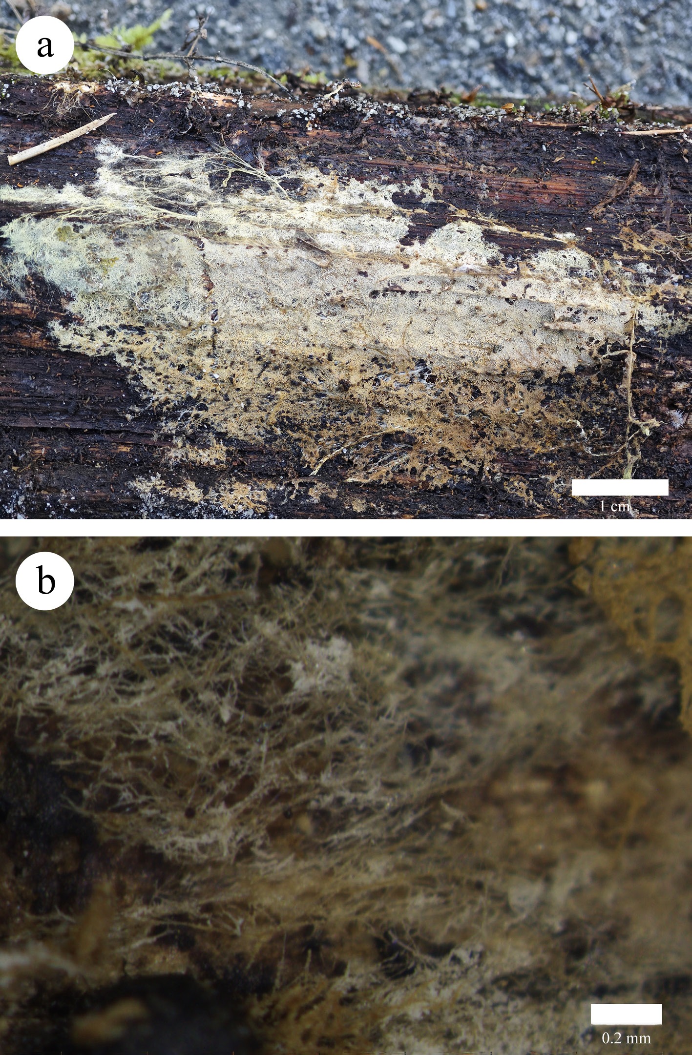
Figure 18.
Basidiomata of Xenasmatella gymnosperma (Dai 32268, holotype). (a) A fresh basidioma. (b) Dry hymenophore.
-
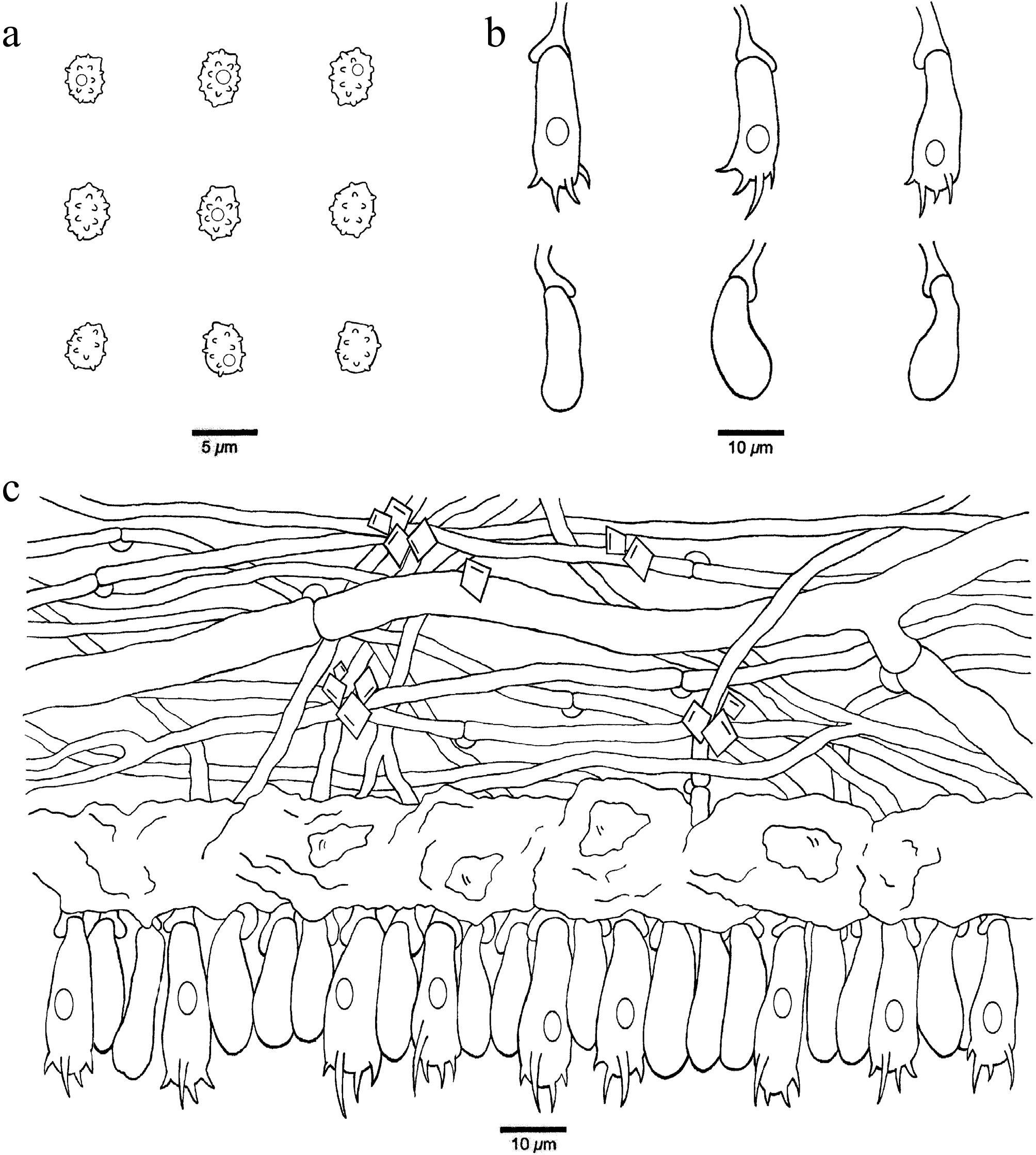
Figure 19.
Microscopic structures of Xenasmatella gymnosperma (Dai 32268, holotype). (a) Basidiospores. (b) Basidia and basidioles. (c) A section of hymenium.
-
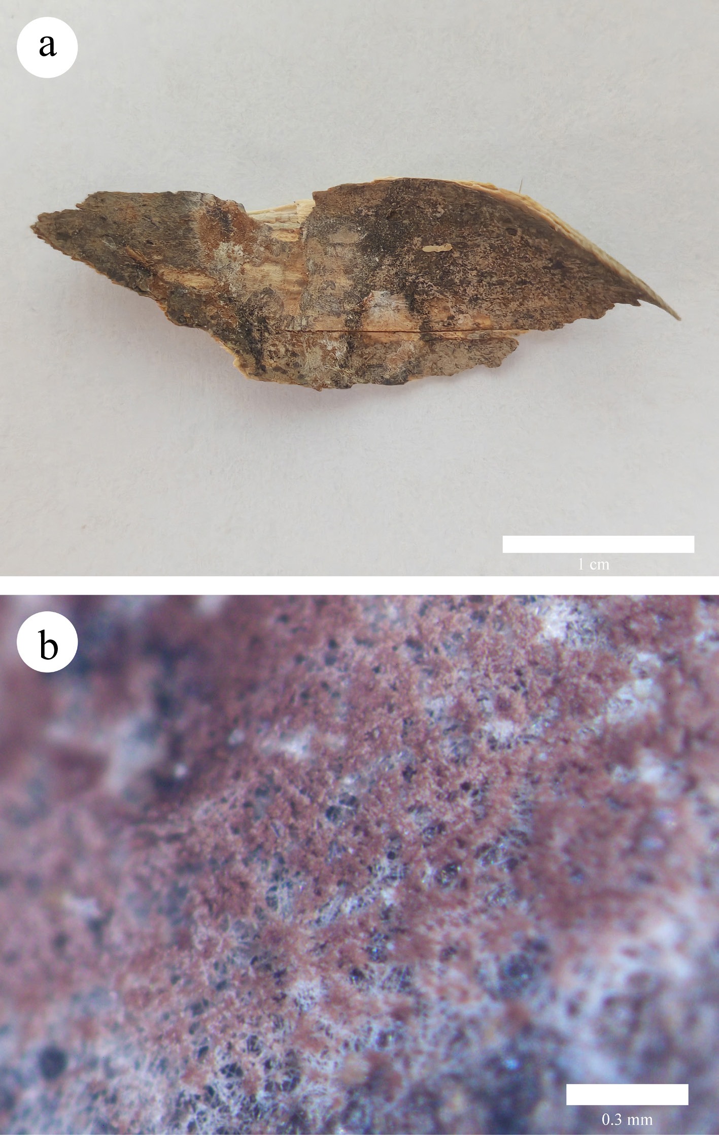
Figure 20.
Basidiomata of Xenasmatella hinnulea (Yuan 3296, holotype). (a) A fresh basidioma. (b) Dry hymenophore.
-
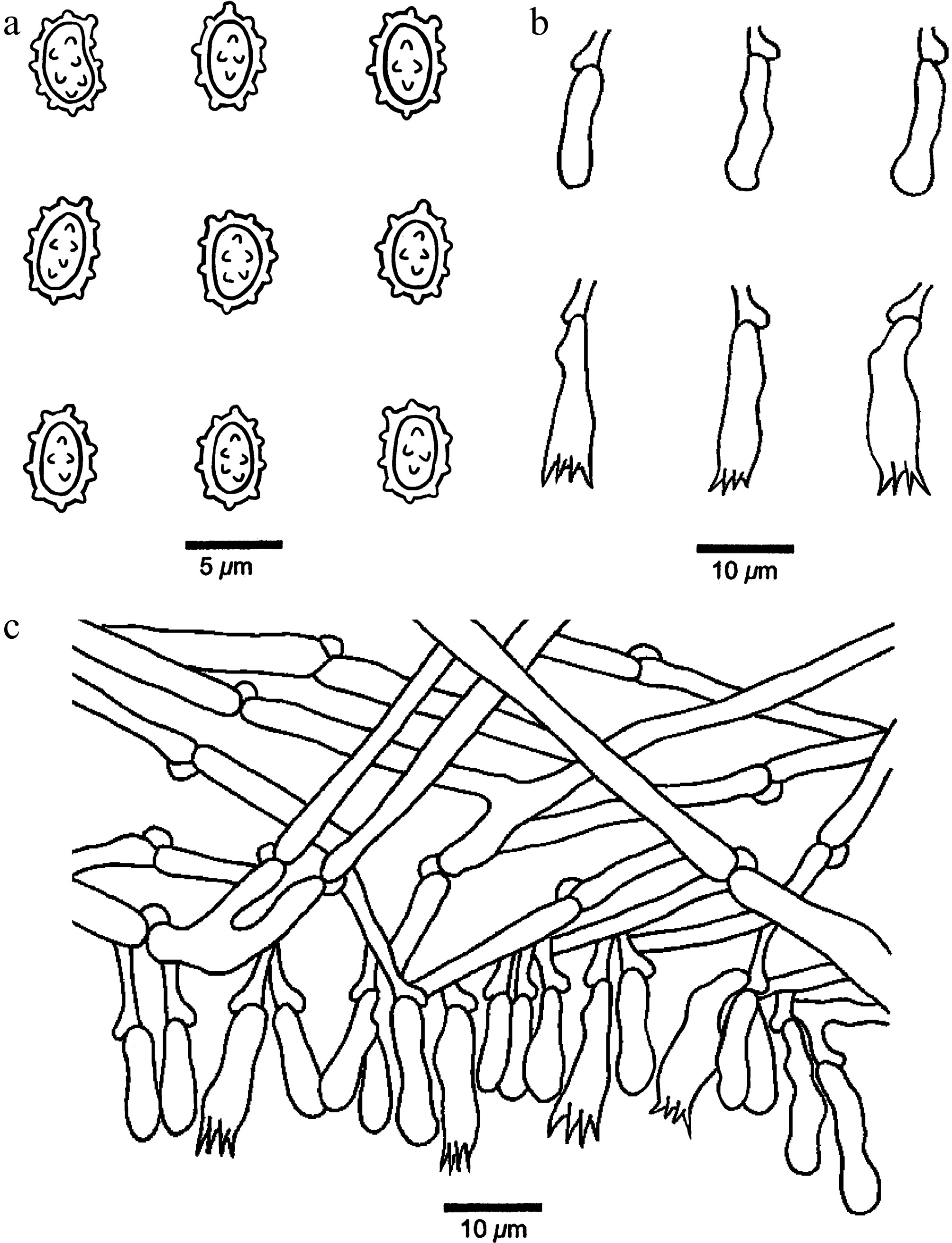
Figure 21.
Microscopic structures of Xenasmatella hinnulea (Yuan 3296, holotype). (a) Basidiospores. (b) Basidia and basidioles. (c) A section of hymenium.
-
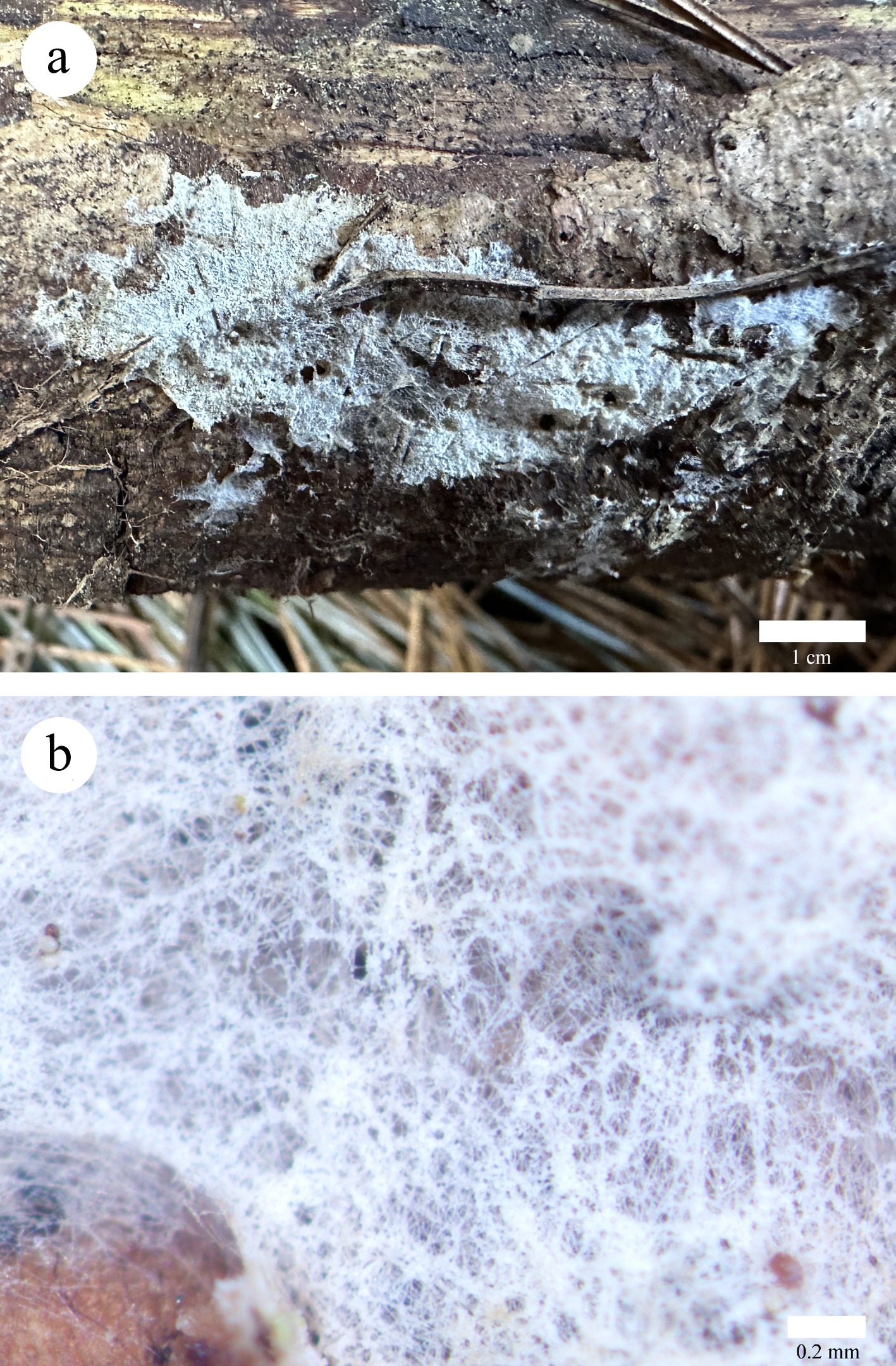
Figure 22.
Basidiomata of Xenasmatella longispinosa (Dai 38498, holotype). (a) A fresh basidioma. (b) Dry hymenophore.
-
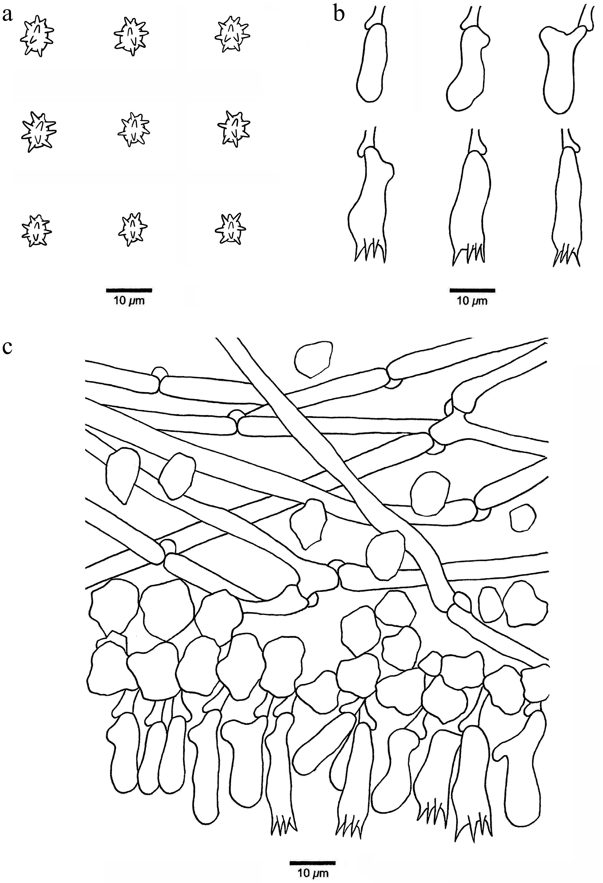
Figure 23.
Microscopic structures of Xenasmatella longispinosa (Dai 38498, holotype). (a) Basidiospores. (b) Basidia and basidioles. (c) A section of hymenium.
-
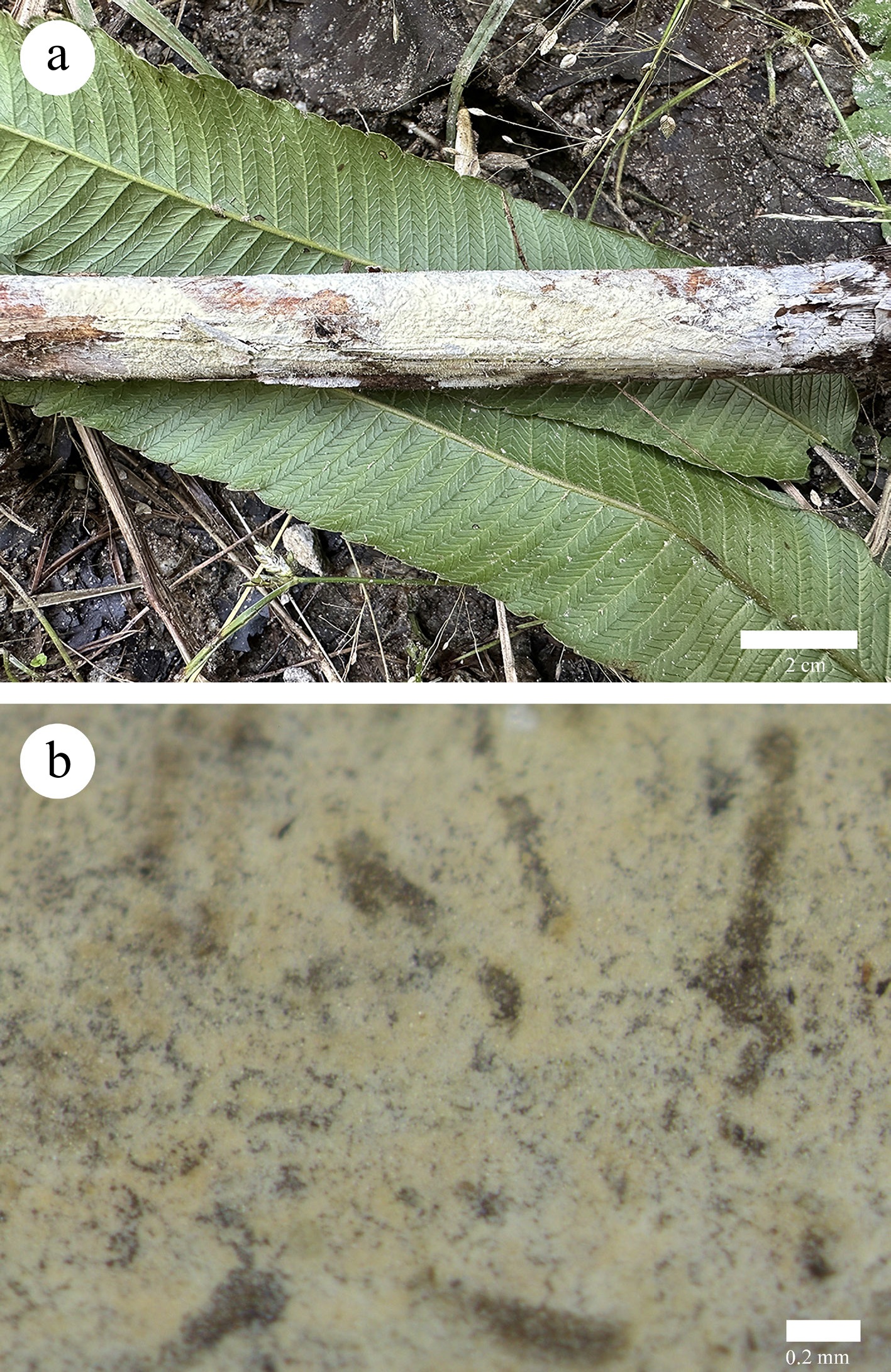
Figure 24.
Basidiomata of Xenasmatella monocotyledonis (Dai 26677, holotype). (a) A fresh basidioma. (b) Dry hymenophore.
-
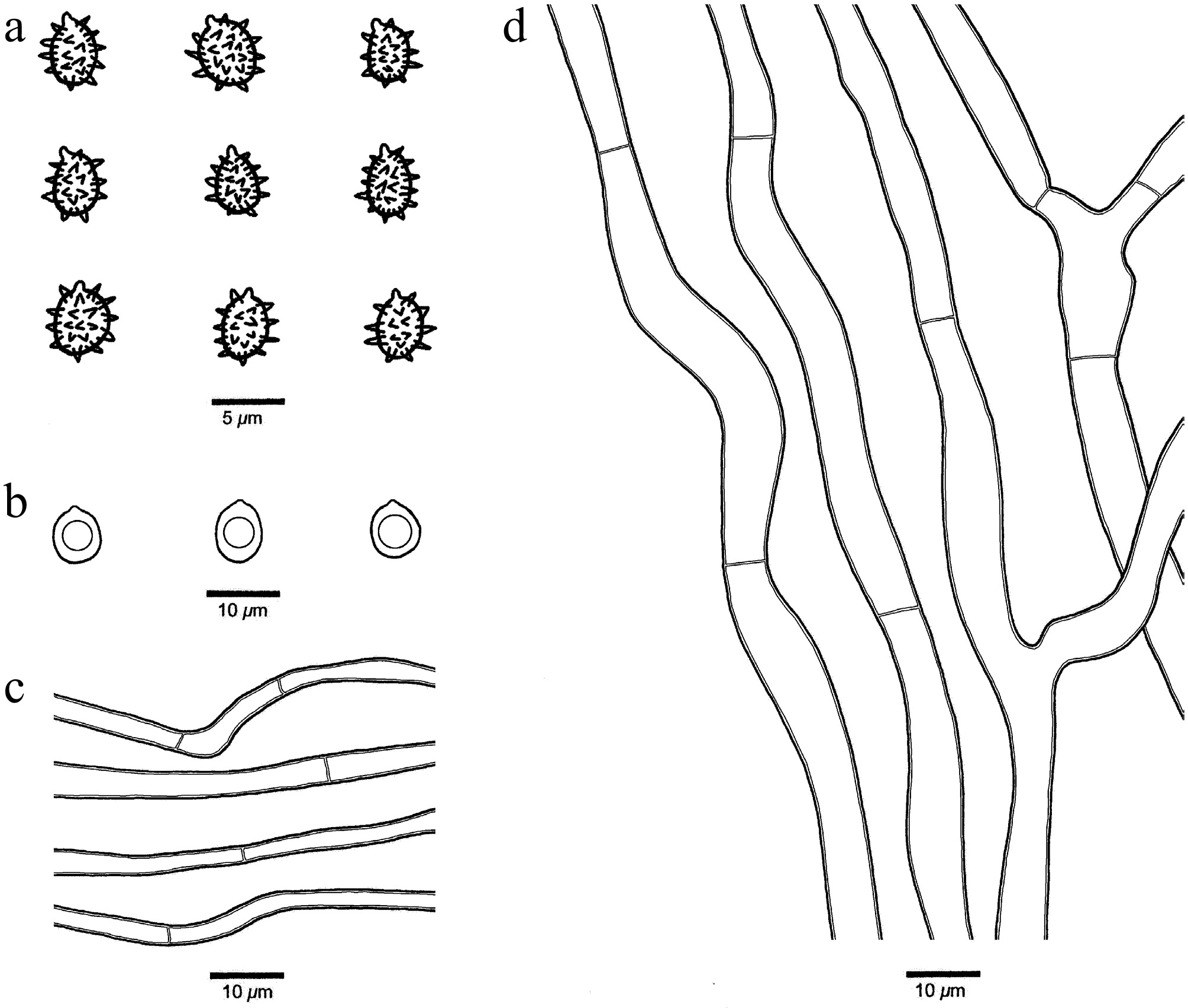
Figure 25.
Microscopic structures of Xenasmatella monocotyledonis (Dai 26677, holotype). (a) Basidiospores. (b) Chlamydospores. (c) Hyphae from subhymenium. (d) Hyphae from subiculum.
-
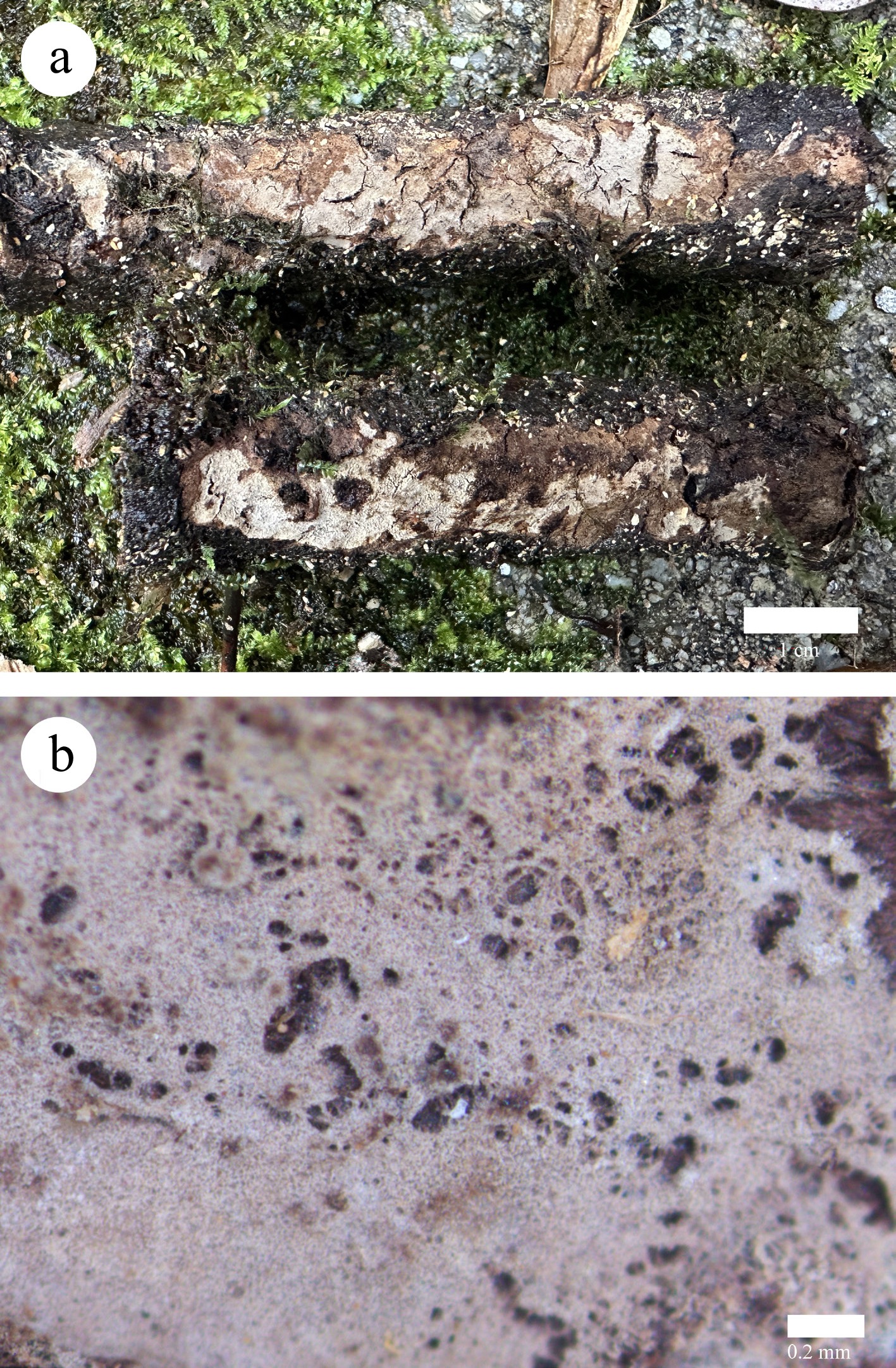
Figure 26.
Basidiomata of Xenasmatella montana (Dai 40267, holotype). (a) A fresh basidioma. (b) Dry hymenophore.
-
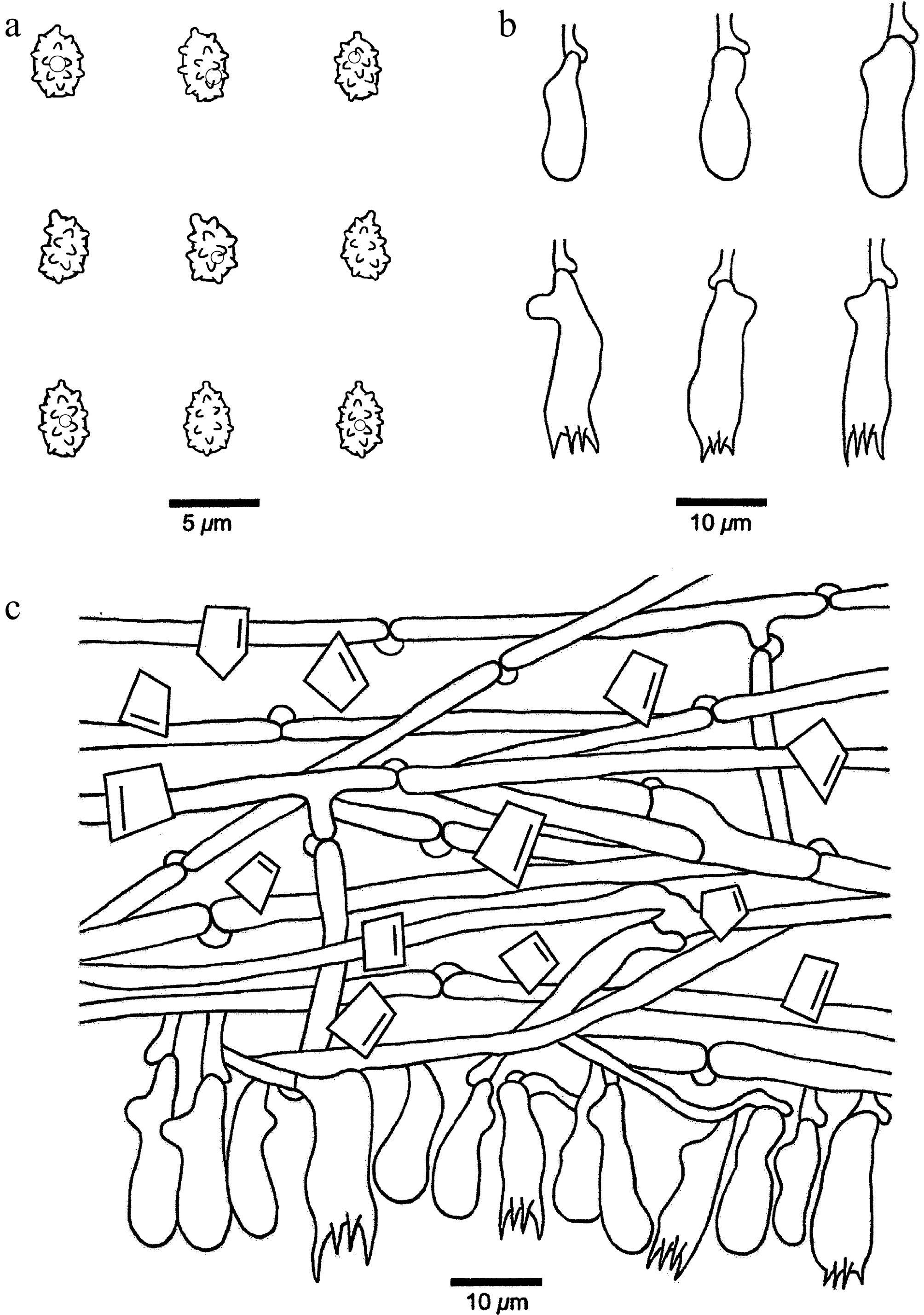
Figure 27.
Microscopic structures of Xenasmatella montana (Dai 40267, holotype). (a) Basidiospores. (b) Basidia and basidioles. (c) A section of hymenium.
-
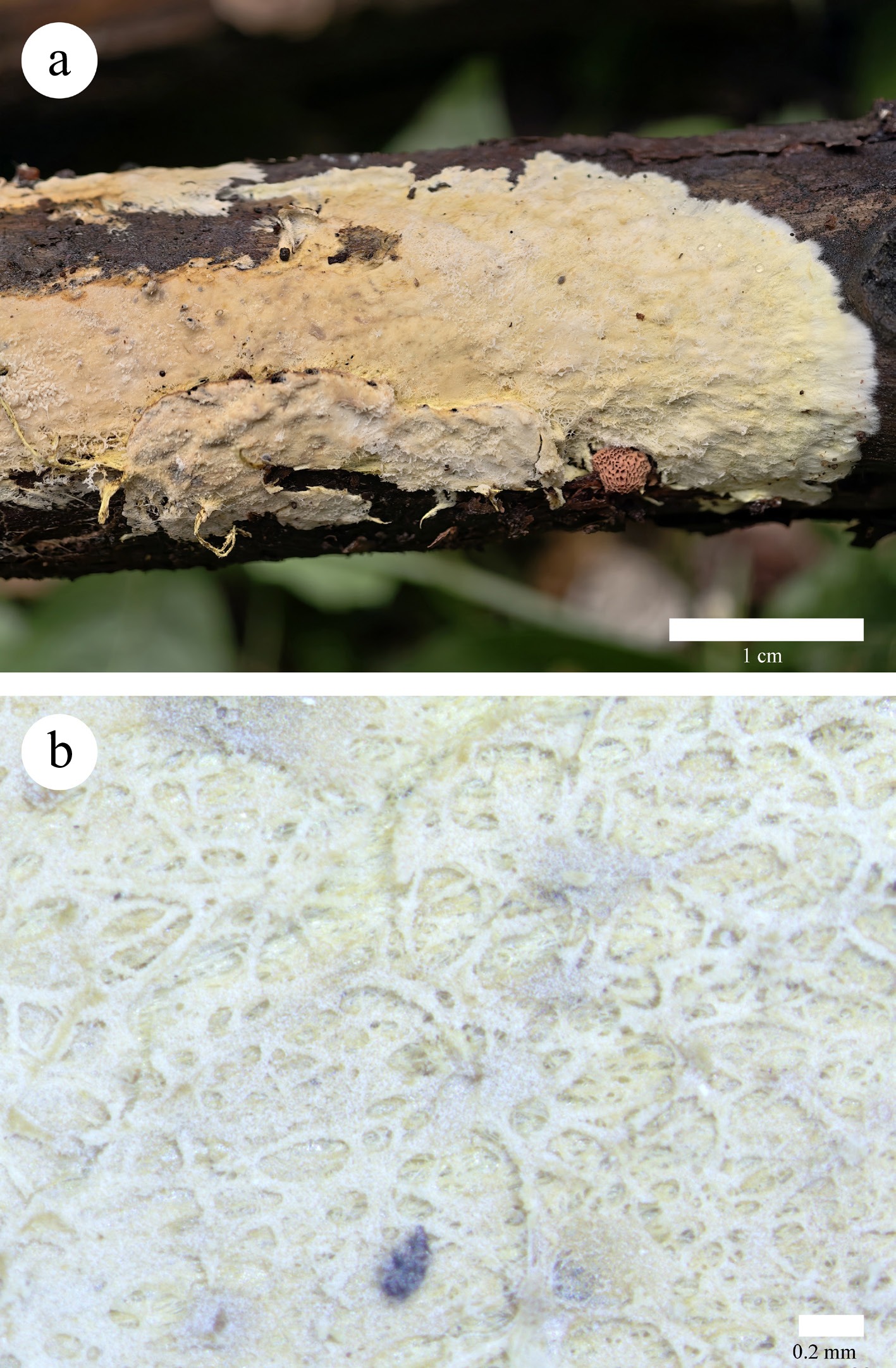
Figure 28.
Basidioma of Xenasmatella tabuliformis (Dai 34854, holotype). (a) A fresh basidioma. (b) Dry hymenophore.
-
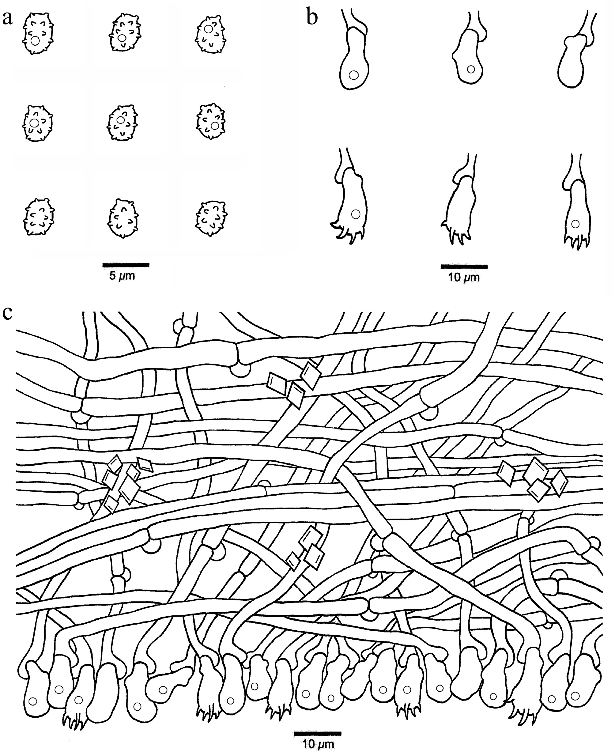
Figure 29.
Microscopic structures of Xenasmatella tabuliformis (Dai 34854, holotype). (a) Basidiospores. (b) Basidia and basidioles. (c) A section of hymenium.
-
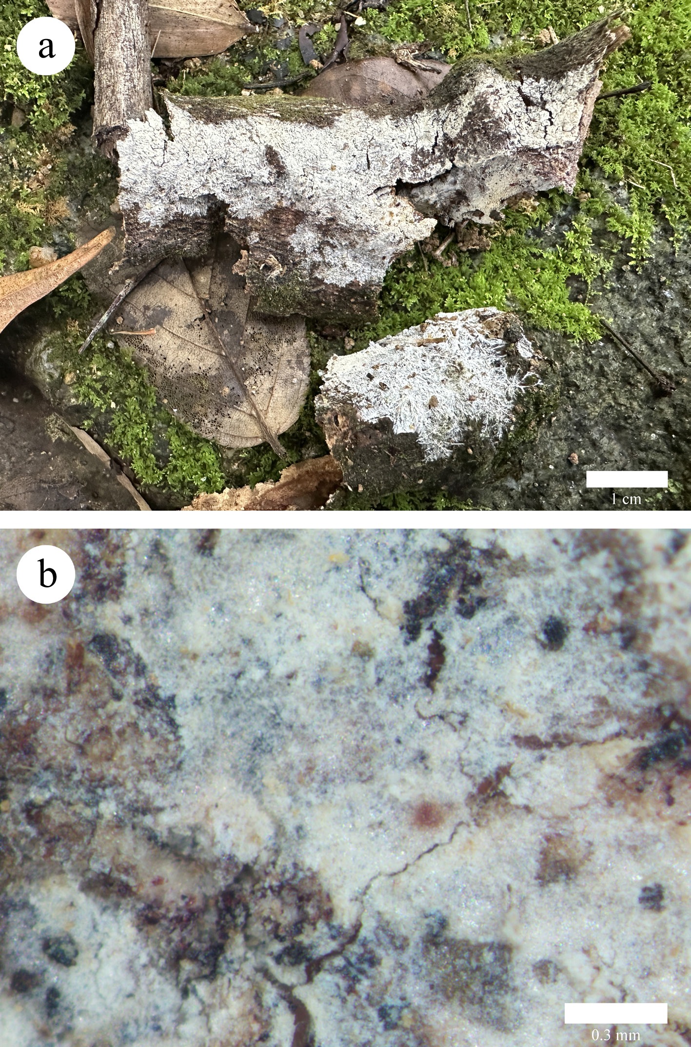
Figure 30.
Basidiomata of Xenasmatella tropica (Dai 36954, holotype). (a) A fresh basidioma. (b) Dry hymenophore.
-
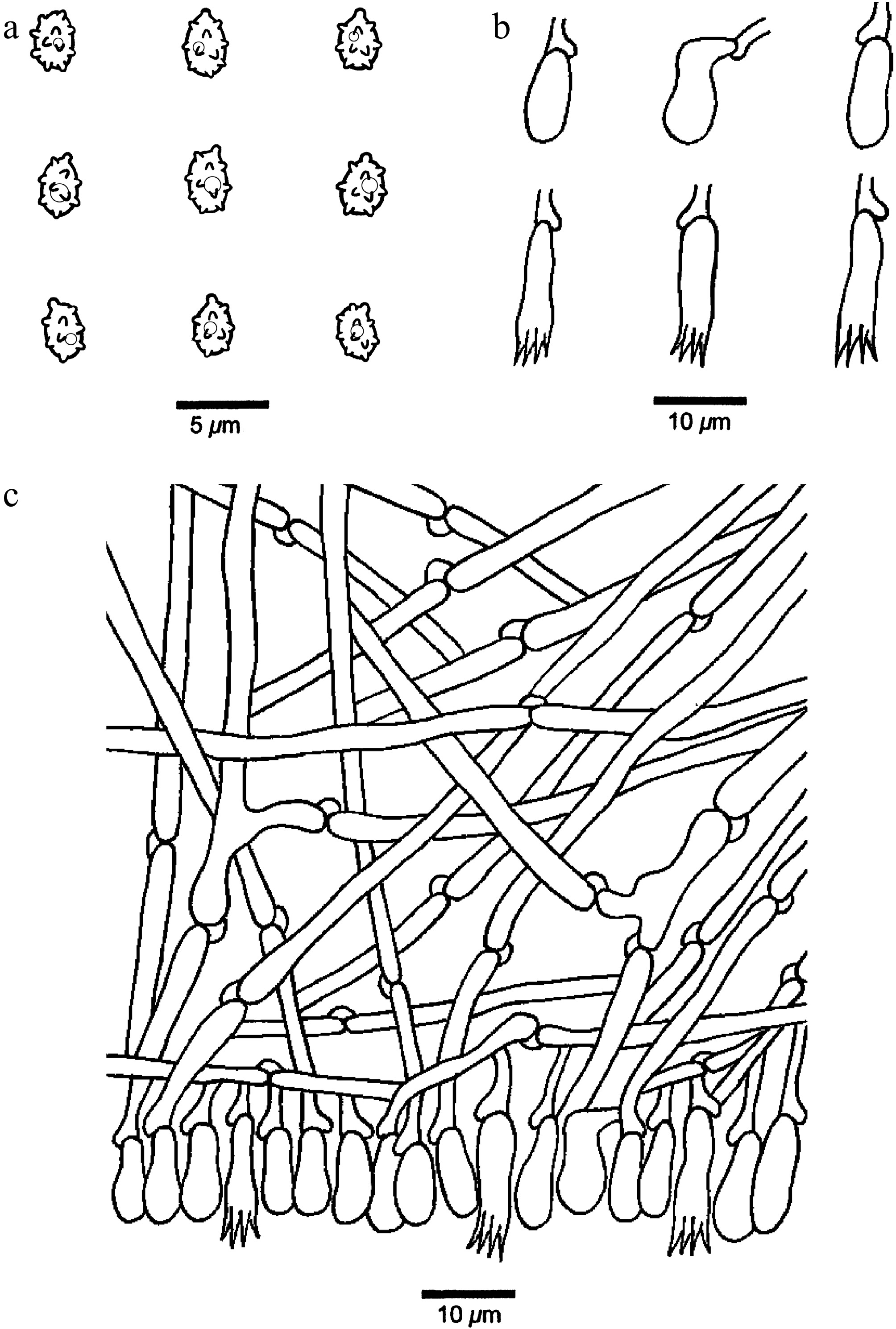
Figure 31.
Microscopic structures of Xenasmatella tropica (Dai 36954, holotype). (a) Basidiospores. (b) Basidia and basidioles. (c) A section of hymenium.
-
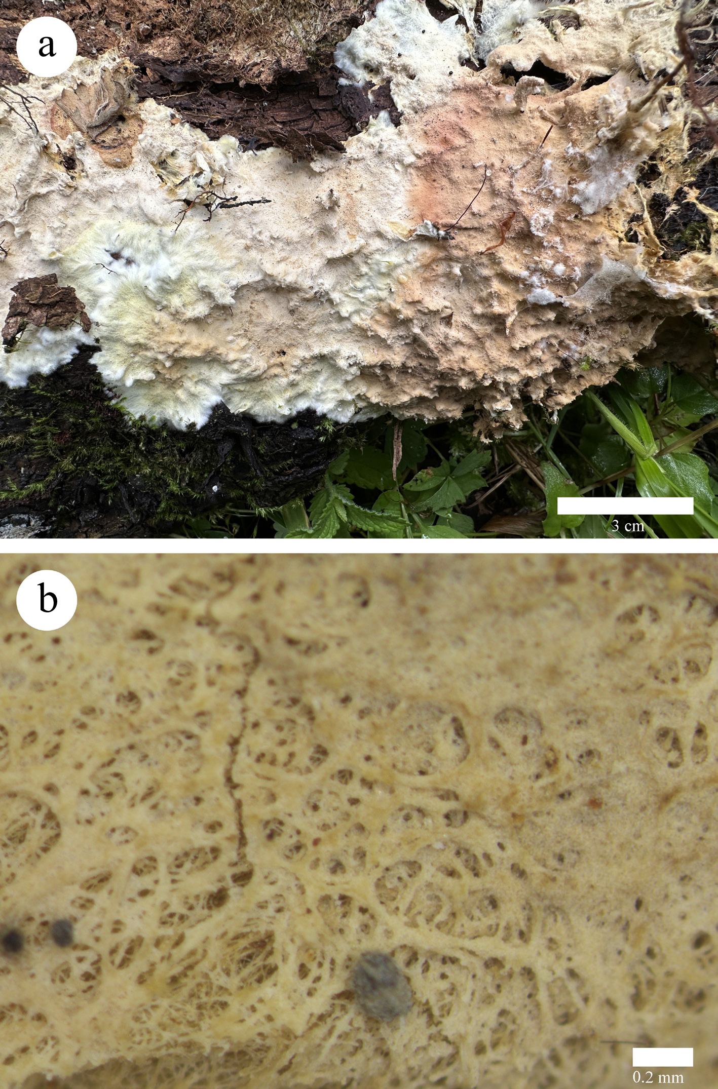
Figure 32.
Basidioma of Xenasmatella versicolor (Dai 26000, holotype). (a) A fresh basidioma. (b) Dry hymenophore.
-
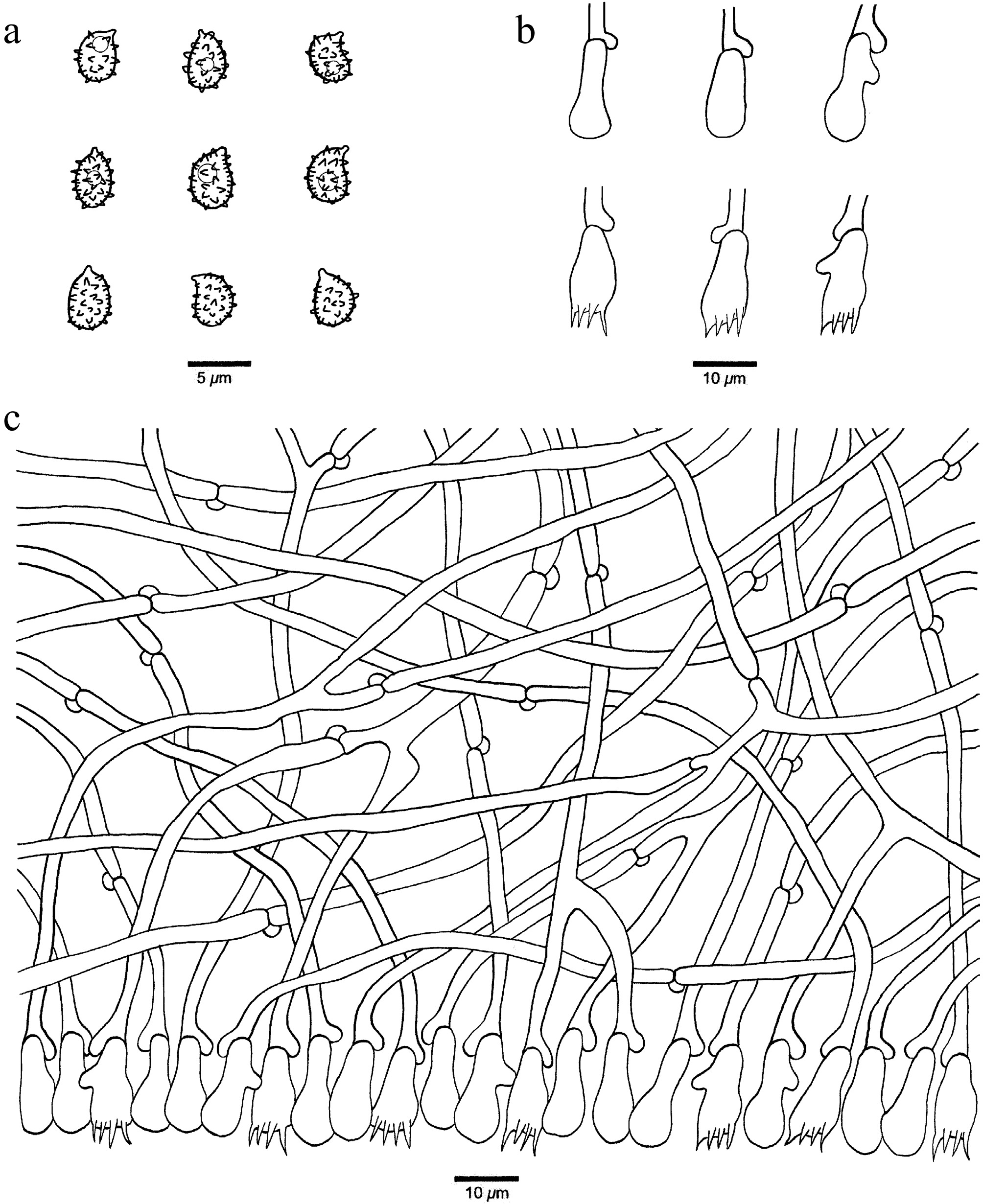
Figure 33.
Microscopic structures of Xenasmatella versicolor (Dai 26000, holotype). (a) Basidiospores. (b) Basidia and basidioles. (c) A section of hymenium.
-
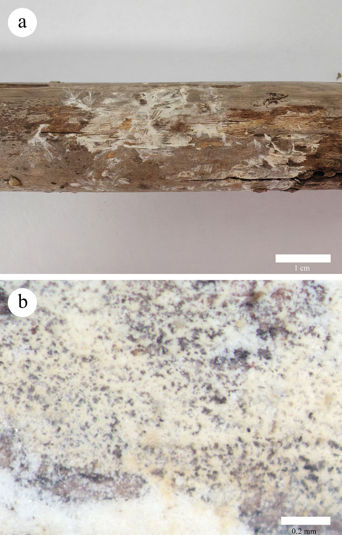
Figure 34.
Basidiomata of Xenasmatella xylina (Dai 36856, holotype). (a) A fresh basidioma. (b) Dry hymenophore.
-
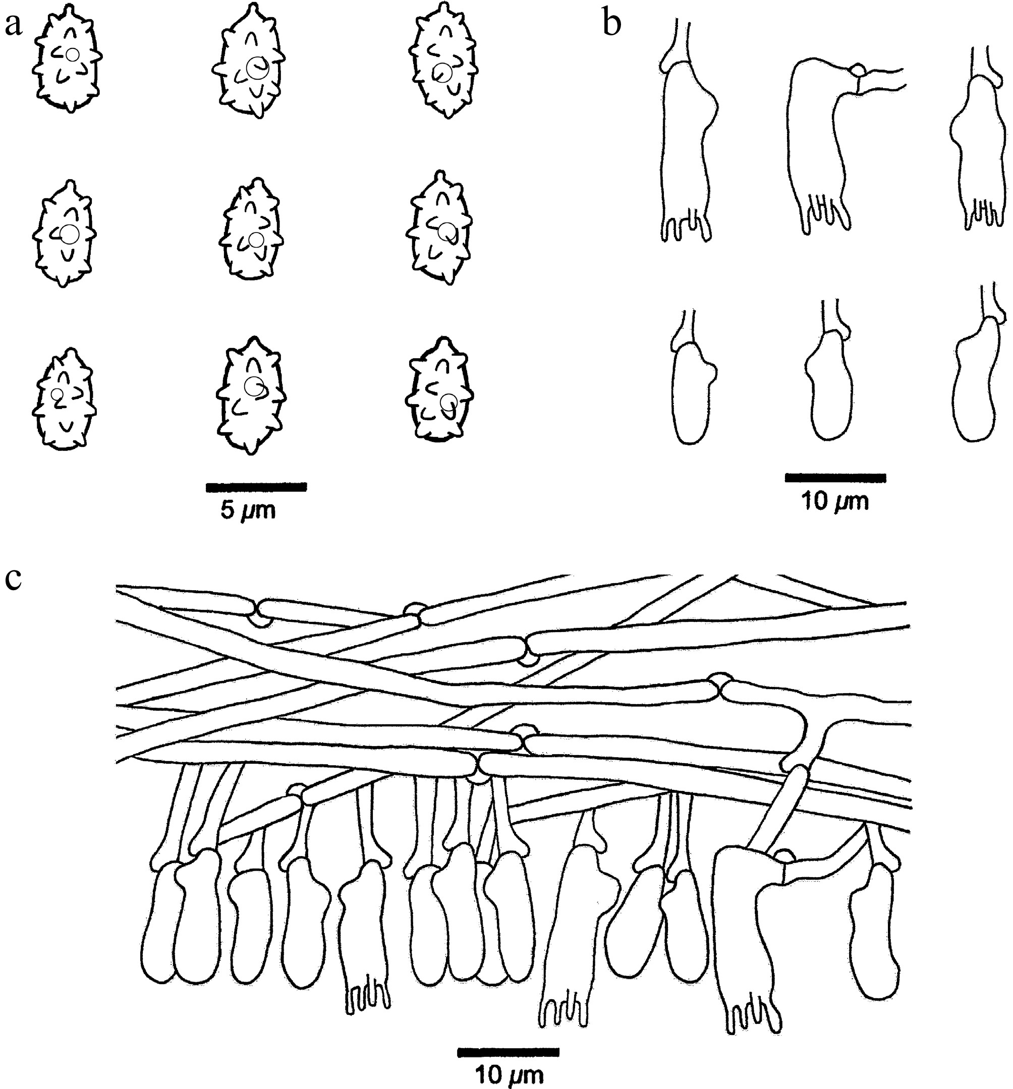
Figure 35.
Microscopic structures of Xenasmatella xylina (Dai 36856, holotype). (a) Basidiospores. (b) Basidia and basidioles. (c) A section of hymenium.
-
1.Rhizomorphspresent 2 1.Rhizomorphsabsent 7 2.Basidiomatacoriaceous 3 2.Basidiomatamembranous 4 3.Hymenophoralsurfacedrab X. conifericola 3.Hymenophoralsurfacewhite X tropica 4.Generativehyphaethick-walled X. rhizomorpha 4.Generativehyphaethin-walled 5 5.Hymenophoralsurfacearachnoidorreticular X. tabuliformis 5.Hymenophoralsurfacebyssaceous 6 6.Basidiospores4.6–6.1µminlength X. abieticola 6.Basidiospores3.4–4.5µminlength X. gymnosperma 7.Basidiosporesthick-walled 8 7.Basidiosporesthin-walled 10 8.Basidiomatamembranous X. hinnulea 8.Basidiomatacoriaceousorsoftcoriaceous 9 9.Basidia18–25µmlong X. alutacea 9.Basidia9–18µmlong X. foraminosa 10.Generativehyphaethick-walledorslightlythick-walled 11 10.Generativehyphaethin-walled 14 11.Hymeniumwithabundantcrystals X. crystallina 11.Hymeniumwithfewcrystalsorabsent 12 12.Basidiosporessubglobosetoglobose X. xinpingensis 12.Basidiosporesellipsoid 13 13.Basidiomatagraytoblack X. nigroidea 13.Basidiomatapalemousegraytoclaybuff,cinnamon X. hjortstamii 14.Hymenophoralsurfacewithtuberculateorgrandinioid 15 14.Hymenophoralsurfacewithouttuberculateorgrandinioid 16 15.Basidiospores5–6×3.5–4.3µm X. jilongensis 15.Basidiospores2.8–3.5×2.5–3µm X. wuliangshanensis 16.Basidiomatacoriaceousorsoftcorky 17 16.Basidiomatamembranousorceraceous 18 17.Hymenophorebluishgray X. caeruleogrisea 17.Hymenophoreverywhite X. candidissima 18.Chlamydosporespresent 19 18.Chlamydosporesabsent 20 19.Generativehyphaewithsimple-septa X. monocotyledonis 19.Generativehyphaebearingclampconnections X. ceracea 20.Basidiawith2sterigmata X. bambusicola 20.Basidiawith4sterigmata 21 21.Basidiomatawithvariablecolors X. versicolor 21.Basidiomatawithoutvariablecolors 22 22.Hymenophoralsurfacewhitetolilac X. tenuis 22.Hymenophoralsurfacewhitetogreyish,whitetocream,creamtobuff,pinkishbuff 23 23.Hymenophoralsurfacegossypineorcottony 24 23.Hymenophoralsurfacesmooth,pruinosetofarinaceous,holey,bassaceoustoreticulate 25 24.Basidiosporessubglobosetoglobose X. gossypina 24.Basidiosporesellipsoid X. xylina 25.Hymenophoralsurfacesmooth X. roseobubalina 25.Hymenophoralsurfacepruinosetofarinaceous,holey,bassaceoustoreticulate 26 26.Hymenophoralsurfacepruinaceoustofarinaceous X. ailaoshanensis 26.Hymenophoralsurfaceholeyorbyssaceoustoreticulate 27 27.Basidiospores5.1–6.2×3.7–4.8µm X. longispinosa 27.Basidiospores3.3–4.1×2.2–3.0µm X. montana
Figures
(35)
Tables
(1)