-
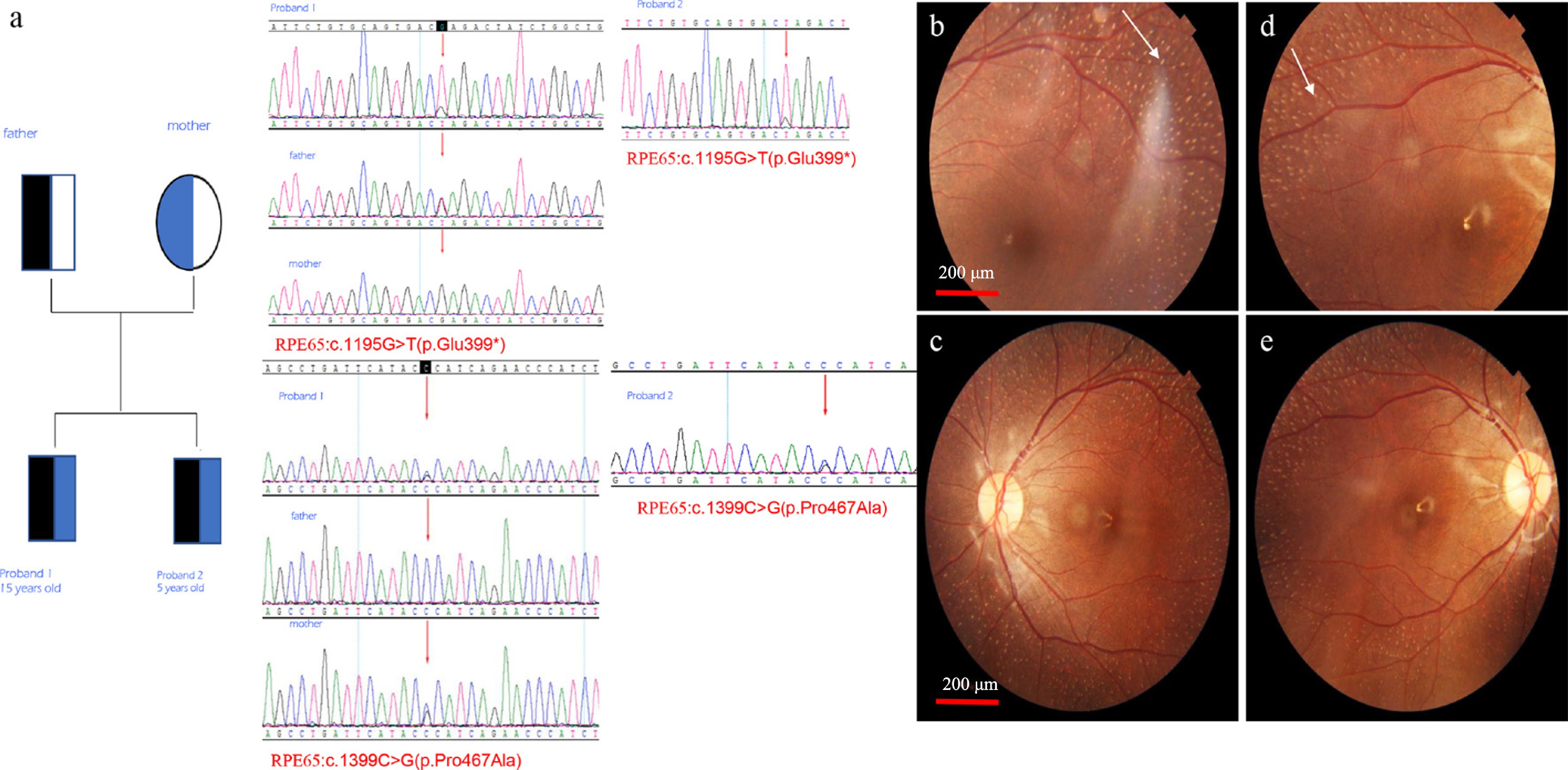
Figure 1.
Compound heterozygous RPE65 mutations (p.Glu399*/p.Pro467Ala) associated with atypical white spot lesions in two Chinese siblings with LCA. (a) Pedigree analysis and Sanger sequencing confirmed compound heterozygosity: p.Glu399* (c.1195G>T) inherited paternally and p.Pro467Ala (c.1399C>G) maternally. (b)–(e) Fundus photographs showing numerous white dot-like lesions distributed throughout the posterior pole, distinct from classic vitelliform maculopathy.
-
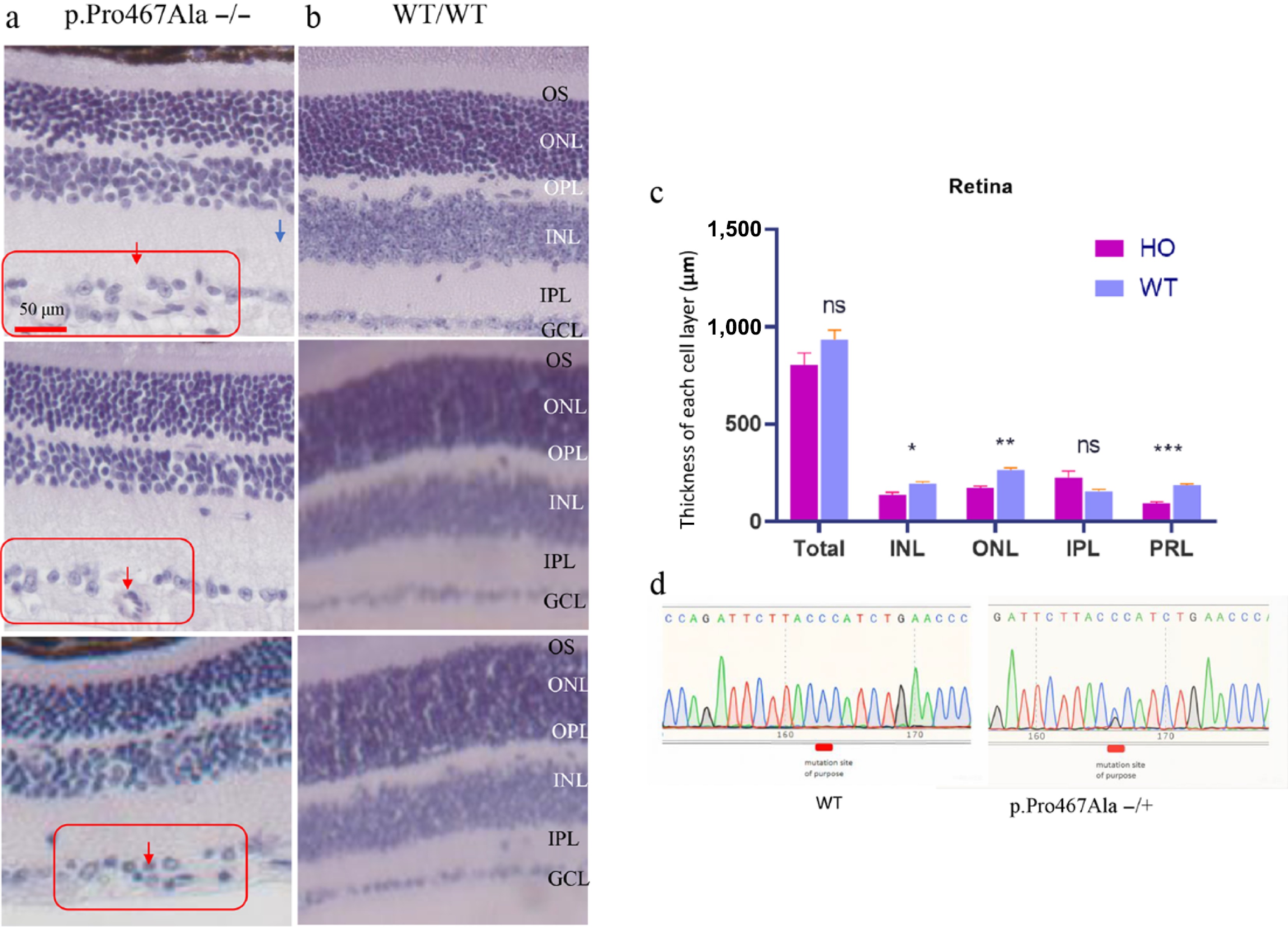
Figure 2.
Toluidine blue-stained retinal cross-sections show retinal degeneration and disorganization of the GCL in Rpe 65 p.Pro467Ala homozygous mice at 12 weeks of age. (a) Representative image of the mutant retina showing distinct areas of cellular disorganization within the GCL, as indicated by the red boxes and red arrows. In contrast, other regions of the GCL exhibit a relatively normal cellular arrangement (blue arrow). Scale bar: 50 µm. (b) Representative image of the WT retina, demonstrating a well-organized and uniformly aligned GCL (n = 6). (c) Quantitative analysis of the thickness of individual retinal layers in both the mutant and control (WT) groups. Data are presented as the mean ± SEM. Statistical significance was determined by unpaired Student's t-tests (* p < 0.05, ** p < 0.01, *** p < 0.001). Scale bars: 50 µm. OS, outer segment; ONL, outer nuclear layer; INL, inner nuclear layer; IPL, inner plexiform layer; OPL, outer plexiform layer; GCL, ganglion cell layer; PRL, photoreceptor layer, which includes both OSs and inner segments (IS); HO, homozygous; OPL, outer plexiform layer. (d) Sanger sequencing confirmed the presence of the c.1399C>G nucleotide substitution at the DNA level.
-
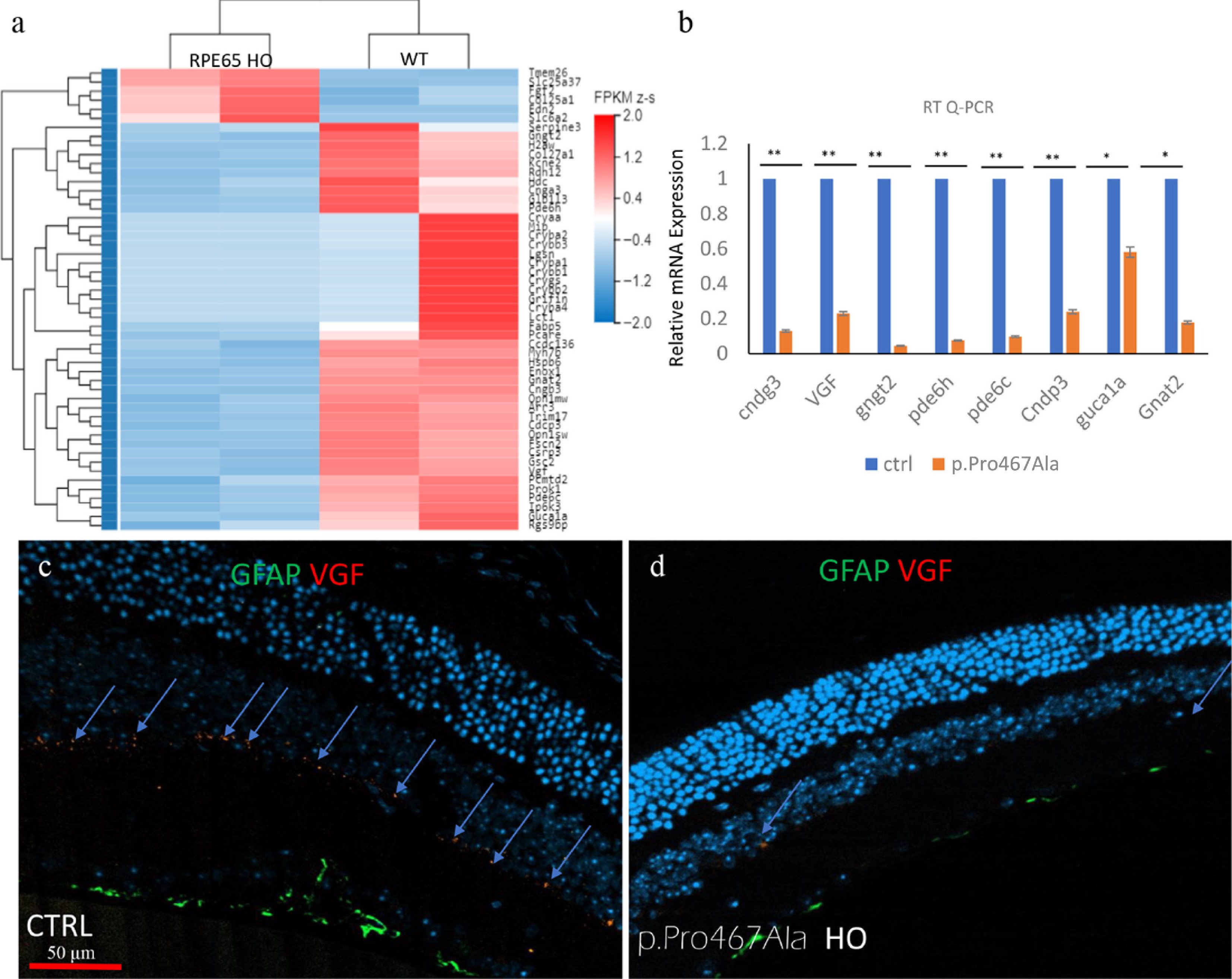
Figure 3.
Transcriptomic analysis reveals downregulation of retinal functional genes and reduced VGF expression in the GCL of Rpe65 p.Pro467Ala homozygous mice. (a) Heatmap of RNA sequencing data showing decreased expression of phototransduction- and neuronal function- related genes, including Vgf, Cndp3, Gngt2, Pde6h, Pde6c, Cnga3, Guca1a, and Gnat2, in mutant retinas. (b) qPCR validation confirmed significant downregulation of these genes, consistent with transcriptome profiling. (c,d) Immunofluorescence staining demonstrates markedly reduced VGF signal (red) predominantly in the INL and in GFAP-positive astrocytic processes surrounding the GCL of (c) wild-type controls compared with (d) mutant mice (nuclei counterstained with DAPI in blue), supporting a glia-derived, non-cell-autonomous role of VGF in GCL pathology.
-

Figure 4.
AQEE-30 treatment ameliorates disorganization of the GCL in p.Pro467Ala homozygous mice. Hematoxylin and eosin (H&E) staining of retinal sections from 8-week-old mice after intravitreal administration of AQEE-30 (50 µM, 2 µL/eye, once every 4 d for 30 d). (a) AQEE-30-treated mutant mice showed improved structural integrity of the GCL. (b) Untreated Rpe65 Pro467Ala homozygous control mice exhibit marked GCL disarray. Scale bar: 50 μm. These findings suggest that the VGF-derived peptide AQEE-30 exerts protective effects on retinal ganglion cells in the context of the RPE65 mutation.
Figures
(4)
Tables
(0)