-
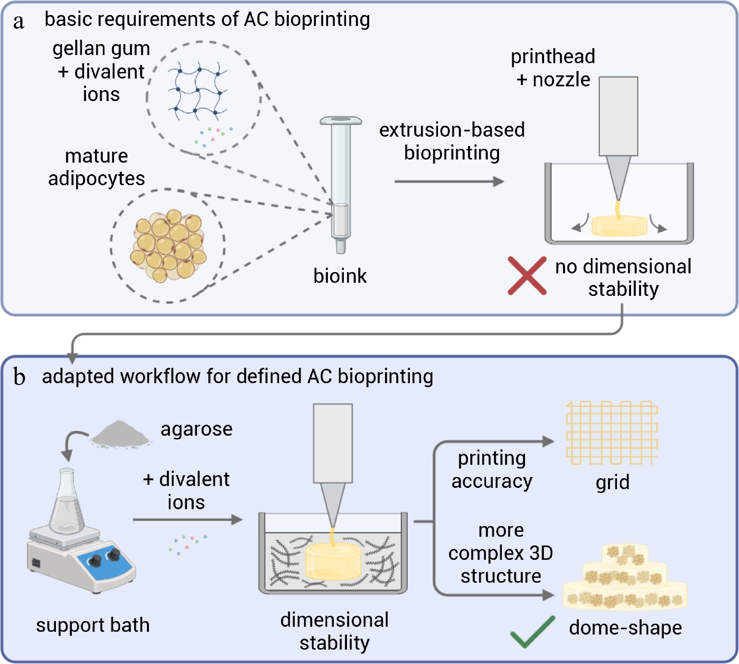
Figure 1.
Schematic overview of the biofabrication strategy for mature adipocyte (AC) bioprinting. (a) Conventional extrusion-based bioprinting of gellan gum-based bioinks containing mature adipocytes lacks dimensional stability due to the softness and buoyancy of the bioink. (b) An adapted workflow employing a yield-stress agarose-based support bath with divalent ions enables stabilized extrusion, resulting in improved printing accuracy demonstrated by the fabrication of defined grid structures and more complex three-dimensional dome-shaped constructs.
-
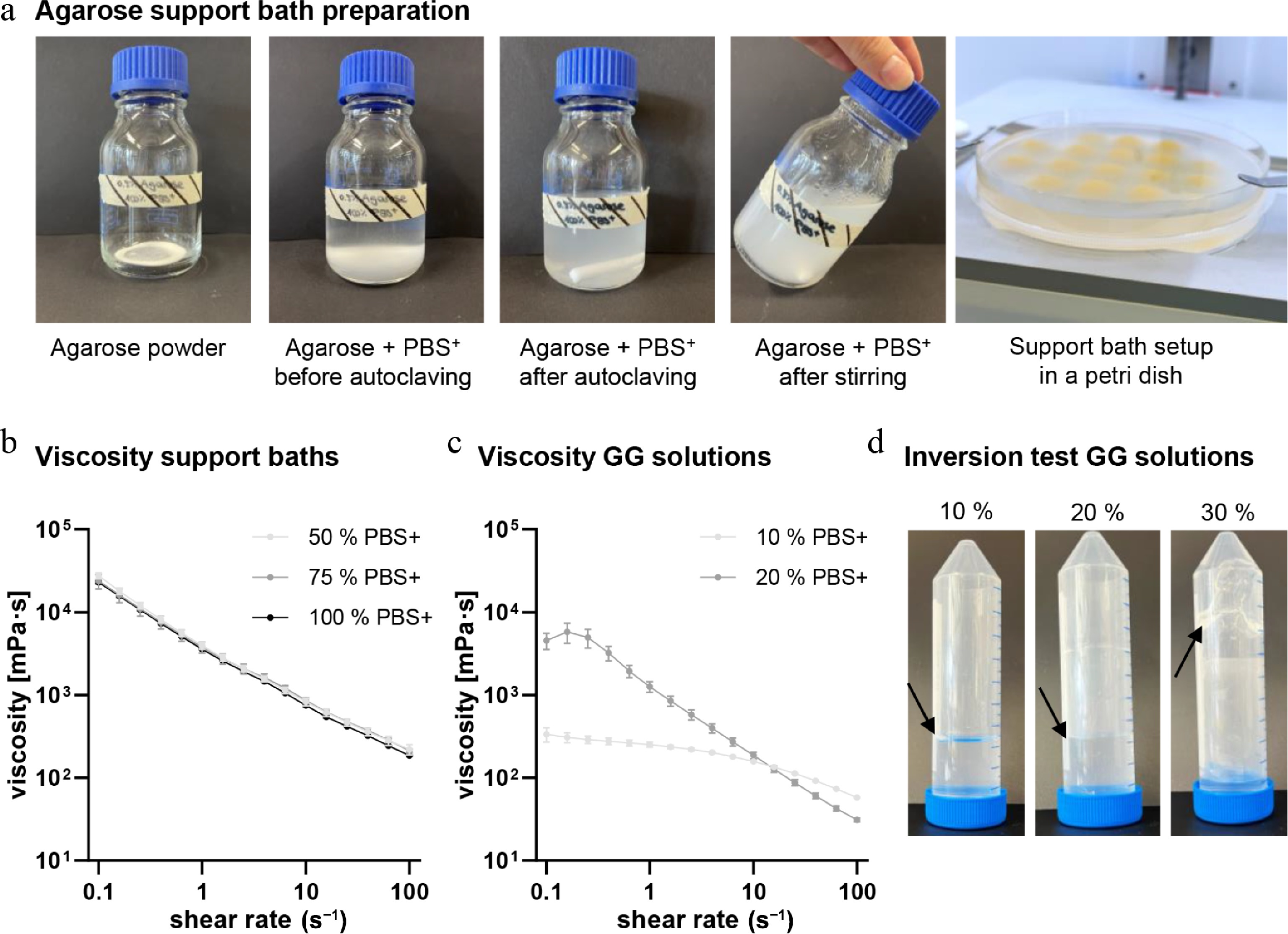
Figure 2.
Support bath preparation and rheological characteristics of agarose support baths and gellan-based bioinks for printing mature adipocytes. (a) Preparation of agarose support bath by mixing agarose with PBS+, autoclaving it, and stirring it overnight. (b) Viscosity of different support bath compositions (50%, 75%, and 100% PBS+) at 10 °C. (c) Viscosity of different acellular GG solutions (10% and 20% PBS+ in stock solution) at 20 °C. (d) Inversion test of different acellular GG solutions (10%, 20%, and 30% PBS+ in stock solution) at room temperature (20 °C). Black arrows indicate the GG solution level after the Falcon tubes were inverted. Each analysis was performed in triplicate. Mean values represent the mean ± standard deviation.
-
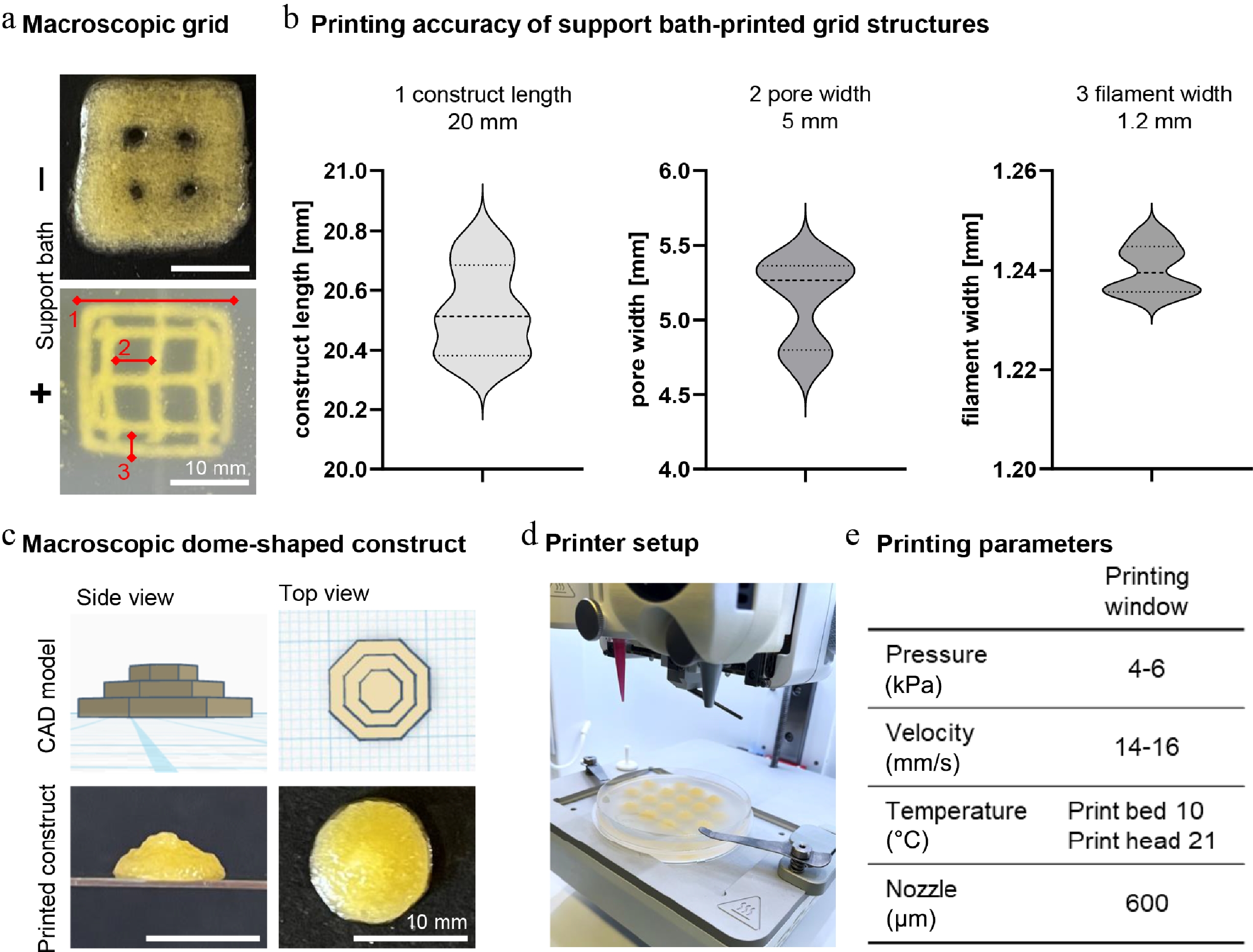
Figure 3.
Bioprinting characteristics and printing protocol. (a) Macroscopic images of grid structures printed without (−) and with (+) support bath; red markings and numbers highlight the analyzed parameters for printing fidelity. Scale bar: 10 mm. (b) Analysis of (1) construct length, (2) pore width, and (3) filament width of support-bath printed grid structures. Design parameters for each measure are indicated above each graph, with a construct length of 20 mm, a pore width of 5 mm, and a filament width of 1.2 mm. (c) Macroscopic pictures of dome-shaped constructs in side and top view with CAD model and support-bath printed and recovered construct. Scale bar: 10 mm. (d) Overview of the printer setup using the BioX printer and a Petri dish as printing vessel. (e) Listed bioprinting windows for printing AC constructs. Mean values represent the mean ± SD of six independent experiments for accuracy measurements of printed grid structures.
-
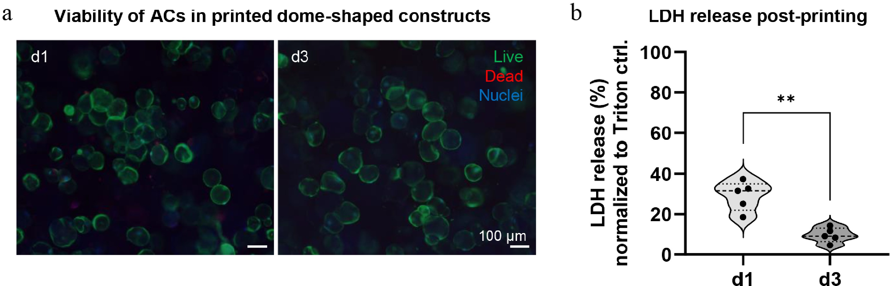
Figure 4.
Viability of mature adipocytes in the printed dome-shaped constructs. (a) Live/dead staining of printed dome-shaped constructs on days 1 and 3 post-printing, living cells in green, dead cells in red, and cell nuclei in blue. Scale bar: 100 µm. (b) Normalized LDH release of printed dome-shaped constructs on days 1 and 3 post-printing, normalized to 1% Triton X-100 control (set to 100%). Data are represented as the mean ± standard deviation, with single values shown as dots from five independent experiments with five different biological donors. ** p ≤ 0.01.
-
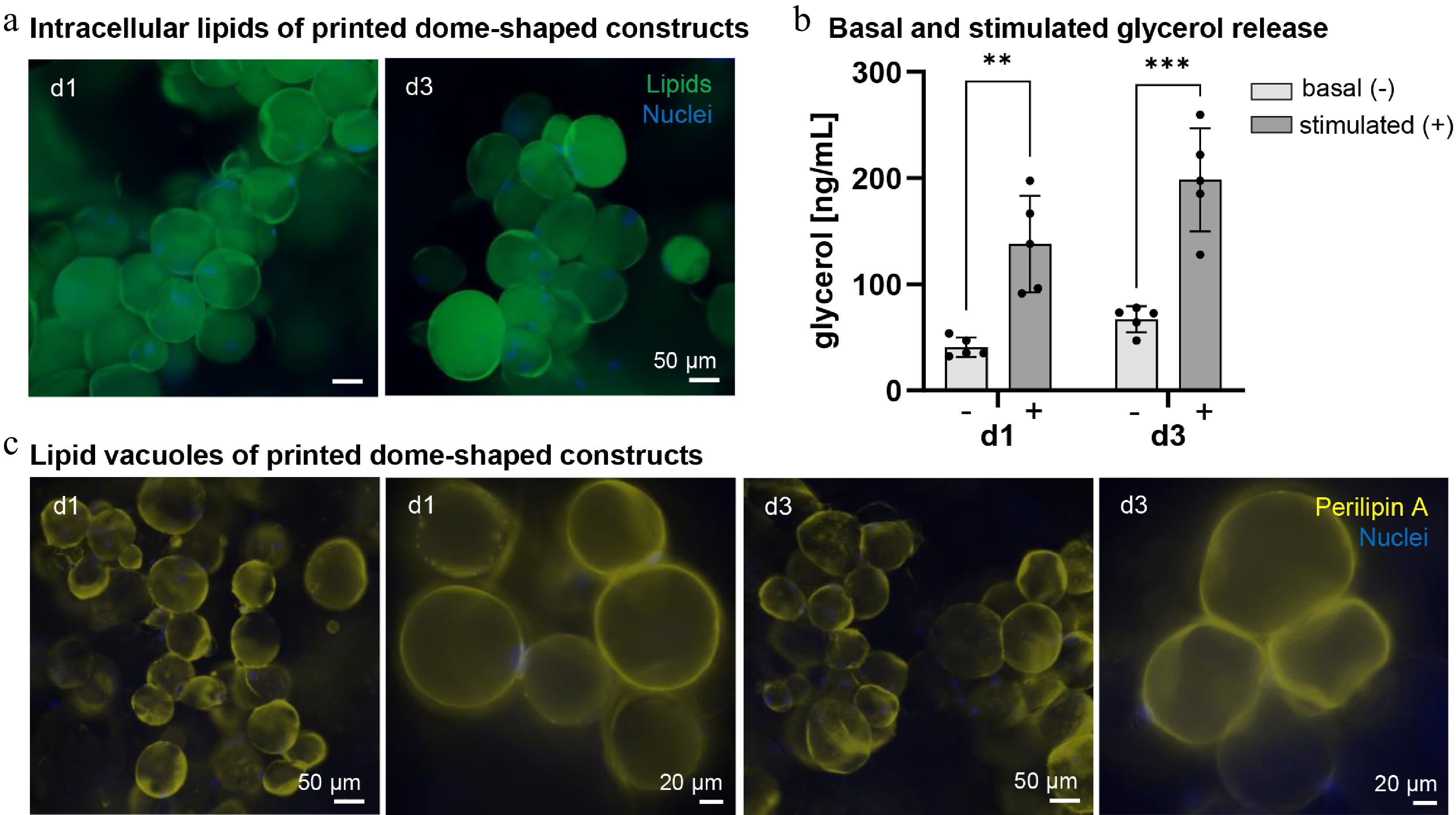
Figure 5.
Morphology and function of mature adipocytes in the printed dome-shaped construct. (a) Intracellular lipid (BODIPY) staining of printed dome-shaped constructs on days 1 and 3, lipids in green, cell nuclei in blue. Scale bar: 50 µm. (b) Basal (−) and isoproterenol-stimulated (10 µM) (+) glycerol release of dome-shaped AC constructs on days 1 and 3 post-printing. (c) Lipid vacuole (perilipin A) staining of printed dome-shaped constructs with two different magnifications (20x and 40x), lipid vacuoles in yellow, nuclei in blue. Scale bars: 50 µm and 20 µm. Mean values are shown as mean ± standard deviation, with individual values as dots from five independent experiments with five different biological donors. ** p ≤ 0.01, *** p ≤ 0.001.
-
Model Printing technique SB SB material Bioink composition Cross-linking Cells used Culture period (d) Research highlights Application Ref. ACs In vitro adipose cell model Extrusion × n.a. GelMA UV-light
(LAP)ACs
ASCs8
15Bioprinting process had no negative effects on differentiation potential or lipid maintenance
Use of ACs resembles the native adipose tissue state moreMore physiological in vitro adipose cell models [11] Vascularized AT model Extrusion ✓ Gellan gum Fibrinogen
collagen microfibersEnzymatic (thrombin) ACs
ASCs
HUVECs7 First documented bioprinting of ACs
Maintenance of AC viability and lipid functionality
Generation of early vascular networks by co-printing with ECsSoft tissue regeneration
(clinical application)[10] Microtissues Autologous fat graft Extrusion × n.a. Fibrinogen
gelatin
HA
PCLEnzymatic (thrombin) Adipose micro-fragments 14 in vitro;
28 in vivoDevelopment of a tissue micronizer for defined fragments
Creation of a viable microfragment-based bioink
Printing of a patient-specific AT graft and in vivo testingPatient-specific volumetric soft tissue reconstruction
(clinical application)[23] Fat graft Extrusion × n.a. Alginate
nanocelluloseIonic
(CaCl2)Lipoaspirate-derived AT 30 Printable lipoaspirate-based bioink containing intact ASCs and ACs
Neovascularization of the implanted graftSoft tissue regeneration
(clinical application)[37] ASCs Life-like AT constructs Stereolithography × n.a. GelMA
HAUV-light
(LAP)ASCs
Fibroblasts
HUVECs27 Multi-material printing platform
3D adipose tissue models with lumenBreast reconstruction (clinical application) [38] Vascularized AT graft Extrusion ✓ Alginate HA
Pluronic F-127
gelatin
PCLIonic
(CaCl2)ASCs 56 in vivo ASC spheroids significantly enhance secretion of angiogenic factors in vivo Soft tissue reconstruction (clinical application) [39] Vascularized adipose cell model Extrusion
FRESH✓
Alginate Collagen
gelatin
alginate
PLLA-PLGAUV-light
(LAP)
Ionic
(CaCl2)ASCs
HAMECs7 Fabrication of thick 3D adipose tissue constructs with perfusable vascular trees Complex, multi-cellular tissue model engineering (clinical application) [40] In vitro densely packed AT model Extrusion ✓ Alginate dECM Ionic
(CaCl2)ASCs
Monocytes28 Highly matured adipose constructs
Recapitulation of obesity-associated pathological changes in vitro (immune component)Obesity research [41] Table 1.
State-of-the-art bioprinting of adipose tissue.
Figures
(5)
Tables
(1)