-
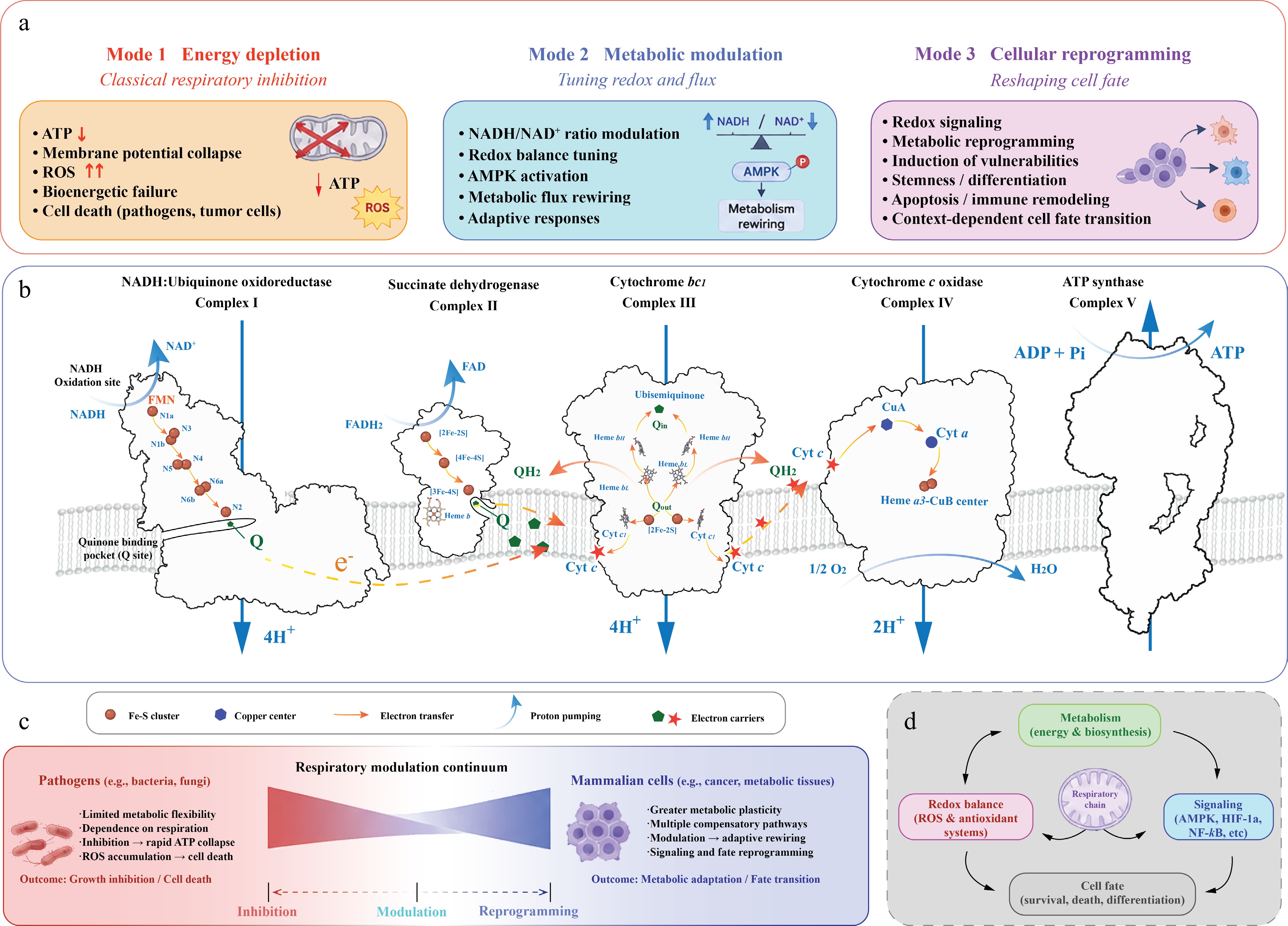
Figure 1.
The respiratory chain as a programmable therapeutic system. (a) Three modes of therapeutic targeting. Small molecules targeting the respiratory chain can be broadly categorized into three functional modes with distinct biological consequences. Mode 1: Energy depletion represents the classical paradigm of respiratory inhibition, in which disruption of electron transfer or proton coupling leads to ATP depletion, membrane-potential collapse, ROS accumulation, and cell death. Mode 2: Metabolic modulation involves partial or adaptive modulation of respiratory activity, leading to altered redox balance, NADH/NAD+ ratio, AMPK activation, and metabolic rewiring without immediate cytotoxicity. Mode 3: Cellular reprogramming extends beyond metabolic control to influence signaling pathways, oxidative stress responses, differentiation states, immune remodeling, and context-dependent cell-fate transitions. CI (NADH:ubiquinone oxidoreductase) transfers electrons from NADH to ubiquinone through a series of iron-sulfur (Fe-S) clusters (N1a, N3, N1b, N4, N5, N6a, N6b, and N2). CII (succinate dehydrogenase) links the tricarboxylic acid cycle to the respiratory chain through FAD-dependent electron transfer to ubiquinone. CIII (cytochrome bc1 complex) mediates the Q cycle and transfers electrons from ubiquinol to cytochrome c, whereas CIV (cytochrome c oxidase) catalyzes oxygen reduction to water. Proton pumping by CI, III, and IV establishes the proton motive force that drives ATP synthesis by CV. (b) Respiratory chain overview. Overview of the mitochondrial respiratory chain illustrating electron transfer and proton translocation across CI–V. Electrons derived from NADH enter through CI, whereas electrons from succinate oxidation enter through CII. Reduced ubiquinone (QH2) transfers electrons to CIII, followed by cytochrome c-mediated transfer to CIV, where molecular oxygen is reduced to water. Coupled proton translocation across the inner mitochondrial membrane generates the proton motive force used by CV for ATP production. (c) Context-dependent biological outcomes. The biological consequences of respiratory modulation differ substantially between pathogens and mammalian systems. Pathogens such as bacteria and fungi often exhibit limited metabolic flexibility and strong dependence on respiration, making them highly susceptible to respiratory inhibition and ATP collapse. In contrast, mammalian cells, particularly cancer and metabolic tissues, possess greater metabolic plasticity and compensatory capacity, allowing respiratory modulation to induce adaptive rewiring, signaling responses, and cellular state transitions rather than immediate lethality. These context-dependent outcomes define a continuum ranging from classical inhibition to metabolic modulation and cellular reprogramming. (d) Respiratory chain as a regulatory hub. The respiratory chain functions as a central regulatory hub coordinating metabolism, redox balance, signaling pathways, and cell-fate determination. Respiratory activity influences biosynthetic metabolism, ROS and antioxidant systems, and signaling pathways including AMPK, HIF-1α, and NF-κB, thereby integrating mitochondrial function with broader cellular regulatory networks controlling survival, differentiation, and stress adaptation.
-
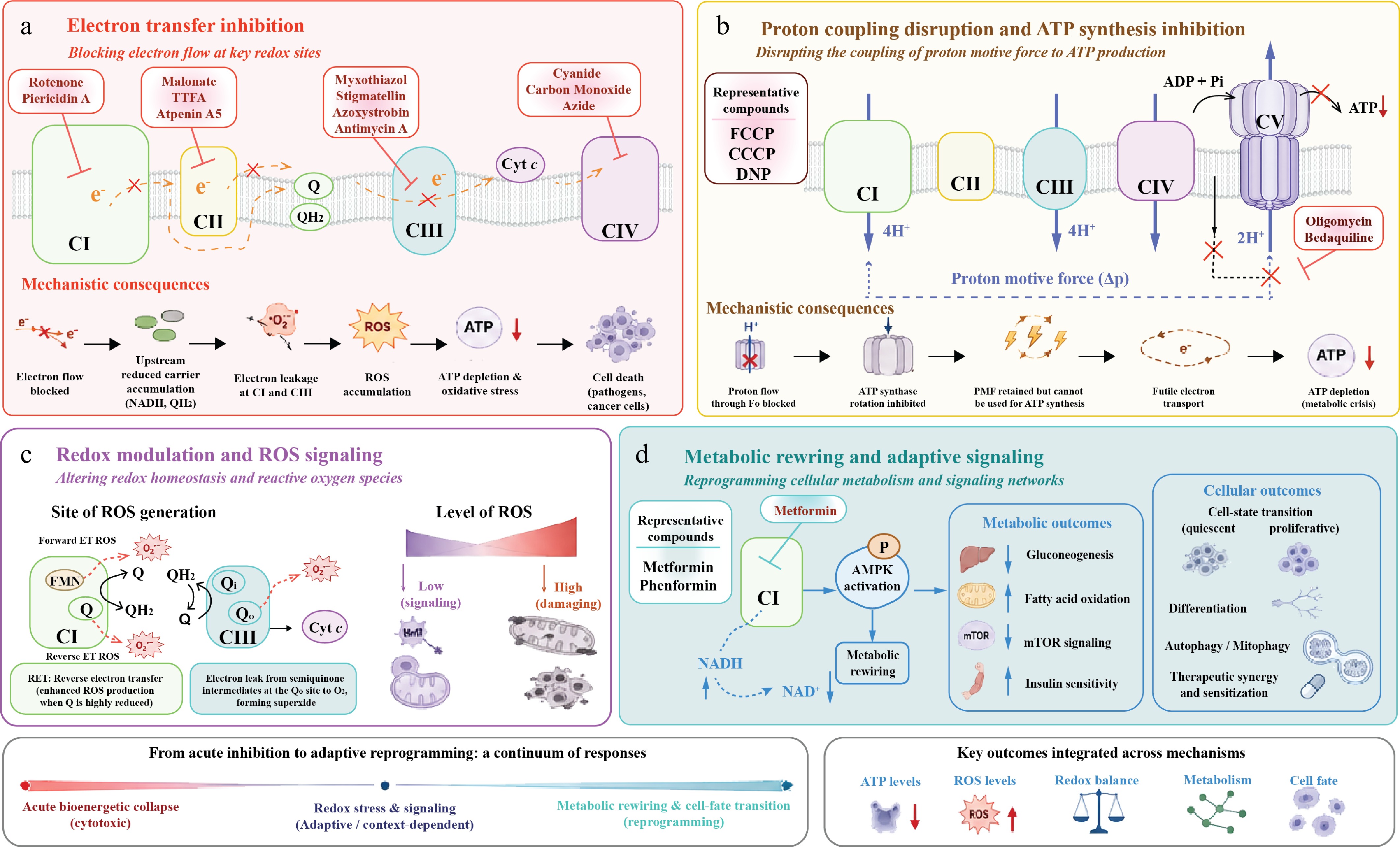
Figure 2.
Mechanisms of small-molecule modulation across the respiratory chain. (a) Electron transfer inhibition. Classical respiratory inhibitors block electron flow at specific redox-active sites within the respiratory chain. Representative inhibitors targeting CI, CII, CIII, and CIV are shown, including Qo-site inhibitors (myxothiazol, stigmatellin, azoxystrobin) and the Qi-site inhibitor antimycin A within CIII. CII inhibitors include substrate-site inhibitors such as malonate and quinone-site inhibitors such as TTFA and Atpenin A5. Inhibition of electron transfer leads to accumulation of upstream reduced carriers, enhanced electron leakage, ROS generation, impaired oxidative phosphorylation, ATP depletion, ultimately resulting in severe energetic stress, impaired cellular viability, or cell death depending on the magnitude and context of inhibition. (b) Proton coupling disruption and ATP synthesis inhibition. Small molecules can disrupt the coupling between proton translocation and ATP production by targeting proton motive force utilization or ATP synthase activity. Representative uncoupling compounds and ATP synthase inhibitors are illustrated. Perturbation of proton coupling compromises ATP generation despite continued or partially preserved respiratory activity, leading to energetic stress and metabolic crisis. (c) Redox modulation and ROS signaling. The respiratory chain functions as a major intracellular source of ROS, particularly at CI and CIII. Forward electron transfer and reverse electron transfer (RET) through CI, as well as semiquinone-mediated electron leakage at the Qo site of CIII, contribute to superoxide production. Moderate ROS levels can function as signaling molecules involved in stress adaptation and metabolic regulation, whereas excessive ROS accumulation induces oxidative damage and mitochondrial dysfunction. (d) Metabolic rewiring and adaptive signaling. Partial respiratory perturbation can induce adaptive metabolic reprogramming rather than acute cytotoxicity. Metformin and phenformin are shown as representative CI-targeting compounds that alter the NADH/NAD+ ratio and activate AMPK, leading to metabolic rewiring and downstream effects on gluconeogenesis, fatty acid oxidation, mTOR signaling, and insulin sensitivity. These changes can further influence cell-state transitions, differentiation, autophagy, mitophagy, and therapeutic sensitization.
-
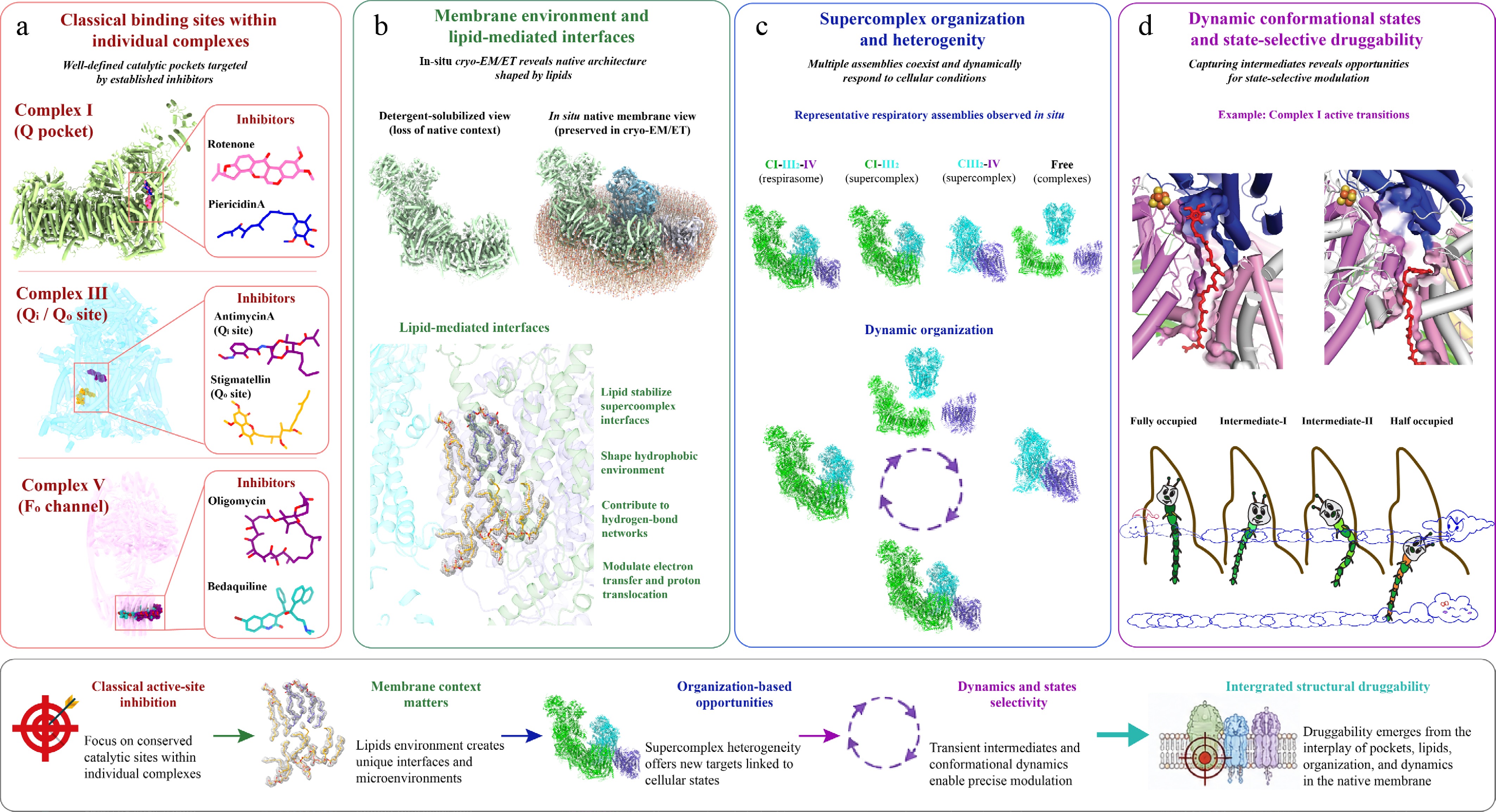
Figure 3.
Structural basis of respiratory chain druggability: from active sites to SC organization. (a) Classical binding sites within individual complexes. Established respiratory inhibitors primarily target conserved catalytic regions involved in electron transfer and proton translocation. Representative inhibitor-binding sites are illustrated for CI ubiquinone-binding pocket, CIII Qi/Qo sites, and the membrane-embedded Fo proton channel of CV. Inhibitors such as rotenone, piericidin A, antimycin A, stigmatellin, oligomycin, and bedaquiline occupy regions overlapping endogenous substrate pathways, thereby blocking electron transfer or proton translocation. Although targeting conserved catalytic centers can achieve potent inhibition, evolutionary conservation often limits selectivity. (b) Membrane environment and lipid-mediated interfaces. In contrast to detergent-solubilized structures, in situ cryo-EM/cryo-ET preserves the native membrane environment and reveals extensive lipid-mediated interactions between respiratory complexes. Structured lipids, including cardiolipin and phospholipids, stabilize SC interfaces, shape hydrophobic microenvironments, and contribute to hydrogen-bonding networks important for electron transfer and proton translocation. These observations highlight that respiratory druggability emerges not only from protein pockets but also from membrane organization and protein-lipid-protein interfaces. (c) SC organization and heterogeneity. Multiple respiratory assemblies coexist within native membranes, including respirasomes, partial SCs, and free complexes. In situ structural analyses reveal that respiratory organization is highly heterogeneous and dynamically responds to metabolic state, substrate availability, and cellular stress. Such organizational plasticity creates context-dependent vulnerabilities and opportunities for selective targeting of specific SC states or assembly-dependent functional behaviors. (d) Dynamic conformational states and state-selective modulation. Respiratory complexes undergo substantial conformational rearrangements during catalysis, generating transient structural intermediates that may serve as selective pharmacological targets. Representative active and deactive conformations of CI are shown to illustrate how substrate occupancy and structural rearrangement reshape catalytic architecture and accessibility. These dynamic states provide opportunities for state-selective modulation, in which small molecules stabilize specific conformations or alter conformational equilibria without necessarily inducing complete respiratory inhibition.
-
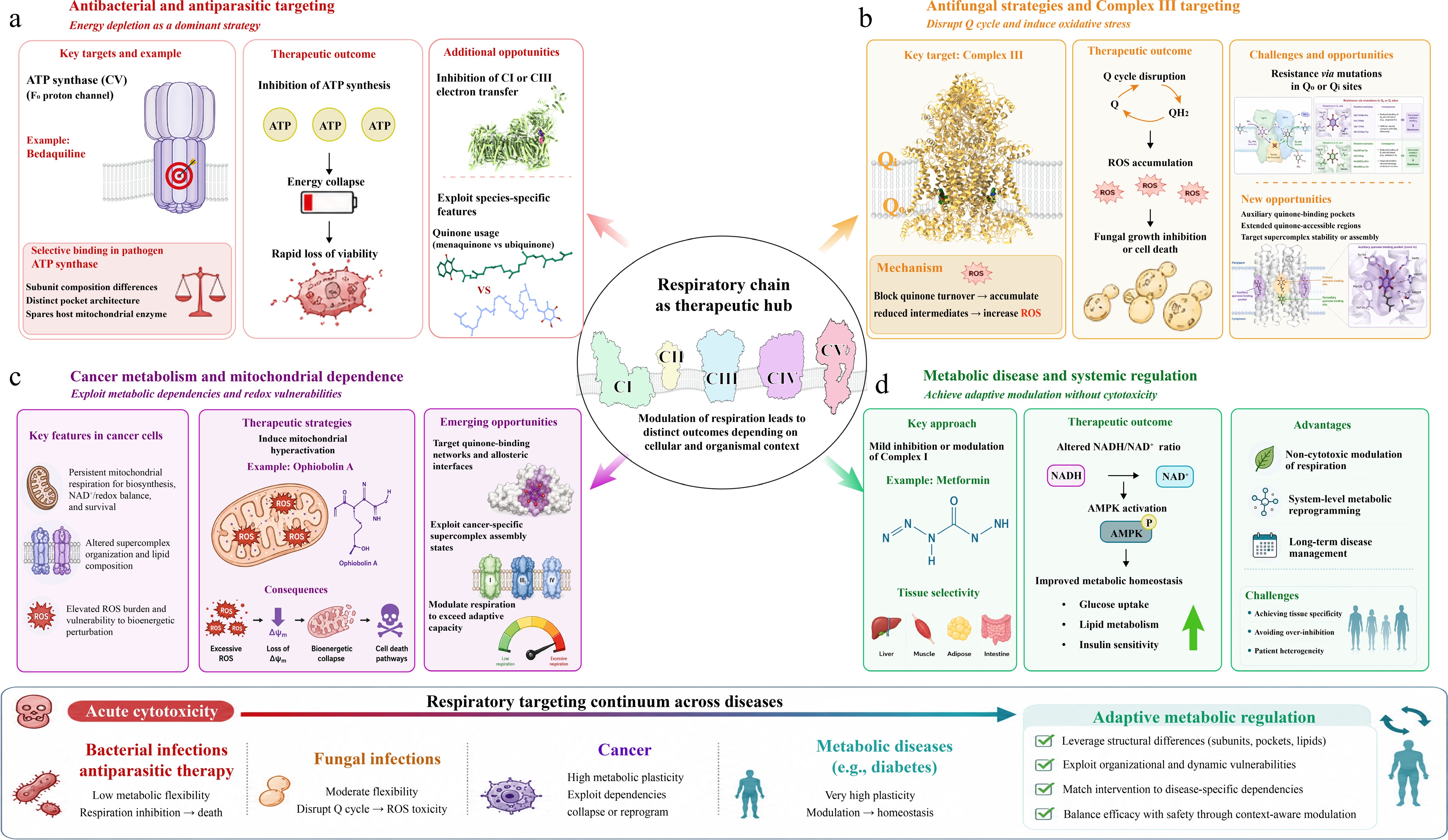
Figure 4.
Therapeutic applications and selectivity across diseases. (a) Antibacterial and antiparasitic targeting. In bacterial and parasitic pathogens, OXPHOS frequently serves as a dominant energy-producing pathway, rendering these organisms highly sensitive to respiratory inhibition. CV represents a major therapeutic target, exemplified by bedaquiline, which selectively inhibits the Fo proton channel of mycobacterial ATP synthase. Structural differences in subunit composition and binding-pocket architecture enable pathogen-selective inhibition while minimizing effects on the host mitochondrial enzyme. Respiratory inhibition leads to rapid ATP depletion, energetic collapse, and loss of viability. Additional opportunities for selective targeting arise from species-specific quinone usage, electron-transfer pathways, and respiratory SC organization. (b) Antifungal strategies and CIII targeting. In fungal pathogens, CIII is a major target of antifungal intervention. Inhibitors acting at the Qo or Qi sites disrupt the Q cycle, block quinone turnover, and promote accumulation of reduced intermediates and ROS, resulting in oxidative stress and fungal growth inhibition. Resistance frequently emerges through mutations in canonical quinone-binding regions. Structural studies further suggest the presence of auxiliary quinone-binding pockets and extended quinone-accessible regions that may provide alternative sites for therapeutic targeting. Modulation of SC stability or assembly may additionally create opportunities for fungal-selective respiratory disruption. (c) Cancer metabolism and mitochondrial dependence. Many tumor cells retain substantial dependence on mitochondrial respiration for biosynthesis, redox homeostasis, and survival despite elevated glycolytic activity. This metabolic reliance creates selective vulnerabilities that can be exploited therapeutically. Compounds such as Ophiobolin A induce mitochondrial hyperactivation, excessive ROS accumulation, membrane-potential disruption, and bioenergetic collapse, ultimately activating cell death pathways. Emerging therapeutic strategies aim to target quinone-binding networks, allosteric interfaces, and cancer-specific SC states in order to selectively perturb tumor metabolism while sparing normal tissues. (d) Metabolic disease and systemic regulation. In metabolic disorders such as type 2 diabetes, respiratory modulation is used not to induce cytotoxicity but to achieve adaptive metabolic regulation. Metformin exemplifies this strategy through mild inhibition or modulation of CI, leading to altered NADH/NAD+ balance, activation of AMPK, and downstream improvement of metabolic homeostasis. These effects contribute to regulation of glucose uptake, lipid metabolism, and insulin sensitivity while avoiding acute energetic collapse. Achieving tissue selectivity and avoiding excessive respiratory inhibition remain major challenges for long-term therapeutic application.
-
Pharmacological category Representative inhibitor ETC target Binding site/
Structural regionMechanism of action Structural effect/
Conformational stateRepresentative structure Quinone-site competitors Piericidin A CI Upper ubiquinone-binding channel near the NDUFS2-ND1 interface Mimics ubiquinone and competitively inhibits electron transfer at the
Q-reduction siteOccupies the upper Q-channel between NDUFS2-Tyr108 and NDUFS2-His59, stabilizing an inhibitor-bound catalytic state 6ZTQ Quinone-site competitors Rotenone CI Ubiquinone-binding cavity extending from the ND1/NDUFS2 region toward the membrane arm Blocks electron transfer from cluster N2 to ubiquinone by occupying the Q-channel Stabilizes an open/deactive-like inhibitor-bound state; rotenone is additionally observed near ND4 in the membrane arm 6ZKM Quinone-site inhibitors Atpenin A5 CII Ubiquinone-binding site in the membrane domain of succinate dehydrogenase Blocks electron transfer from succinate/FAD/Fe–S relay to ubiquinone by occupying the quinone-binding pocket Stabilizes an Atpenin A5-bound inhibited state of porcine mitochondrial CII 3AEE Quinone-site inhibitors TTFA CII Ubiquinone-binding site in the membrane domain of succinate dehydrogenase Blocks electron transfer from the FAD/Fe–S relay to ubiquinone by occupying the quinone-binding pocket Stabilizes a TTFA-bound inhibited state of avian mitochondrial CII 6MYP Qi-site blockers Antimycin A1 CIII Qi site within cytochrome b near heme bH Blocks quinone reduction at the Qi site and interrupts the Q cycle Occupies the Qi pocket and prevents quinone/quinol exchange, stabilizing a
Qi-inhibited state of
cytochrome bc11NTK Rieske-domain locking Qo-site inhibitors Stigmatellin CIII Qo site of cytochrome b near the Rieske [2Fe–2S] protein interface Mimics quinol binding at the Qo site and blocks bifurcated electron transfer Forms a hydrogen-bonding interaction with the Rieske His residue, locking the Rieske domain in the b-position and stabilizing a Qo-inhibited state 1PP9 Qo-site inhibitors Myxothiazol CIII Proximal Qo site of cytochrome b Blocks quinol oxidation at the Qo site and interrupts bifurcated electron transfer Occupies the proximal Qo pocket; unlike stigmatellin, it does not lock the Rieske domain tightly at the Qo site 1SQP ATP synthase inhibitors Oligomycin A CV Fo proton channel at the interface between the c-ring and subunit a Blocks proton translocation through the Fo motor and inhibits rotary ATP synthesis Immobilizes the membrane rotor machinery and stabilizes a proton-conduction-blocked state of ATP synthase 6CP3
6CP5ATP synthase inhibitors Bedaquiline CV Mycobacterial ATP synthase Fo c-ring/rotor region Selectively inhibits proton-driven rotary catalysis in mycobacterial ATP synthase Captures bedaquiline-saturated ATP synthase in rotational state 1 7JG8 Table 1.
Structural and mechanistic basis of respiratory chain inhibition.
-
Compound Chemical
structureTarget complex Primary indication Development stage Key pharmacological features Metformin 
CI Type 2 diabetes; oncology (investigational) Approved Weak but clinically relevant CI modulator; activates AMPK and induces metabolic reprogramming with an excellent safety profile. IACS-010759 
CI Relapsed/refractory AML; advanced solid tumors Phase I Potent and selective OXPHOS inhibitor developed through structure-guided optimization; preferentially targets OXPHOS-dependent tumors. IM156 
CI Advanced solid tumors Phase I Second-generation biguanide with improved potency, pharmacokinetics,
and oral bioavailability compared with metformin.EVT-701 
CI Solid tumors Phase I Highly selective CI inhibitor with an improved therapeutic window and potent activity against OXPHOS-dependent tumors. BAY 87-2243 
CI Solid tumors Phase I (terminated) Potent inhibitor of mitochondrial respiration that suppresses HIF signaling under hypoxic conditions and exhibits antitumor activity. Bedaquiline 
CV (ATP synthase) Multidrug-resistant tuberculosis Approved Selective bacterial ATP synthase inhibitor with minimal activity toward mammalian ATP synthase; first-in-class respiratory-chain antibiotic. Telacebec (Q203) 
CIII (QcrB) Tuberculosis Phase II/III Highly selective inhibitor of mycobacterial cytochrome bc1 complex with potent activity against drug-resistant
M. tuberculosis.AML, acute myeloid leukemia; AMPK, AMP-activated protein kinase; OXPHOS, oxidative phosphorylation. Table 2.
Representative respiratory-chain-targeting therapeutics with clinical or translational potential.
Figures
(4)
Tables
(2)