-
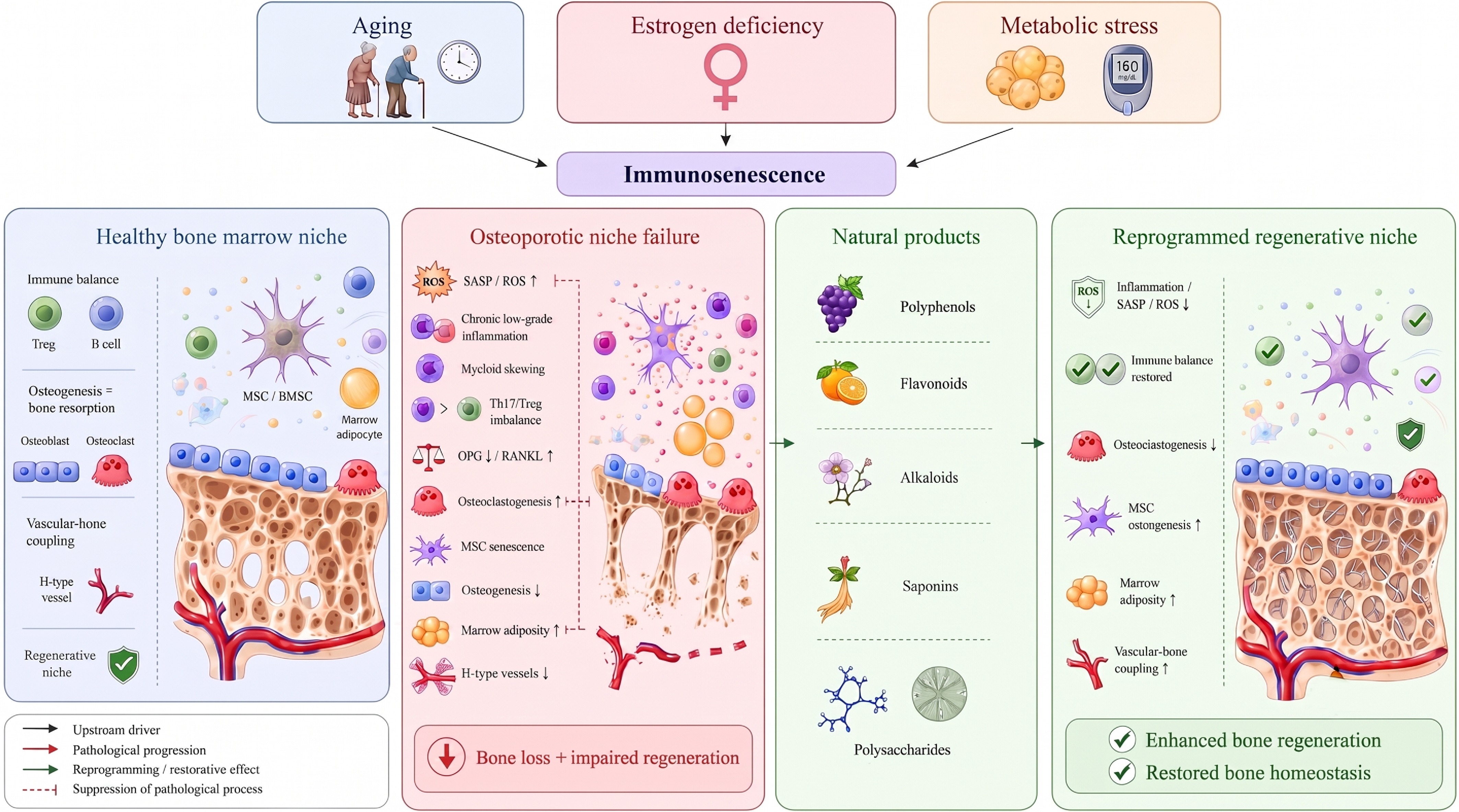
Figure 1.
Immunosenescence-associated dysfunction of the bone marrow niche in osteoporosis and potential natural product-mediated reprogramming. In a healthy bone marrow niche, immune cells, mesenchymal stem cells (MSC)/BMSC, osteoblasts, osteoclasts, H-type vessels, and appropriate levels of bone marrow adiposity collectively maintain immune homeostasis, vascular-bone coupling, and balanced bone remodeling. Aging, estrogen deficiency, and metabolic stress may converge on immunosenescence and dysfunction of the bone marrow immune niche, thereby contributing to chronic low-grade inflammation, elevated senescence-associated secretory phenotype (SASP)/reactive oxygen species (ROS), myeloid skewing, Th17/regulatory T-cell (Treg) imbalance, disruption of the B-cell osteoprotegerin (OPG)/RANKL axis, MSC senescence, increased bone marrow adiposity, and reduced H-type vessels, ultimately leading to bone loss and regenerative dysfunction. Natural products promote the reprogramming of the pathological bone marrow niche into a regeneration-supportive niche by modulating multiple nodes, including inflammatory responses, immune homeostasis, MSCs' osteogenic potential, bone marrow adiposity, and vascular-bone coupling.
-
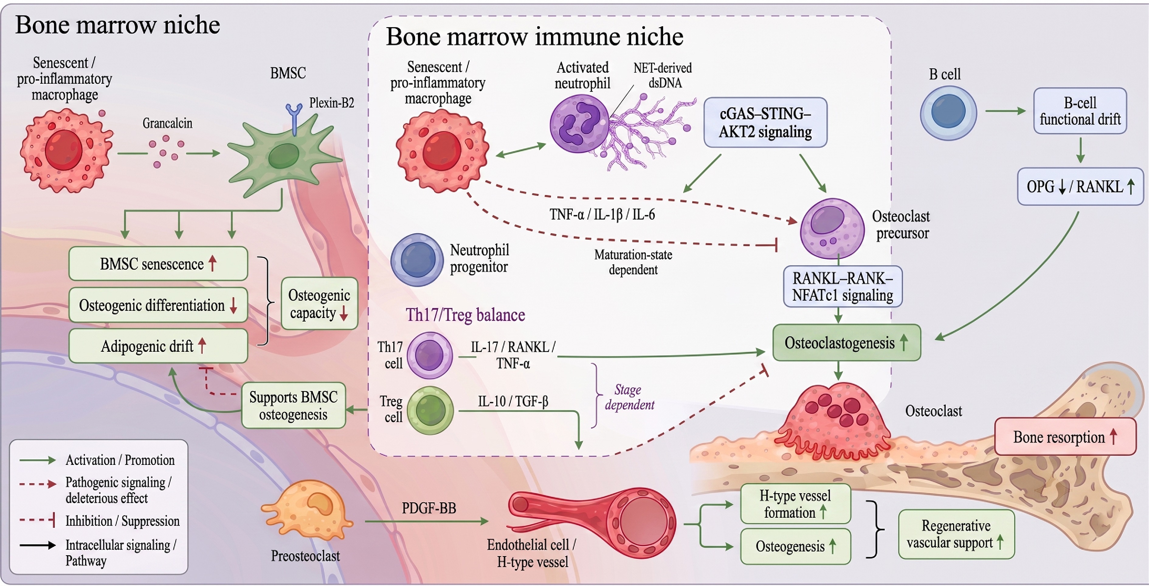
Figure 2.
Cellular interaction map of immunosenescence-associated bone marrow niche destabilization. Senescent or proinflammatory macrophages promote osteoclastogenesis through tumor necrosis factor alpha (TNF-α), interleukin (IL)-1β, and IL-6 signaling, while impairing BMSCs' osteogenic differentiation and promoting cellular senescence and adipogenic drift through the grancalcin-Plexin-B2 axis. Activated neutrophils release neutrophil extracellular trap (NET)-derived double-stranded DNA (dsDNA), which activates cGAS-STING-AKT2 signaling in macrophages and osteoclast precursors, thereby amplifying inflammatory macrophage activation and osteoclastogenesis; in contrast, neutrophil progenitors may restrain osteoclast formation in a maturation-state-dependent manner. Th17-derived IL-17, RANKL, and TNF-α, together with B-cell functional drift characterized by reduced OPG and increased RANKL, enhance osteoclast differentiation and bone resorption. Conversely, Treg-derived IL-10 and TGF-β suppress osteoclastogenesis and support BMSC-related osteogenesis, although these effects may vary with the stage of bone remodeling or repair. In parallel, preosteoclast-derived PDGF-BB promotes H-type vessel formation and osteogenesis, supporting regenerative vascular-bone coupling.
-
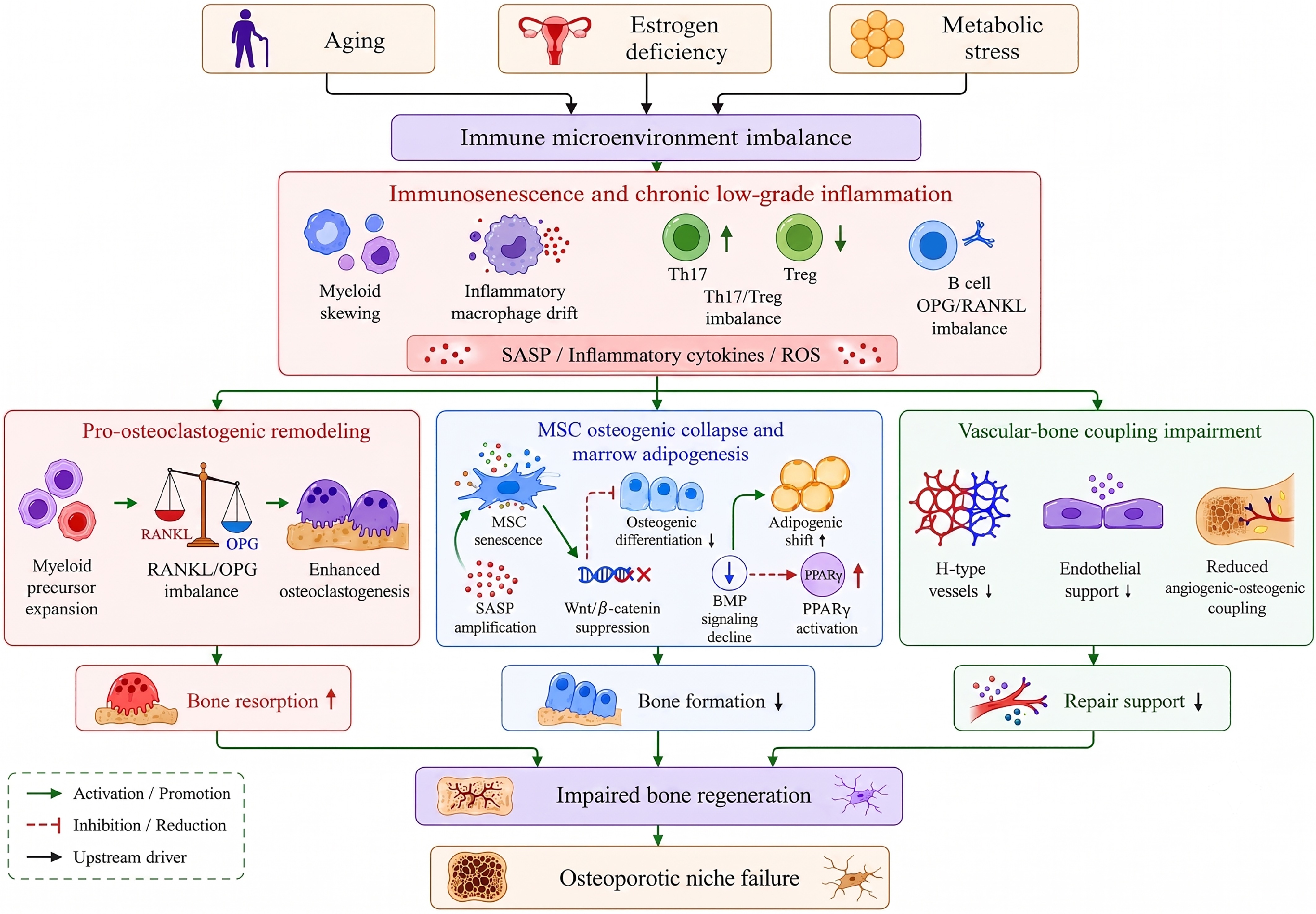
Figure 3.
Mechanistic cascade from immune microenvironmental imbalance to regenerative failure in osteoporosis. Aging, estrogen deficiency, and metabolic stress may converge on an imbalanced immune microenvironment, which further manifests as immunosenescence and chronic low-grade inflammation, accompanied by myeloid skewing, inflammatory macrophage drift, Th17/Treg imbalance, and disruption of the B-cell OPG/RANKL axis. Subsequently, pathological signals diverge into three major mechanistic modules: Pro-osteoclastogenic remodeling, MSCs' osteogenic collapse with bone marrow adiposity, and impaired vascular-bone coupling. Pro-osteoclastogenic remodeling leads to increased bone resorption. MSC senescence, Wnt/β-catenin suppression, BMP signaling decline, and PPARγ activation contribute to reduced bone formation, and the loss of H-type vessels together with impaired endothelial support results in insufficient reparative support.
-
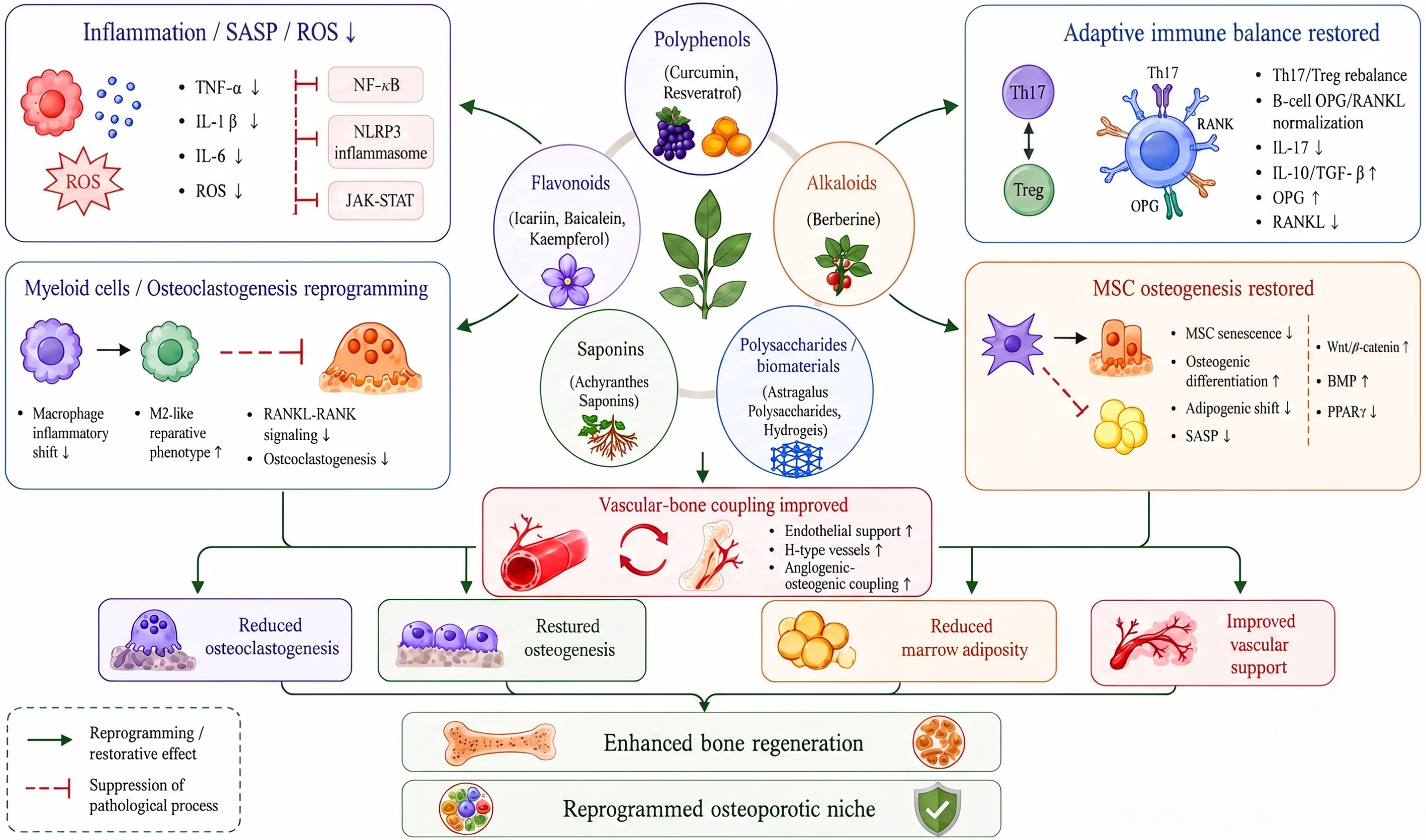
Figure 4.
Natural product-mediated reprogramming network of the bone marrow immune niche in osteoporosis. Natural products derived from diverse chemical classes converge on several microenvironmental regulatory modules, including inflammatory control, immune cell remodeling, restoration of MSCs, metabolic regulation, and vascular-bone coupling. Their major effects include suppressing chronic inflammation, SASP, and ROS; reducing pro-inflammatory mediators such as TNF-α, IL-1β, and IL-6; and modulating inflammatory amplification pathways, including NF-κB, the NLRP3 inflammasome, and JAK-STAT signaling. They may also reshape myeloid cell states, attenuate RANKL-RANK signaling and osteoclastogenesis, restore Th17/Treg balance and the B-cell OPG/RANKL axis, promote MSCs' osteogenic differentiation, inhibit MSCs' senescence and bone marrow adiposity, and improve endothelial support, H-type vessel formation, and vascular-bone coupling.
-
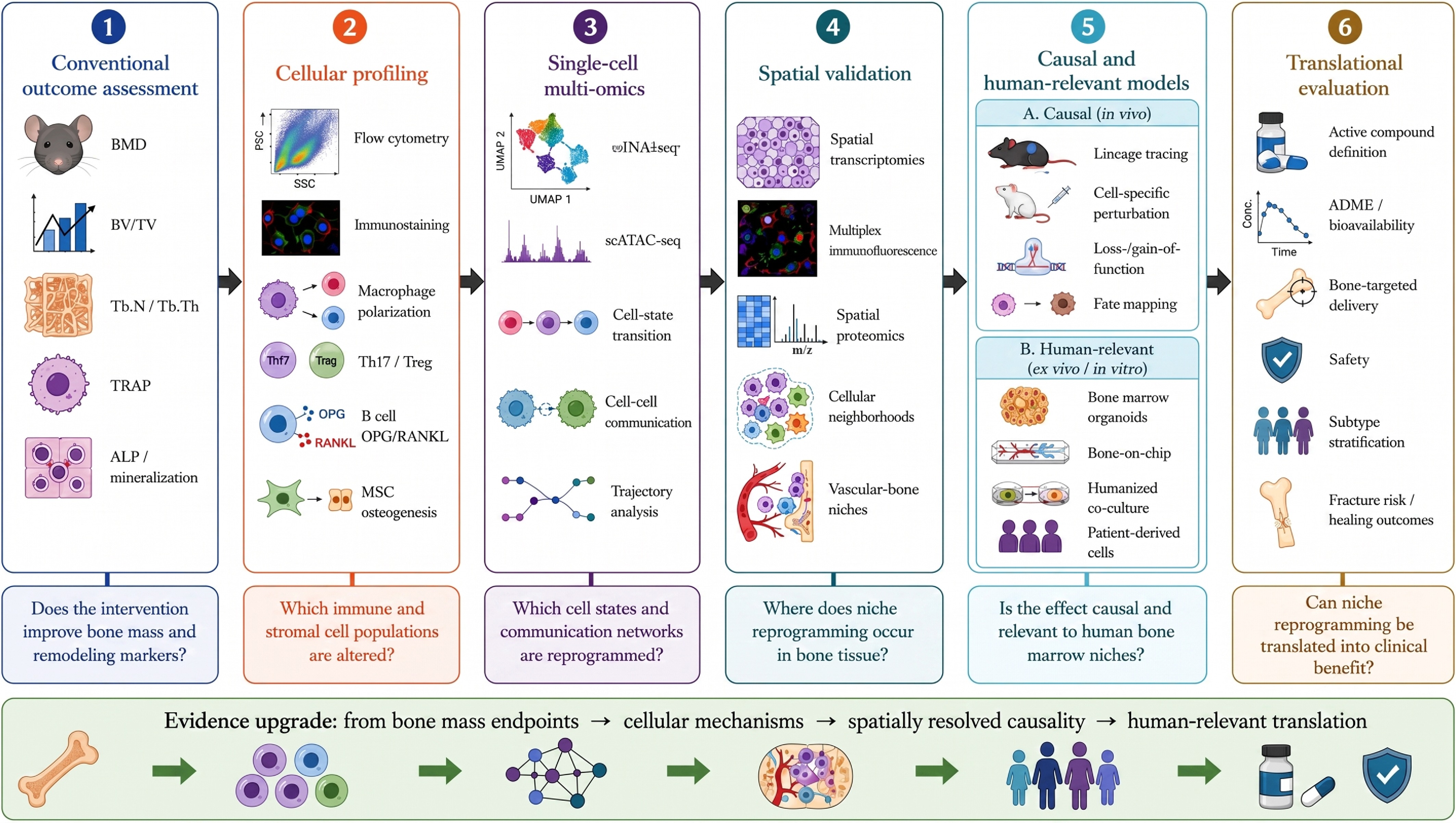
Figure 5.
Technical roadmap for validating natural product-mediated reprogramming of the bone marrow niche. Traditional studies usually assess endpoints such as bone mass, trabecular parameters, TRAP staining, ALP activity, and mineralized nodules to determine whether an intervention improves bone remodeling. Cellular profiling using flow cytometry, immunostaining, and functional assays can further evaluate macrophage polarization, Th17/Treg balance, the B-cell OPG/RANKL axis, and MSC osteogenesis. Single-cell multi-omics can reveal cell state transitions, lineage trajectories, and cell-cell communication, whereas spatial omics and high-dimensional imaging can localize microenvironmental reprogramming within tissue niches. Lineage tracing, functional perturbation, organoids, bone marrow chips, and humanized co-culture models provide causal and human-relevant evidence. Integrating active compound definition, ADME/bioavailability, bone-targeted delivery, safety, patient stratification, and fracture/healing outcomes may move natural product-based osteoporosis research from endpoint observation toward mechanistic precision and clinical translation.
-
Cell/structural unit Trends of change Molecule signaling axis Impact on bone remodeling Impact on tissue regeneration Intervention focus on natural products Hematopoietic stem/progenitor cells Myeloid shift occurs, with an increase in mononuclear/myeloid precursors and a decrease in lymphoid generation. Epigenetic drift, DNA damage, ROS, inflammatory factors, myeloid-biased transcriptional program. Enhance the sources of mononuclear macrophages and osteoclast precursors to promote intensified bone resorption. Impair the immune cell renewal capacity required for immune homeostasis and tissue repair. Amelioration of HSCs' aging through antioxidant, anti-inflammatory, and immunometabolic regulation. Monocytes/osteoclast precursors Myeloid progenitors undergo amplification and exhibit enhanced sensitivity to stimuli such as RANKL, TNF-α, and IL-1β, leading to intensified differentiation into osteoclasts. RANKL-RANK-NFATc1, TNF-α, IL-1β, NF-κB, MAPK, c-Fos Increased osteoclast formation, enhanced bone resorption, and a shift in bone turnover toward resorption. Excessive bone resorption disrupts the trabecular bone structure, reducing the stability of the callus after a fracture. Inhibition of the RANKL/RANK/NFATc1 axis regulates osteoclast differentiation. Macrophages/osteocytes Under aging and inflammatory conditions, cells exhibit a pro-inflammatory phenotype with an increase in M1-like macrophages, the number or function of osteocytes declines, hindering the resolution of inflammation, senescent macrophages secrete grancalcin, which inhibits osteogenesis in BMSCs. TNF-α, IL-1β, IL-6, NF-κB, NLRP3, p38 MAPK, grancalcin-Plexin-B2 Promote the activation of osteoclast precursors and enhance immune-osteoclast coupling; reduce the supportive role of osteogenic macrophages in bone formation. Inflammation fails to resolve promptly, leading to inhibition of MSCs' osteogenesis and a shift in fracture repair from a repair-promoting state to a persistent inflammatory state. Induce the transformation of macrophages from M1 to a M2/pro-focal repair phenotype. Neutrophils Excessive activation of neutrophils leads to increased release of NETs, amplifying sterile inflammation. NETs, extracellular DNA, histones, myeloperoxidase (MPO), elastase, ROS, NLRP3 Indirectly promotes osteoclast formation, exacerbates local inflammation and tissue damage. Injury to endothelial cells and MSC function, interfering with vascularization and bone repair processes. Antioxidant effects, inhibited NET formation, and reduced inflammatory microbody activation. T-cell/Th17-Treg axis The initial T-cell repertoire shrinks, while effector/memory T-cells and senescent T-cells accumulate, leading to an increase in Th17 cells and a decline in the quantity or function of Tregs. IL-17, TNF-α, IFN-γ, RANKL, IL-10, TGF-β, STAT3, Foxp3 Th17 cells promote osteolysis through IL-17, RANKL, and TNF-α, whereas Treg deficiency weakens antiosteolytic effects and immune tolerance. Immunological tolerance and inflammatory resolution are impaired, with deterioration of the MSCs' osteogenic microenvironment Restore the Th17/Treg balance and reduce T-cell-derived RANKL and inflammatory cytokines. B-cells B-cell production declines, resulting in reduced homeostatic OPG support, though some B-cells may transition to a RANKL-expressing and pro-inflammatory secretory state. OPG, RANKL, TNF-α,
G-CSFThe decline in OPG and the increase in RANKL jointly relieve the inhibition of osteoclastogenesis, thereby promoting bone resorption. From a bone protective regulator to an inflammatory amplifier and osteoclast synergizer, compromising the regenerative niche Regulate the B-cells' OPG/RANKL balance to suppress the pro-inflammatory B-cell state. Bone marrow mesenchymal stem/progenitor cells Signs include aging, decreased proliferation, inhibited osteogenic differentiation, enhanced adipogenic differentiation, and increased release of SASP. p16, p21, ROS, SASP, Wnt/β-catenin, BMP/Smad, PPARγ, C/EBPα, Plexin-B2 The reduction in osteoblast-derived cells leads to decreased bone formation, while the differentiation of MSCs toward the adipocyte lineage further depletes the pool of osteogenic progenitor cells. Delayed initiation of osteogenesis, impaired fracture healing and reduced capacity for bone defect regeneration. Promote MSCs' commitment to the osteogenic lineage and inhibit adipogenesis. Senescent osteocyte/osteoblast lineage Osteocytes and osteoblasts undergo aging and functional decline. Levels of RANKL, sclerostin, and SASP factors increase, leading to reduced osteogenic support capacity. RANKL, OPG, sclerostin, IL-6, TNF-α, damage-associate molecular patterns (DAMPs), Wnt/β-catenin, Nrf2/HO-1 Elevated RANKL promotes osteolysis, whereas sclerostin inhibits Wnt-mediated osteogenesis, leading to further imbalance between bone formation and bone resorption. Decreased deposition and mineralization quality of the bone matrix, with impaired mechanical properties and repair quality of bone tissue. Antioxidant, antiapoptotic, inhibits abnormal expression of sclerostin/RANKL and activates the osteogenic pathway. Bone marrow adipocytes The expansion of bone marrow adipose tissue leads to adipocytes secreting adipokines, free fatty acids, ROS, and inflammatory mediators, which subsequently form a metabolic-inflammatory positive feedback loop with immune cells. IL-6, RANKL, adiponectin, free fatty acids, ROS, PPARγ, C/EBPα Promote pro-inflammatory transformation and osteogenesis of myeloid cells, while inhibiting osteogenic differentiation of MSCs. The bone marrow cavity transitions from an osteogenesis-supportive environment to a fatty and inflammatory environment, reducing bone repair efficiency. Inhibit MSCs' adipogenic differentiation; reduce lipid toxicity and metabolic inflammation. Vascular endothelial cells/H-type vessels Inflammation and oxidative stress induce endothelial aging, leading to impaired angiogenesis and a reduction in H-type vessels. VEGF, HIF-1α, Notch, CD31, TNF-α, ROS Vascular-bone coupling decreases, resulting in reduced nutrient, oxygen supply, and paracrine support to osteoblasts and MSCs. Insufficient vascularization in the fracture healing area results in restricted osteogenic space and regenerative dynamics. Improve vascular-bone coupling and the local repair microenvironment Table 1.
The cellular axis of immune microenvironment disruption in osteoporosis.
-
Microenvironment target Representative compound Target cells Molecular pathway Evidence source Ref. Chronic inflammation, oxidative stress, and osteocyte-immune cell interaction Curcumin Macrophages, T-cells, osteoblasts, osteoclasts, BMSCs Improving abnormal interactions among macrophages, T-cells, and osteocytes through anti-inflammatory and antioxidant effects Cell and animal model [106] Macrophage polarization, metabolic inflammation, osteogenesis-osteoclast coupling Berberine Macrophages, BMSCs, osteoclasts, osteoblasts Reducing TNF-α and IL-6 levels, increasing IL-10 levels, and promoting the transformation of macrophages toward the anti-inflammatory M2 phenotype. Animal model [107] Macrophage polarization and chronic inflammatory amplification Isoeugenol Bone marrow macrophages, osteoclast precursors Regulation of bone marrow macrophage M1 polarization via p38 MAPK and arachidonic acid metabolic pathways ameliorates chronic inflammation-mediated immune dysregulation. Cell and animal model [108] STING-dependent inflammation, chronic inflammatory microenvironment RTA-408 Macrophages, BMSCs, osteoblasts, and osteoclast-related cells Inhibit STING-dependent NF-κB signaling to mitigate microenvironmental dysregulation induced by chronic inflammation. Cell and animal model [109] Inflammatory bodies, Th17/Treg balance, oxidative stress Baicalein, icariin T-cells, macrophages, BMSCs, osteoclast precursors Regulate NLRP3 inflammasome activation, Th17/Treg balance, and antioxidant pathways to enhance its targeted modulation of immune cells and microenvironment responsiveness. Cell model [110] Oxidative stress, ferroptosis, immune cell homeostasis Chlorogenic acid, protocatechuic acid Osteoblasts, BMSCs, immune cells, osteoclast-related cells Clear ROS, alleviate oxidative stress, and inhibit ferroptosis associated with immune dysregulation, thereby maintaining the homeostasis of immune cells and osteocytes in the bone microenvironment. Cell and animal model [111] T-cell-RANKL axis, osteogenesis Resveratrol Activated T-cells, osteoclast precursors, BMSCs Inhibit the secretion of RANKL by activated T-cells, thereby blocking the RANKL/RANK signaling pathway and reducing osteoclast differentiation and bone resorption. Animal model [112] Oxidative stress, cellular senescence, local inflammation Salvianolic acid A Osteoblasts, BMSCs, immune cells Eliminate free radicals to reduce intracellular oxidative stress and achieve microenvironment remodeling Cell model [113] Osteoblast apoptosis, oxidative stress, and impaired osteogenic differentiation Naringenin Osteoblasts, BMSCs Activation of the Nrf2/HO-1 signaling pathway inhibits osteoblast apoptosis and alleviates impaired osteoblast differentiation. Cell model [114] Osteoclast differentiation and inflammatory bone resorption Homoisoflavonoid derivative 5g Osteoclast precursor cells, osteoclasts Reduce the activation of the ERK1/2 and IκBα/NF-κB signaling pathways, thereby blocking bone loss caused by abnormal osteoclast activity. Cell model [115] Aging of BMSCs, mitochondrial autophagy, and restoration of osteogenic function Kaempferol BMSCs, osteoblasts Targeting Sp1 to activate FUNDC1-mediated mitochondrial autophagy for improving BMSCs' senescence in postmenopausal osteoporosis Cell and animal model [116] Th17 cells, systemic immune homeostasis, osteoclastogenesis Chloroquine Th17 cells, Tregs, osteoclast precursors, and bone marrow immune cells Reduce the proportion of Th17 cells promoting osteoclastogenesis in the bone marrow; enhance protective cytokines such as IFN-γ, IL-4, and IL-10; and restore systemic immune homeostasis. Cell and animal model [117] Th17/Treg balance, inflammatory bone erosion Nuciferine T-cells, synovial/inflammatory microenvironment cells, osteoclast-related cells Improve collagen-induced bone erosion; reduce pro-inflammatory cytokines; and serum immunoglobulins (Ig)G, IgG1, and IgG2a; and restore the Th17/Treg balance in rats. Cell and animal model [118] RANKL-induced osteoclastogenesis Kirenol Osteoclast precursor cells, osteoclasts Inhibition of the NFATc1 and Cav-1 signaling pathways suppresses RANKL-induced osteoclastogenesis and prevents ovarianectomy-induced osteoporosis Cell and animal model [119] RANKL triggering signal, osteoclast formation Corosolic acid Osteoclast precursor cells, osteoclasts Modulate RANKL triggering signals to inhibit osteoclast formation-induced bone loss Cell and animal model [120] Abnormal osteoclastogenesis and bone resorption Holothurin A, Echinoside A Osteoclast precursor cells, osteoclasts Targeting CB1 and MKP-1 to inhibit the expression of factors such as c-fos and NFATC1 improves abnormal bone resorption Animal model [121] ER/RANK/NFATc1 axis, OVX-associated immune microenvironment Achyranthes saponin Osteoclast precursors, osteoclasts, and bone marrow immune cells Regulation of the RER/RANK/NFATc1 signaling pathway improves the bone marrow immune niche, thereby inhibiting OVX-induced osteoporosis Cell and animal model [122] Vessel-bone coupling, H-type angiogenesis, estrogen-deficient bone loss Harmine Osteoclast precursors, endothelial cells, osteoblasts Promote the secretion of PDGF-BB by osteoclast precursors, induce H-type angiogenesis, improve bone vascular-immune coupling, and alleviate bone loss caused by estrogen deficiency. Cell and animal model [123] ROS, NF-κB/NFATc1, osteoclast differentiation Dictamnine Osteoclast precursor cells, osteoclasts Regulate the ROS, NF-κB, and NFATc1 signaling pathways to inhibit RANKL-induced osteoclast differentiation Cell and animal model [124] SIRT1, NF-κB acetylation, ROS activity Crebanine Osteoclasts, osteoblasts, BMSCs, inflammatory cells Targeting Sirt1 to interfere with NF-κB acetylation and ROS activity improves bone loss Cell and animal model [125] Intestinal-bone-immune axis, systemic inflammation, short-chain fatty acids Dendrobium officinale polysaccharides Gut microbiota, intestinal epithelial cells, osteoblast/osteoclast-related cells Reshapes short-chain fatty acid (SCFA)-producing microbiota, restores intestinal barrier integrity and butyrate/isovalerate levels, reduces systemic inflammation, and activates Wnt/β-catenin signaling; fecal microbiota transplantation reproduces the osteoprotective effect. Animal model [126] Osteogenesis, bone marrow fat accumulation, and immune microenvironment repair Dendrobium polysaccharides and Scutellaria baicalensis polysaccharides Osteoclasts, BMSCs, bone marrow adipocytes, immune cells Inhibits osteoclastogenesis in OVX rats, regulates lipid accumulation in the bone marrow cavity, and exerts a reparative effect on the bone marrow immune niche. Animal model [127] Local immune microenvironment, macrophage function, bone repair Astragalus polysaccharide hydrogels Macrophages, BMSCs, osteoblasts, vascular endothelial cells Regulate macrophages' function and optimize the local immune microenvironment to promote bone repair. Cell model [128] Bone-targeted delivery, aging metabolism, and the immune microenvironment Self-assembled nanoparticles with well-defined oligosaccharide CS4-NP BMSCs, bone marrow immune cells, osteoclasts, osteoblasts Achieve targeted delivery of active ingredients to significantly enhance bone mass in the OVX model while improving the local aging metabolism and immune microenvironment. Cell and animal model, human observational studies [129] Table 2.
Natural products redefining microenvironmental targets and effects.
Figures
(5)
Tables
(2)