-
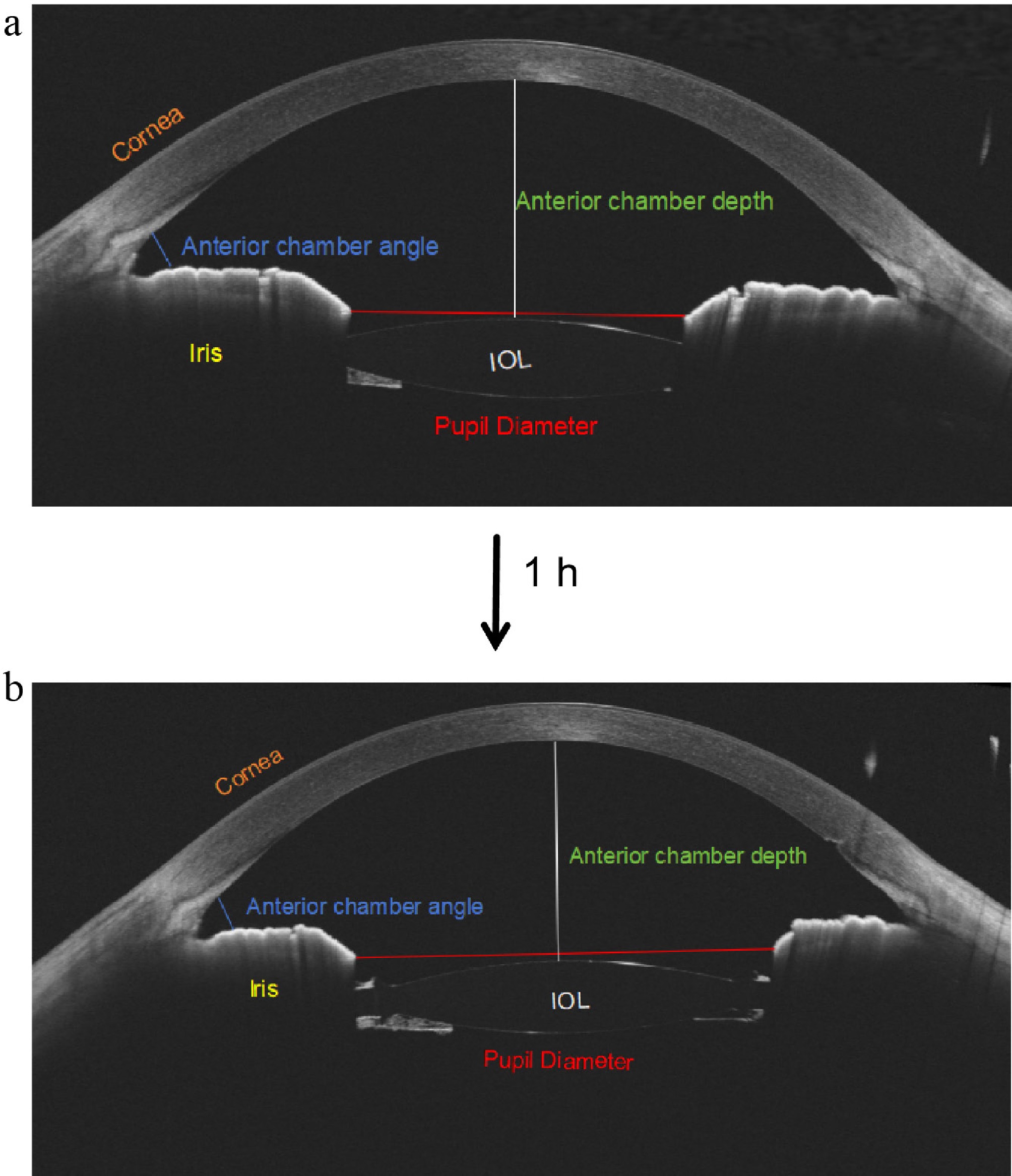
Figure 1.
Changes in the anterior structure of the AS-OCT image. (a) The AS-OCT parameters at baseline. (b) The pupil's diameter (red line) increased and the anterior chamber angle area (blue line) decreased slightly without changes in the other parameters after 1 h.
-
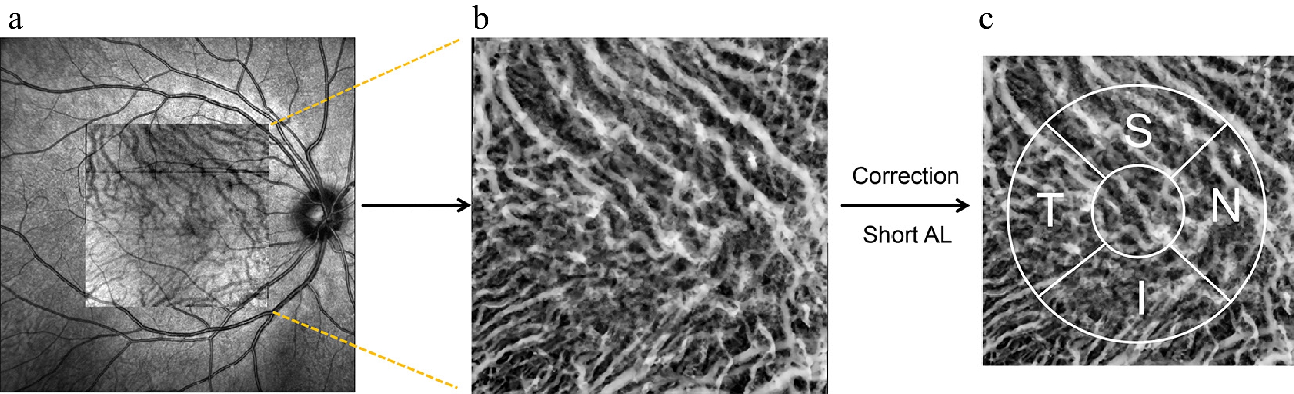
Figure 2.
Illustration of analyzing OCTA image parameters. (a) OCTA scan region of 6 × 6 mm. (b) Original en face OCTA image of the choroidal vascularity index image. (c) The 3-mm ETDRS grid on the magnified OCTA choroidal vascularity index image corrected with a short axial length (AL).
-
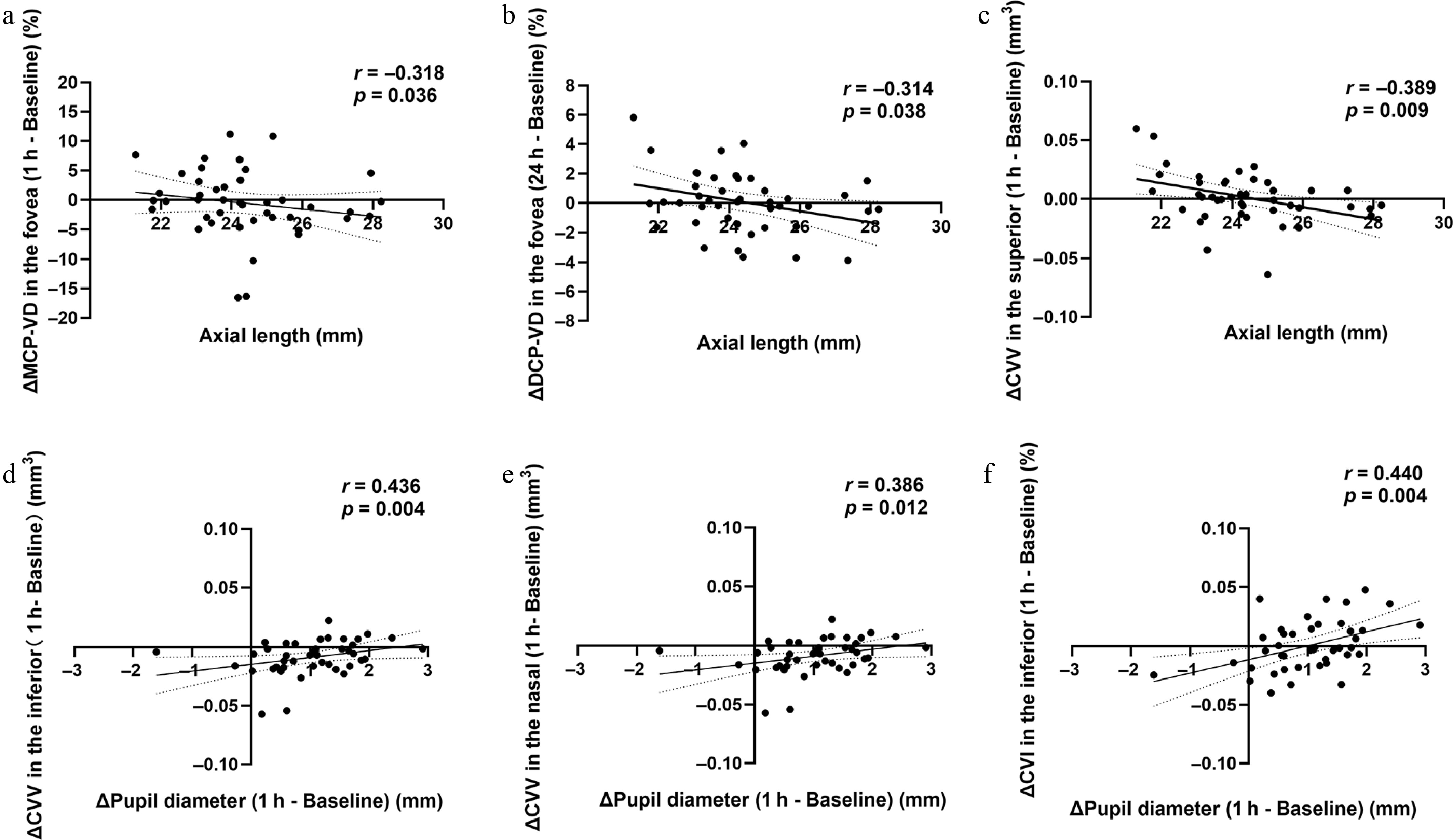
Figure 3.
The correlations of AL and the change in PD with the posterior parameters at 1 and 24 h. Correlations were assessed using partial correlation analysis, with p < 0.05 considered statistically significant.
-
Variable Value (mean ± SD) Sex (%) Male (20, 68.96%) Female (9, 31.04%) Age (years) 10.52 ± 2.17 SE (D) −3.47 ± 1.60 AL (mm) 24.38 ± 1.68 IOP (mmHg) 15.88 ± 1.71 CCT (mm) 0.53 ± 0.14 ACD (mm) 4.24 ± 0.40 PD (mm) 4.38 ± 0.74 SE, spherical equivalent; AL, axial length; IOP, intraocular pressure; CCT, central corneal thickness; ACD, anterior chamber depth; PD, pupil diameter. Table 1.
Demographic and clinical characteristics.
-
Variable Mean ± SD P P1 P2 P3 Baseline 1 h 24 h IOP (mmHg) 15.88 ± 1.71 16.73 ± 2.03 15.70 ± 2.06 < 0.001*** 0.002** 0.614 0.004** CCT (mm) 0.53 ± 0.14 0.52 ± 0.17 0.53 ± 0.14 0.331 0.641 0.065 0.550 ACD (mm) 4.24 ± 0.40 4.20 ± 0.39 4.21 ± 0.40 0.526 0.448 0.567 0.875 PD (mm) 4.38 ± 0.74 5.12 ± 0.84 4.10 ± 0.87 < 0.001*** < 0.001*** 0.032* < 0.001*** TISA-N(mm2) 0.23 ± 0.08 0.20 ± 0.07 0.23 ± 0.09 0.021* 0.079 0.987 0.060 TISA-T(mm2) 0.29 ± 0.14 0.24 ± 0.06 0.28 ± 0.09 0.020* 0.071 0.583 < 0.01** IOP, intraocular pressure; CCT, central corneal thickness; ACD, anterior chamber depth; PD, pupil diameter; TISA-N, trabecular iris space area – nasal; TISA-T, trabecular iris space area – temporal. P, p-value among the three groups; P1, p-value for the comparison between baseline and 1 h after receiving medication; P2, for the baseline versus 24 h after the medication; P3, for 1 h versus 24 h after medication. * p < 0.05. ** p < 0.01, *** p < 0.001. Repeated-measures ANOVA or the Friedman test was used, as appropriate. Table 2.
Comparative analysis of the anterior segment parameters across three time points.
-
Variable Mean ± SD P P1 P2 P3 Baseline 1 h 24 h RT (µm) Fovea 260.72 ± 19.85 261.09 ± 20.69 261.70 ± 21.51 0.653 0.887 0.746 0.894 Para-S 343.76 ± 18.11 344.30 ± 18.42 342.08 ± 22.59 0.551 > 0.999 > 0.999 0.907 Para-T 327.80 ± 15.58 328.15 ± 16.00 326.58 ± 19.58 0.328 0.723 0.497 > 0.999 Para-I 337.19 ± 16.13 337.29 ± 15.98 335.37 ± 20.92 0.649 > 0.999 > 0.999 > 0.999 Para-N 342.30 ± 21.70 342.47 ± 21.74 340.24 ± 25.72 0.978 > 0.999 > 0.999 > 0.999 GCC (µm) Fovea 43.14 ± 8.63 42.81 ± 8.64 44.00 ± 10.29 0.853 > 0.999 > 0.999 > 0.999 Para-S 121.49 ± 10.48 121.58 ± 11.16 120.37 ± 13.67 0.913 > 0.999 > 0.999 > 0.999 Para-T 107.04 ± 9.01 106.82 ± 9.01 107.45 ± 9.39 0.138 0.758 0.403 0.149 Para-I 121.56 ± 8.49 121.48 ± 8.06 121.76 ± 8.82 0.570 0.970 0.750 0.555 Para-N 117.85 ± 9.36 118.15 ± 9.46 118.26 ± 9.79 0.496 0.784 0.618 0.784 RNFL (µm) Fovea 14.74 ± 1.39 14.47± 1.38 14.21 ± 1.51 0.178 > 0.999 0.264 0.407 Para-S 29.21 ± 3.89 29.82 ± 3.44 29.48 ± 3.57 0.328 > 0.999 0.723 0.497 Para-T 19.60 ± 1.92 19.61 ± 1.62 19.66 ± 1.72 0.960 > 0.999 0.964 0.973 Para-I 29.89 ± 3.77 30.19 ± 2.82 30.30 ± 3.74 0.913 > 0.999 > 0.999 > 0.999 Para-N 24.91 ± 2.75 25.30 ± 2.34 25.31 ± 3.76 0.744 > 0.999 > 0.999 > 0.999 GCIPL (µm) Fovea 28.40 ± 8.06 28.34 ± 8.21 29.78 ± 9.72 0.136 0.972 0.303 0.269 Para-S 91.91 ± 7.98 91.76 ± 8.40 92.11 ± 8.55 0.084 > 0.999 0.210 0.129 Para-T 87.34 ± 7.85 87.21 ± 7.99 87.74 ± 8.09 0.394 0.951 0.660 0.268 Para-I 91.37 ± 6.51 91.28 ± 6.23 91.52 ± 6.97 0.808 0.959 0.945 0.873 Para-N 92.68 ± 8.13 92.85 ± 8.03 92.99 ± 8.23 0.494 > 0.999 > 0.999 0.723 RT, retinal thickness; GCC, ganglion cell complex; RNFL, retinal nerve fiber layer; GCIPL, ganglion cell–inner plexiform layer; Para-S, parafovea superior; Para-I, parafovea inferior; Para-N, parafovea nasal; Para-T, parafovea temporal. P, p-value among the three groups; P1, p-value for the comparison between the baseline and 1 h after receiving the medication; P2, p-value for the baseline versus 24 h after receiving the medication; P3, p-value for 1 h versus 24 h after receiving the medication. p < 0.05 was considered statistically significant. Repeated-measures ANOVA or the Friedman test was used, as appropriate. Table 3.
Retinal thickness and retinal neural parameters at baseline, 1 and 24 h after atropine instillation.
-
Variable Mean ± SD P P1 P2 P3 Baseline 1 h 24 h SCP VD (%) Fovea 9.58 ± 3.57 9.18 ± 3.38 9.29 ± 4.06 0.605 0.632 0.683 0.974 Para-S 46.24 ± 9.05 45.05 ± 9.54 44.78 ± 10.34 0.695 > 0.999 > 0.999 > 0.999 Para-T 41.10 ± 6.84 38.62 ± 7.04 39.43 ± 7.50 0.090 0.031* 0.366 0.774 Para-I 45.54 ± 8.39 44.47 ± 7.47 44.33 ± 8.39 0.695 > 0.999 > 0.999 > 0.999 Para-N 42.78 ± 8.67 41.29 ± 8.35 41.84 ± 9.60 > 0.999 > 0.999 > 0.999 > 0.999 MCP VD (%) Fovea 19.66 ± 5.72 19.09 ± 6.00 19.06 ± 7.17 0.742 0.784 0.751 0.999 Para-S 31.29 ± 10.05 30.79 ± 10.55 29.43 ± 10.53 0.052 0.770 0.046* 0.592 Para-T 36.51 ± 6.32 35.25 ± 6.67 34.97 ± 7.33 0.595 > 0.999 > 0.999 > 0.999 Para-I 30.86 ± 7.47 28.78 ± 8.27 29.19 ± 9.24 0.468 0.723 > 0.999 > 0.999 Para-N 36.19 ± 7.25 34.42 ± 6.78 34.04 ± 10.46 0.270 0.723 0.367 > 0.999 DCP VD (%) Fovea 2.66 ± 2.41 2.41 ± 2.28 2.74 ± 3.04 0.928 > 0.999 > 0.999 > 0.999 Para-S 7.02 ± 4.44 7.45 ± 4.61 6.45 ± 4.42 0.533 > 0.999 > 0.999 0.836 Para-T 8.27 ± 5.66 7.87 ± 5.08 7.80 ± 4.82 0.740 0.834 0.775 0.993 Para-I 5.65 ± 4.12 4.93 ± 3.59 4.48 ± 3.20 0.014* 0.330 0.012* 0.603 Para-N 7.95 ± 4.79 7.00 ± 4.74 7.23 ± 4.38 0.408 0.548 > 0.999 > 0.999 FAZ (mm2) 0.31 ± 0.11 0.32 ± 0.11 0.32 ± 0.12 0.744 > 0.999 > 0.999 > 0.999 SCP VD, superficial capillary plexus vessel density; MCP VD, middle capillary plexus vessel density; DCP VD, deep capillary plexus vessel density; Para-S, parafovea superior; Para-I, parafovea inferior; Para-N, parafovea nasal; Para-T, parafovea temporal. FAZ, foveal avascular zone. P, p-value among the three groups; P1, p-value for the comparison between the baseline and 1 h after administration of the medication; P2, p-value for the baseline versus 24 h after administration of the medication; P3, p-value for 1 h versus 24 h after administration of the medication. * p < 0.05. Repeated measures ANOVA or the Friedman test was used, as appropriate. Table 4.
Comparison of the vessel density of the superficial, middle, and deep capillary plexus at baseline, 1 and 24 h after receiving the medication.
-
Variable Mean ± SD P P1 P2 P3 Baseline 1 h 24 h ChT (µm) Fovea 325.00 ± 84.05 318.40 ± 77.40 323.10 ± 77.32 0.060 0.407 > 0.999 0.057 Para-S 305.00 ± 102.50 302.5 ± 101.60 302.80 ± 96.81 0.551 > 0.999 > 0.999 0.907 Para-T 334.30 ± 79.72 326.90 ± 73.14 331.70 ± 72.91 0.016* 0.129 > 0.999 0.017* Para-I 320.80 ± 88.28 310.50 ± 80.33 317.10 ± 83.66 0.032* 0.129 > 0.999 0.043* Para-N 299.70 ± 83.33 291.40 ± 78.54 294.10 ± 73.58 0.019* 0.023* > 0.999 0.099 CCF (mm2) Fovea 0.61 ± 0.05 0.60 ± 0.05 0.61 ± 0.03 0.529 > 0.999 0.859 > 0.999 Para-S 1.16 ± 0.12 1.14 ± 0.13 1.17 ± 0.10 0.542 > 0.831 > 0.872 > 0.534 Para-T 1.19 ± 0.09 1.16 ± 0.11 1.18 ± 0.08 0.815 > 0.999 > 0.999 > 0.999 Para-I 1.18 ± 0.11 1.18 ± 0.15 1.20 ± 0.25 0.983 > 0.999 > 0.999 > 0.999 Para-N 1.14± 0.10 1.11 ± 0.13 1.12 ± 0.16 0.431 > 0.999 0.723 0.859 CVV (mm3) Fovea 0.11 ± 0.04 0.10 ± 0.03 0.11 ± 0.04 0.073 0.087 > 0.999 0.295 Para-S 0.21 ± 0.06 0.21 ± 0.05 0.21 ± 0.06 0.049* 0.043* 0.859 0.497 Para-T 0.22 ± 0.07 0.21 ± 0.06 0.22 ± 0.07 0.060 0.407 > 0.999 0.057 Para-I 0.21 ± 0.08 0.20 ± 0.07 0.20 ± 0.08 0.029* 0.072 0.890 0.018* Para-N 0.19 ± 0.08 0.18 ± 0.07 0.19 ± 0.08 < 0.001*** 0.001** 0.493 0.011* CSV (mm3) Fovea 0.11 ± 0.03 0.11 ± 0.03 0.11 ± 0.03 0.420 0.661 > 0.999 0.934 Para-S 0.22 ± 0.05 0.22 ± 0.06 0.22 ± 0.04 0.649 > 0.999 > 0.999 > 0.999 Para-T 0.23 ± 0.06 0.23 ± 0.06 0.23 ± 0.05 0.049* 0.497 0.859 0.043* Para-I 0.22 ± 0.07 0.21 ± 0.06 0.22 ± 0.06 0.110 0.407 > 0.999 0.129 Para-N 0.20 ± 0.06 0.20 ± 0.06 0.19 ± 0.05 0.897 > 0.999 > 0.999 > 0.999 CVI Fovea 0.48 ± 0.05 0.48 ± 0.05 0.48 ± 0.05 0.788 0.839 0.995 0.831 Para-S 0.49 ± 0.05 0.48 ± 0.04 0.48 ± 0.05 0.443 0.388 0.768 > 0.839 Para-T 0.48 ± 0.05 0.48 ± 0.05 0.48 ± 0.04 0.728 0.758 0.988 0.812 Para-I 0.48 ± 0.06 0.48 ± 0.06 0.48 ± 0.05 > 0.999 > 0.999 > 0.999 > 0.999 Para-N 0.48 ± 0.06 0.48 ± 0.07 0.48 ± 0.07 0.090 0.081 0.992 0.208 ChT, choroidal thickness; CCF, choriocapillaris flow; CVV, choroidal vascular volume; CSV, choroidal stromal volume; CVI, choroidal vascularity index; Para-S, parafovea superior; Para-I, parafovea inferior; Para-N, parafovea nasal; Para-T, parafovea temporal. P, p-value among the three groups; P1, p-value for the comparison between baseline and 1 hour after receiving the medication; P2, p-value for baseline versus 24 h after receiving the medication; P3, p-value for 1 h versus 24 h after receiving the medication. * p < 0.05, ** p < 0.01,*** p < 0.001. Repeated measures ANOVA or the Friedman test was used, as appropriate. Table 5.
Choroidal parameters at baseline, 1 and 24 h after atropine instillation.
Figures
(3)
Tables
(5)