-
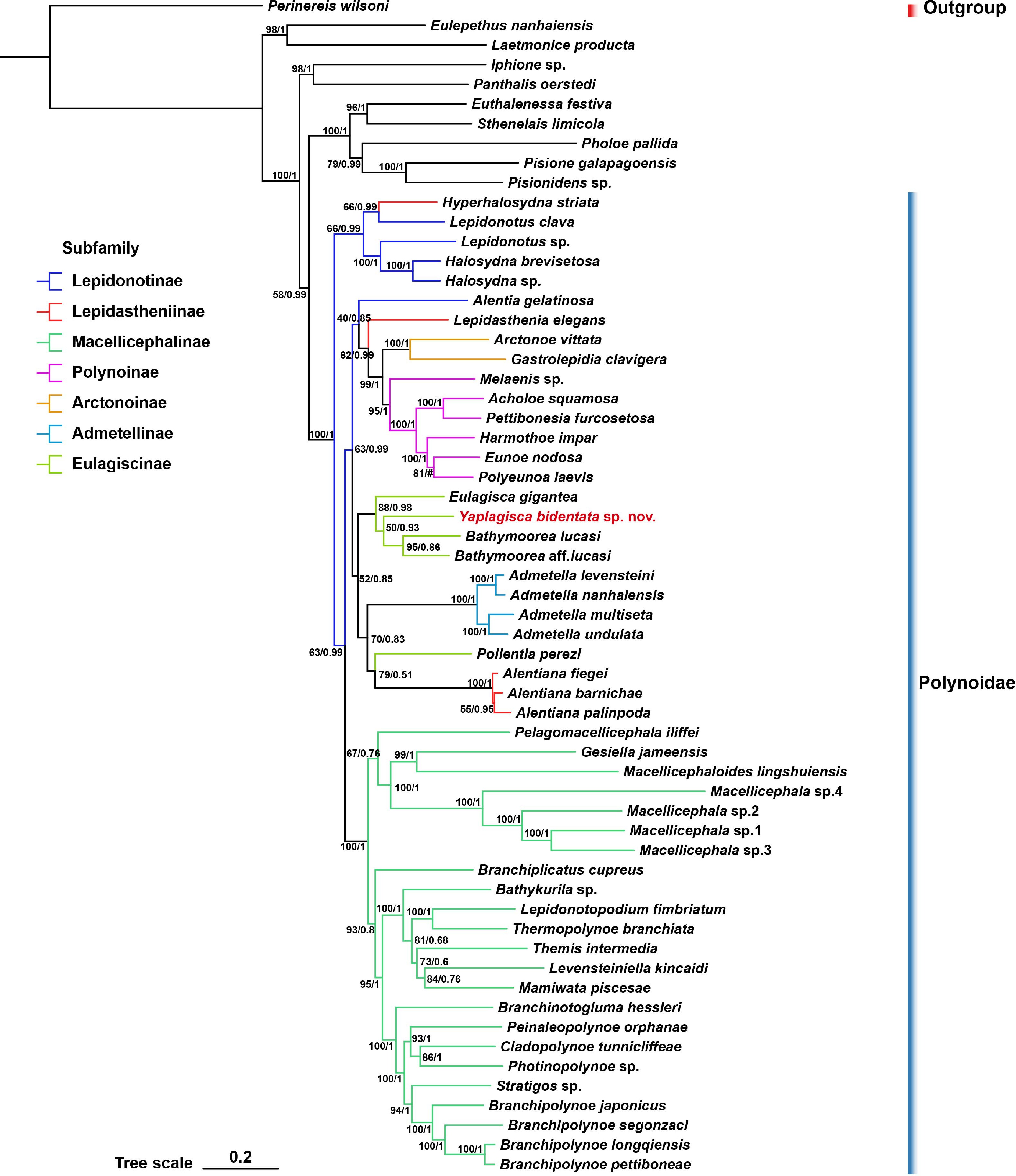
Figure 1.
Phylogenetic relationships inferred from the concatenated 17-gene matrix. The topology shown is the maximum-likelihood (ML) tree. Node values indicate ML ultrafast bootstrap support (left) and Bayesian posterior probability (BI; right). Different branch colors denote different subfamilies of Polynoidae. The hash sign marks the node at which the ML and BI topologies were incongruent.
-
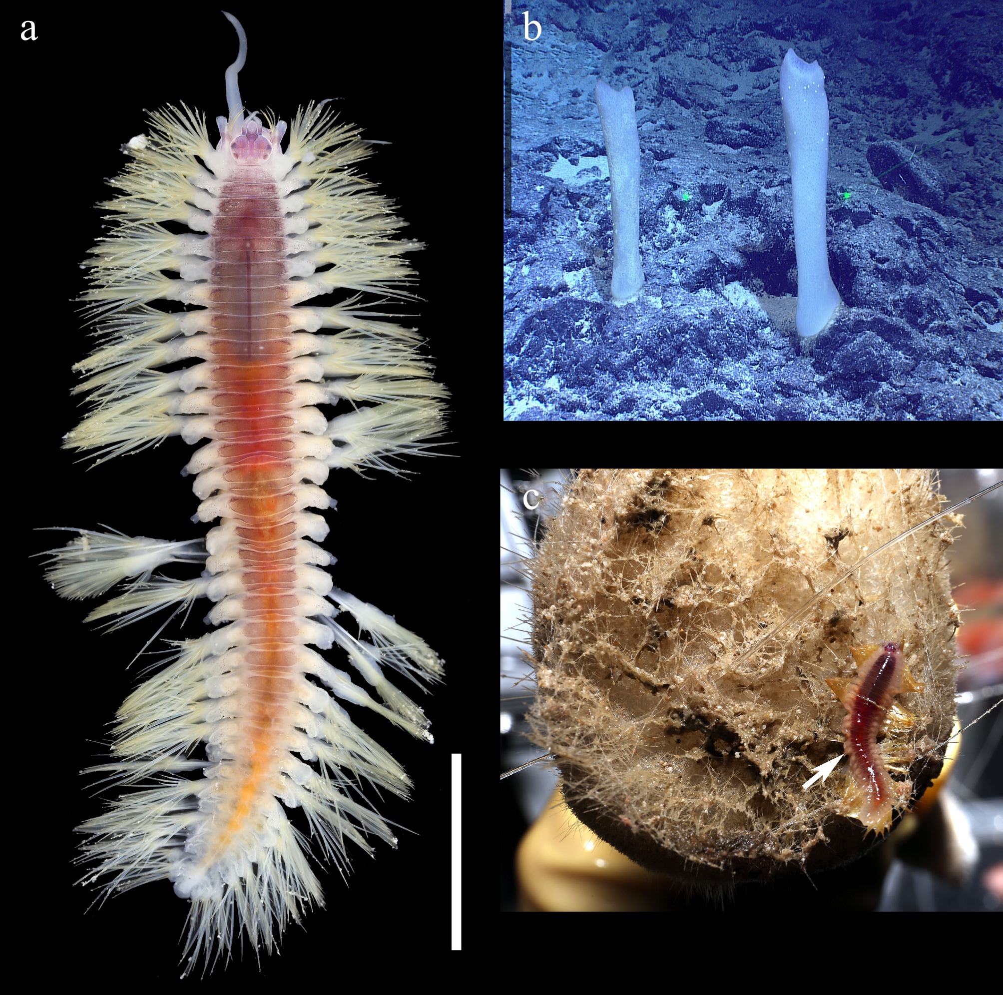
Figure 2.
Yaplagisca bidentata gen. et sp. nov. (a) Dorsal view of living holotype. (b) In situ image of putative host sponge Poliopogon distortus Gong & Li, 2018. (c) Holotype found on the surface of P. distortus after ROV recovery. The distance between the paired laser beams in the in situ image (b) is 33 cm. Scale bar: (a) 5 mm.
-
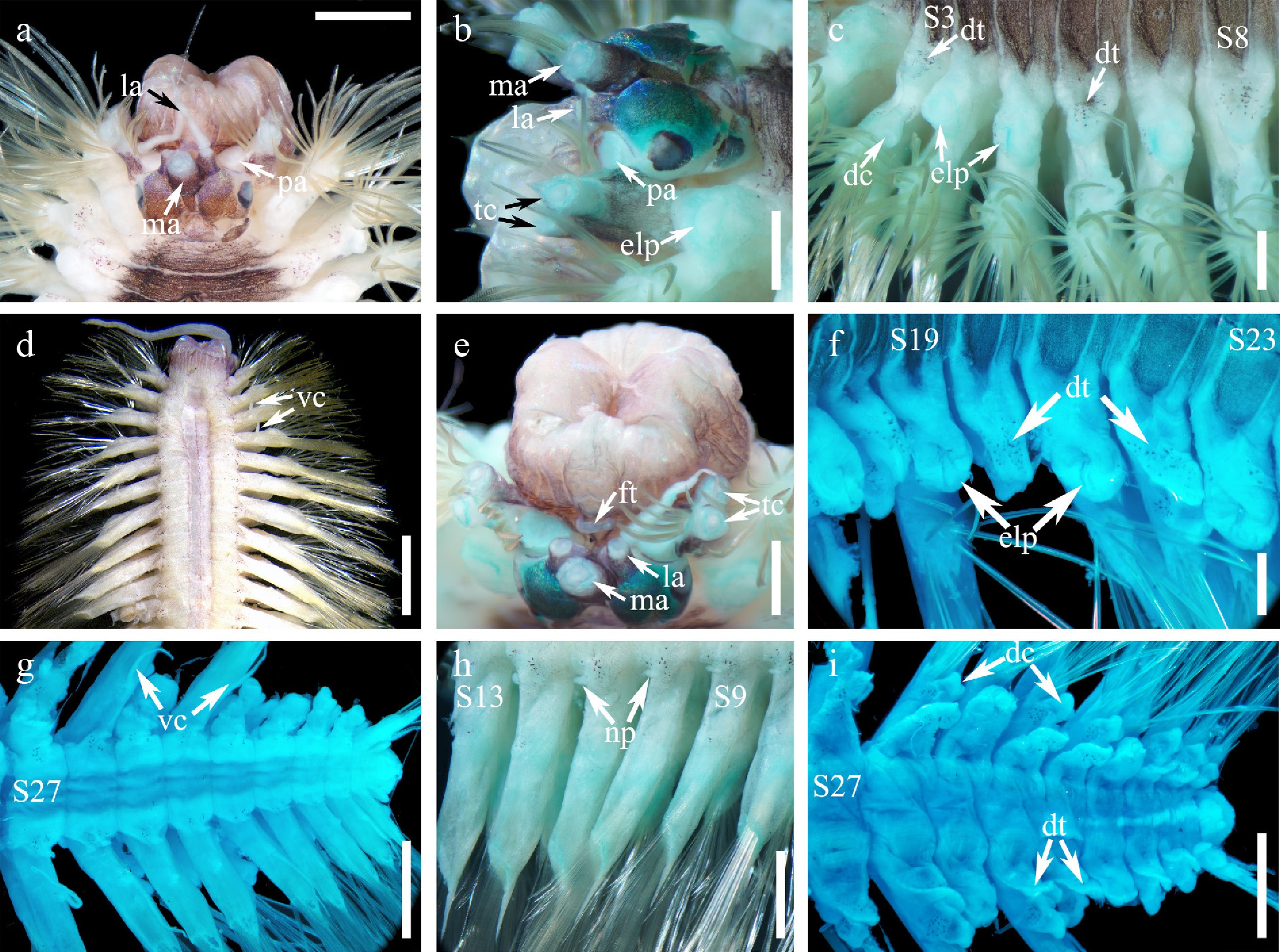
Figure 3.
Yaplagisca bidentata gen. et sp. nov., details of the anterior, median and posterior body regions. (a) Dorsal view of prostomium and anterior segments. (b) Left dorsolateral view of prostomium and anterior segments. (c) Left dorsolateral view of segments 2 to 8, showing dorsal tubercles and elytrophores. (d) Ventral view of anterior body region. (e) Oblique anterodorsal view of prostomium, showing facial tubercle below ceratophore of median antenna. (f) Left dorsolateral view of segments 17–23, showing dorsal tubercles, dorsal cirrophores and elytrophores. (g) Ventral view of posterior body region. (h) Left ventrolateral view of segments 8 to 13, showing nephridial papillae. (i) Dorsal view of posterior body region. dc: dorsal cirrophore; dt: dorsal tubercles; elp: elytrophores; ft: facial tubercle; la: lateral antenna or ceratophore of lateral antenna; ma: ceratophore of median antenna; np: nephridial papillae; pa: palpophore; S3: segment 3; tc: tentacular cirrophore; vc: ventral cirrus. Scale bars: (d) 2 mm, (a, g–i) 1 mm, and (b, c, e, f) 0.5 mm.
-
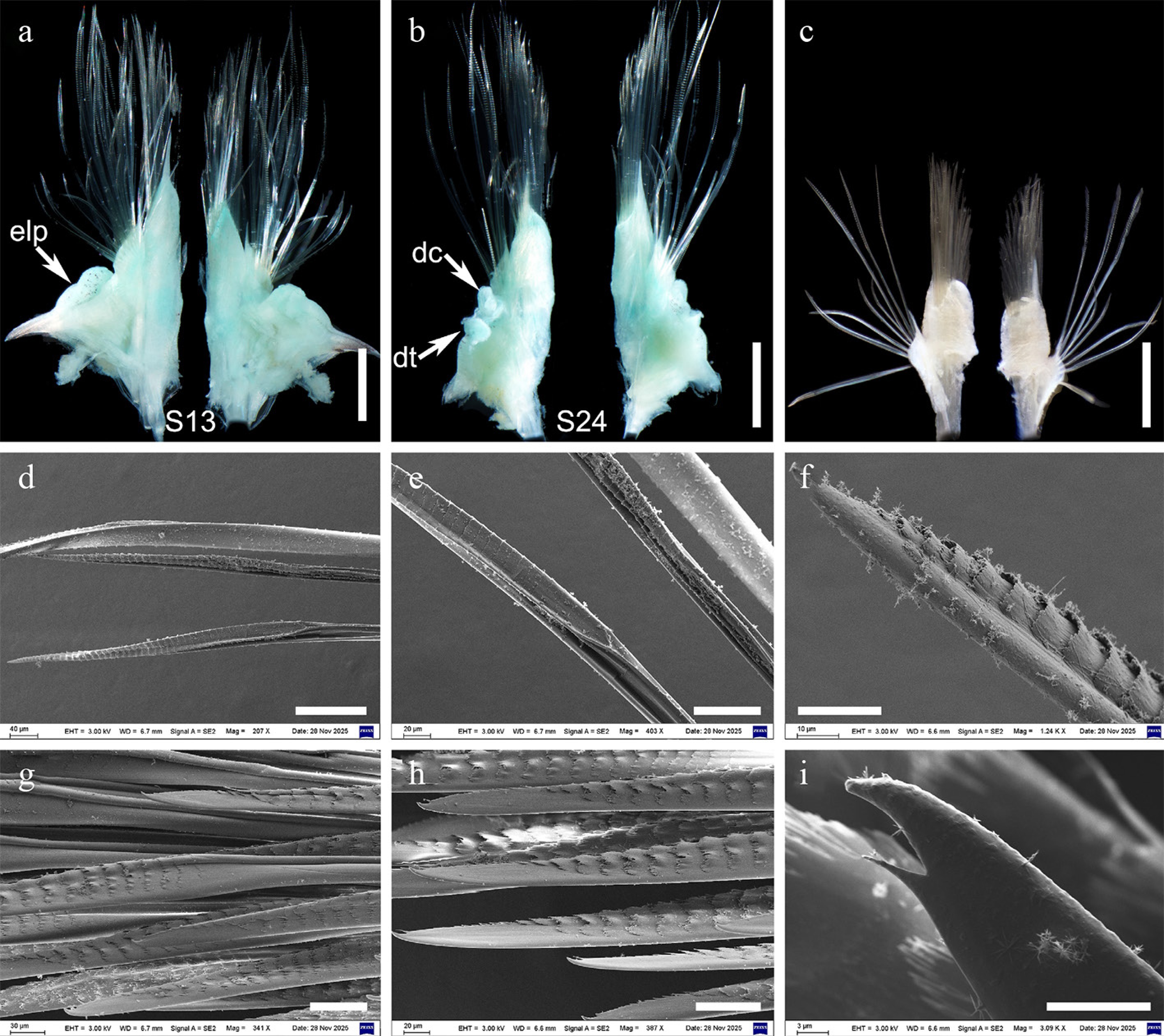
Figure 4.
Yaplagisca bidentata gen. et sp. nov., parapodia and chaetae. (a) Left parapodium from segment 13, anterior view (left) and posterior view (right). (b) Right parapodium from segment 24, posterior view (left) and anterior view (right). (c) Left posterior parapodium, unstained, anterior view (left) and posterior view (right). (d) General view of notochaetae. (e) Detail of notochaeta, showing basal part of spinous region. (f) Detail of notochaeta, showing transverse rows of spines and a pointed tip. (g) Neurochaetae from the middle part of the neurochaetal fascicle. (h) Neurochaetae from the lower part of the neurochaetal fascicle. (i) Distal tip of neurochaeta, showing bidentate apex. dc: dorsal cirrophore; dt: dorsal tubercles; elp: elytrophores; S13: segment 13; S24: segment 24. Scale bars: (a–c) 1 mm, (d) 100 μm, (e, g, h) 50 μm, (f) 20 μm, and (i) 10 μm.
-
Character Eulagisca Pareulagisca Bathymoorea Kristianides Pollentia Yaplagisca gen. nov. Number of segments up to 41 up to 37 up to 33 38 26–27 36 Number of elytral pairs 15 16 13–14 15 13 15 Eyes two pairs two pairs one pair of ocular areas or absent/reduced two pairs absent two pairs Facial tubercle distinct weak distinct absent absent distinct Chaetae on tentacular segment present present, single chaeta absent or present (emended concept) absent present present Acicular lobe on tentacular segment present absent absent or present (emended concept) present present present Nuchal fold present present absent absent absent absent Dorsal tubercles bulbous or nodular slightly bulbous conspicuous, inflated to lamelliform inconspicuous inconspicuous conspicuous; bulbous/nodular anteriorly and medially, lamelliform posteriorly Elytral ornamentation variable, often with papillae/tubercles/vesicles smooth without tubercles or papillae poorly known in intact material; genus diagnosed mainly from body and parapodia marginal papillae, microtubercles and macrotubercles on surface smooth or with microtubercles
along external edgeall elytra detached in holotype Ventral lamellae absent absent absent present absent absent Types of notochaetae one type two types one type two types one type one type Types of neurochaetae one type one type one type two types two types one type Neurochaetal distal tip pointed, entire or weakly hooked, depending on species pointed, with or without minute secondary tooth pointed, entire not bidentate superior tridentate; inferior lanceolate/pectinate bidentate Dorsal pigmentation variable poorly known weak or poorly known poorly known absent except
minute specklesconspicuous reddish to orange-brown median band Notes: (1) Characters of Bathymoorea summarized from Pettibone[9], Bonifácio & Menot[15], and Periasamy et al.[29]; (2) Pollentia treated here as a valid genus of Eulagiscinae following current usage; (3) Condition of elytral surface in Yaplagisca bidentata gen. et sp. nov. remains unknown because all elytra were detached from the holotype. Table 1.
Comparative morphological characters of the six genera of Eulagiscinae.
Figures
(4)
Tables
(1)