-
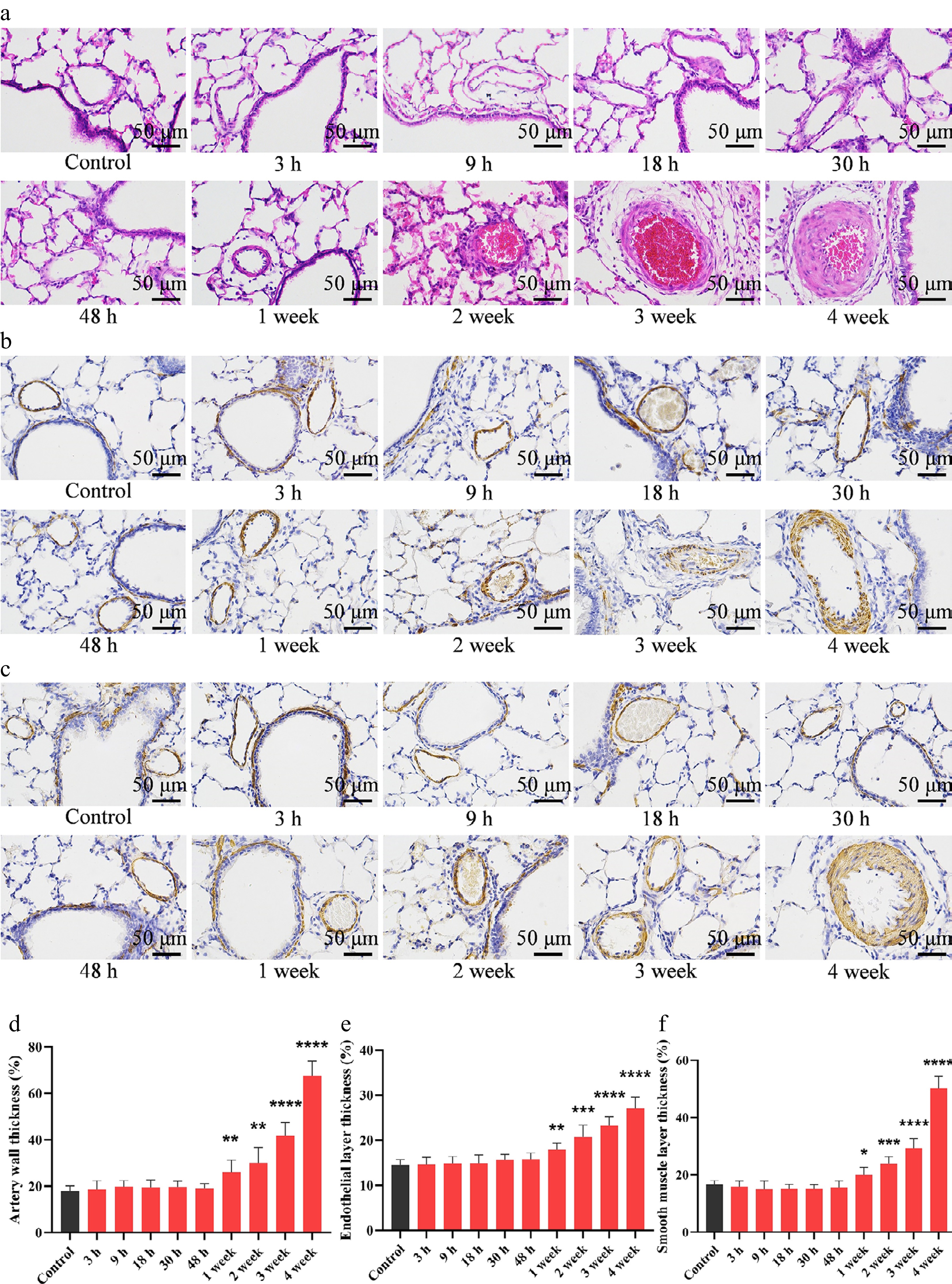
Figure 1.
MCT exposure triggers pulmonary vascular remodeling. (a) Representative images of H&E-stained lung sections from MCT-treated rats, showing prominent thickening of the pulmonary arterial wall. Representative immunohistochemical staining for (b) vWF and (c) α-SMA in lung tissues, highlighting changes in the endothelial and medial smooth muscle layers, respectively. Quantitative analysis of (d) total pulmonary arterial wall thickness, (e) vWF-positive endothelial layer thickness, and (f) α-SMA-positive medial smooth muscle layer thickness. Data are presented as mean ± SD (n = 3). * P < 0.05, ** P < 0.01, *** P < 0.001, **** P < 0.0001 compared with the control group (unpaired Student's t-test). Scale bar = 50 μm.
-
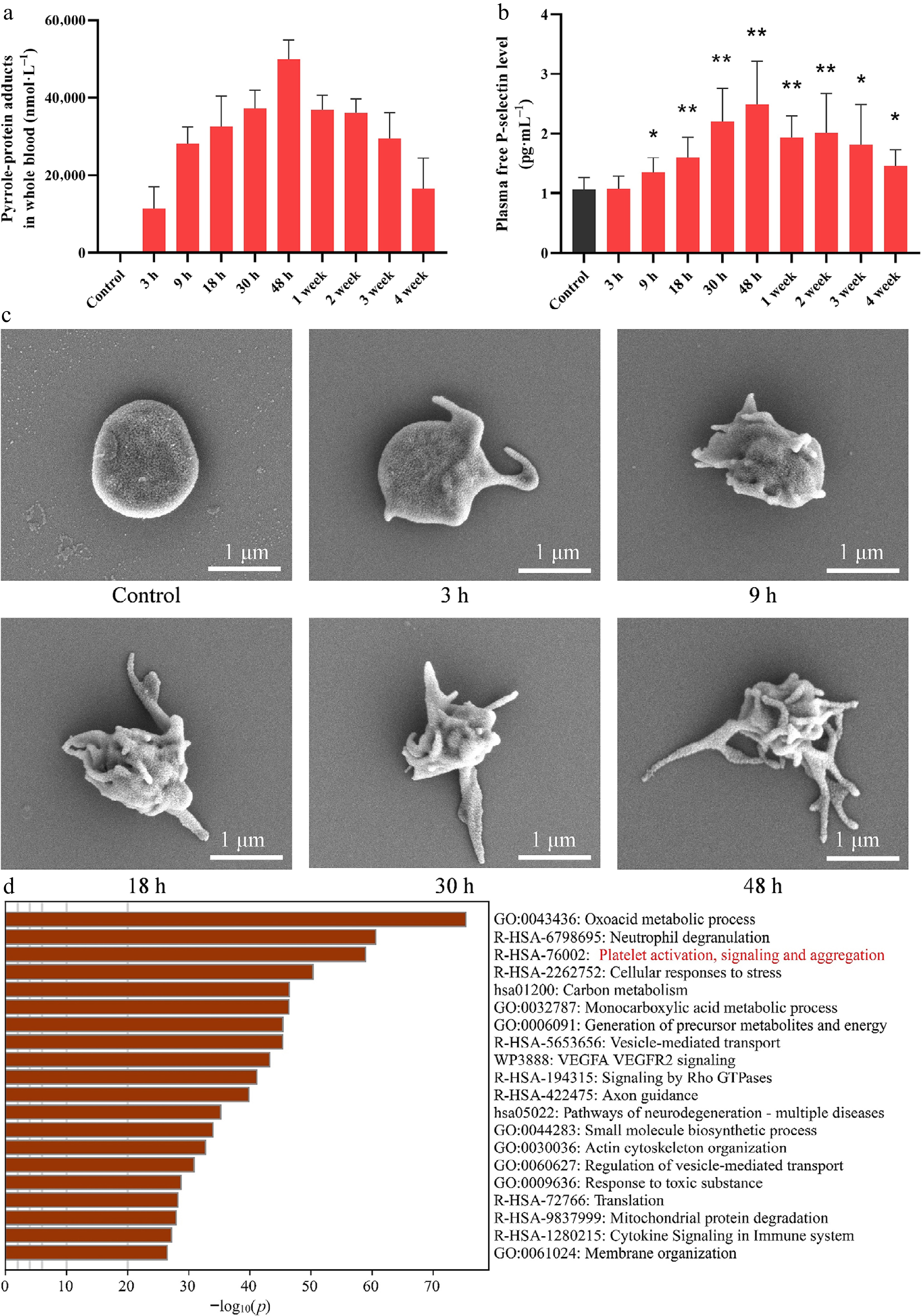
Figure 2.
MCT metabolites form covalent adducts with blood proteins and trigger platelet activation. (a) Levels of pyrrole-protein adducts in whole blood following MCT exposure. (b) Plasma free P-selectin concentrations following MCT exposure, reflecting time-dependent platelet activation. (c) Representative scanning electron microscopy images of platelets isolated from control and MCT-exposed rats at different time points. Compared with the smooth, discoid morphology of control platelets, platelets from MCT-exposed rats progressively exhibited morphological features of activation, including shape irregularity and the formation of increasingly prominent pseudopodia. Scale bar = 1 μm. (d) Metascape pathway enrichment analysis of DHP-adducted proteins identified by untargeted proteomic mass spectrometry in platelets from MCT-exposed rats, revealing significant enrichment of pathways associated with platelet activation, signaling, and aggregation. For (a) and (b), data are presented as mean ± SD (n = 5). * P < 0.05, ** P < 0.01 vs. the control group (unpaired Student's t-test).
-
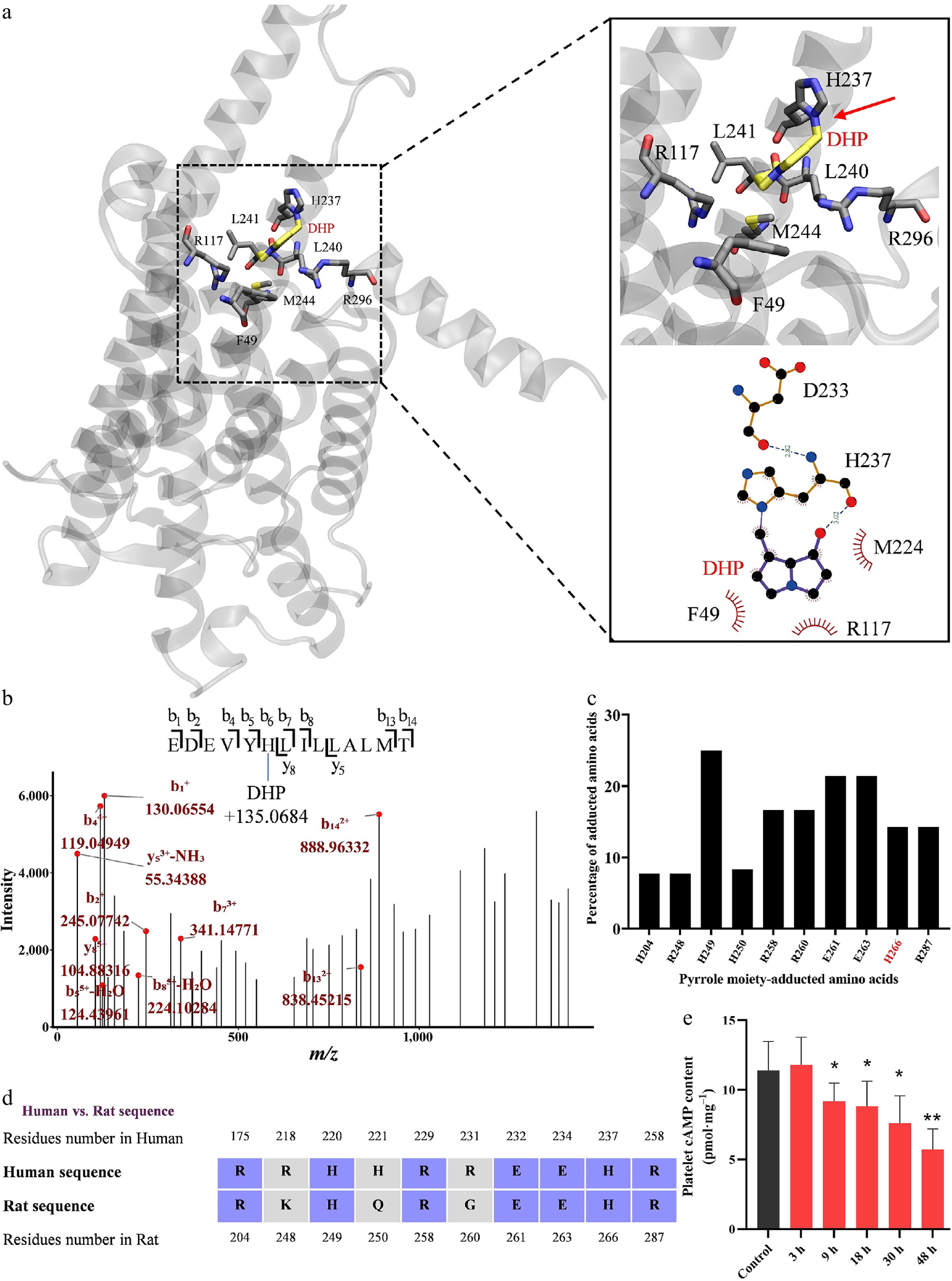
Figure 3.
The active MCT metabolite dehydropyrrolizidine (DHP) directly binds to the IP receptor, as demonstrated by molecular docking and proteomic analysis. (a) Molecular docking suggests a stable interaction between DHP and residue H237 of the human IP receptor (corresponding to H266 in rats). (b) Proteomic mass spectrometry analysis of the IP receptor purified from platelets of MCT-exposed rats identifies covalent modification of specific amino acid residues by DHP. A representative mass spectrum confirms DHP adduction to residue H266 (b6), corresponding to human H237. (c) Summary of DHP-modified residues identified across the IP receptor protein. (d) Sequence alignment of rat and human IP receptors showing that the DHP-modified residue H266 in rats is conserved in humans. (e) Decreased cAMP levels in platelets from MCT-exposed rats further support DHP-mediated inhibition of the IP receptor. Data are presented as mean ± SD (n = 5). * P < 0.05, ** P < 0.01 vs. the control group (unpaired Student's t-test).
-
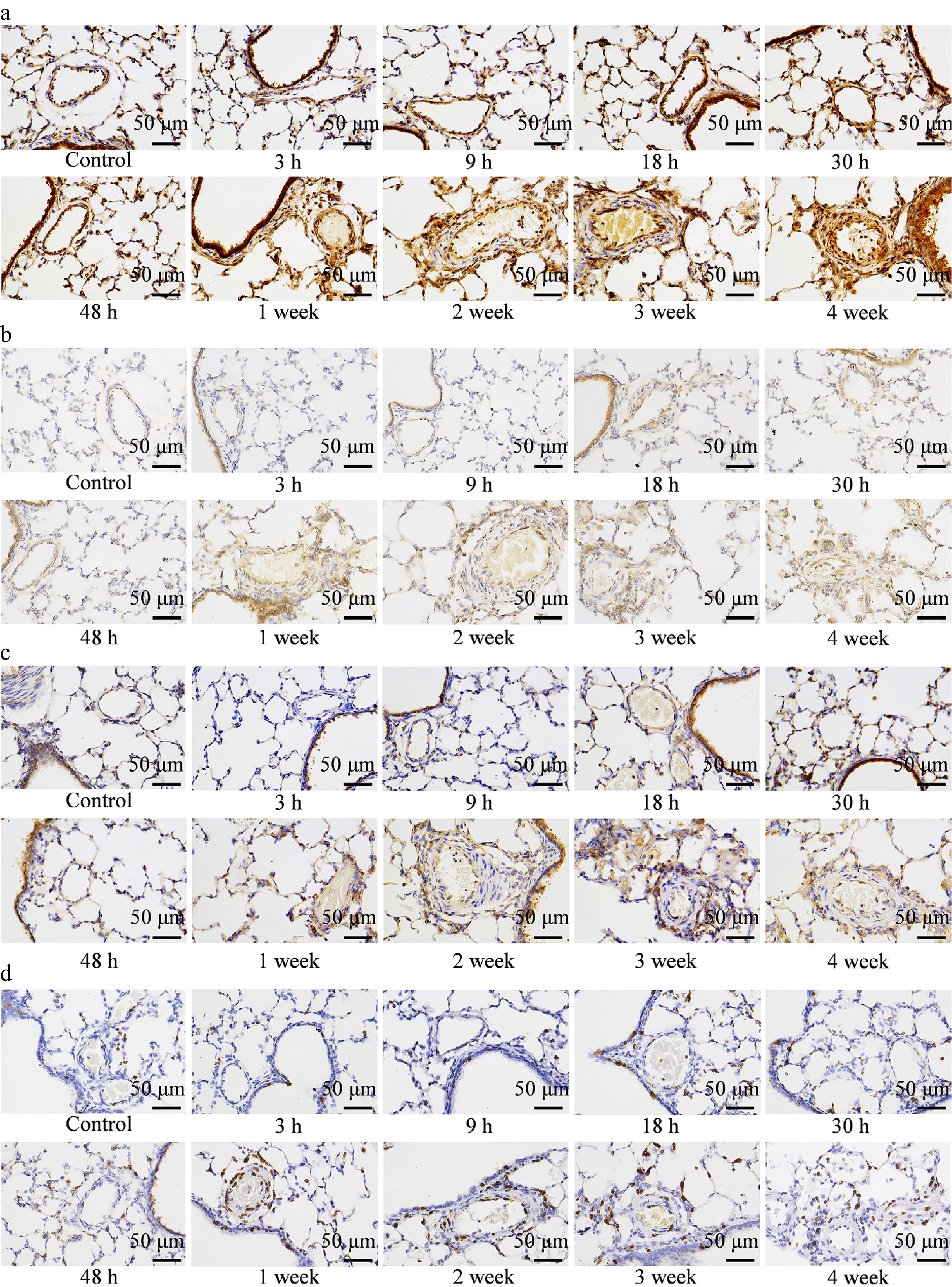
Figure 4.
MCT-induced platelet activation promotes PDGF-B accumulation and activates the PDGFR/PI3K signaling pathway in lung tissues. (a) Representative images of IHC staining for PDGF-B, showing increased PDGF-B expression in pulmonary arteries of MCT-exposed rats. Representative images of (b) p-PDGFR and (c) p-PI3K expression in lung tissues, indicating activation of the PDGFR/PI3K cascade. (d) Ki67 staining in lung sections, demonstrating enhanced cellular proliferation in response to PDGFR/PI3K pathway upregulation. Scale bar = 50 μm.
-
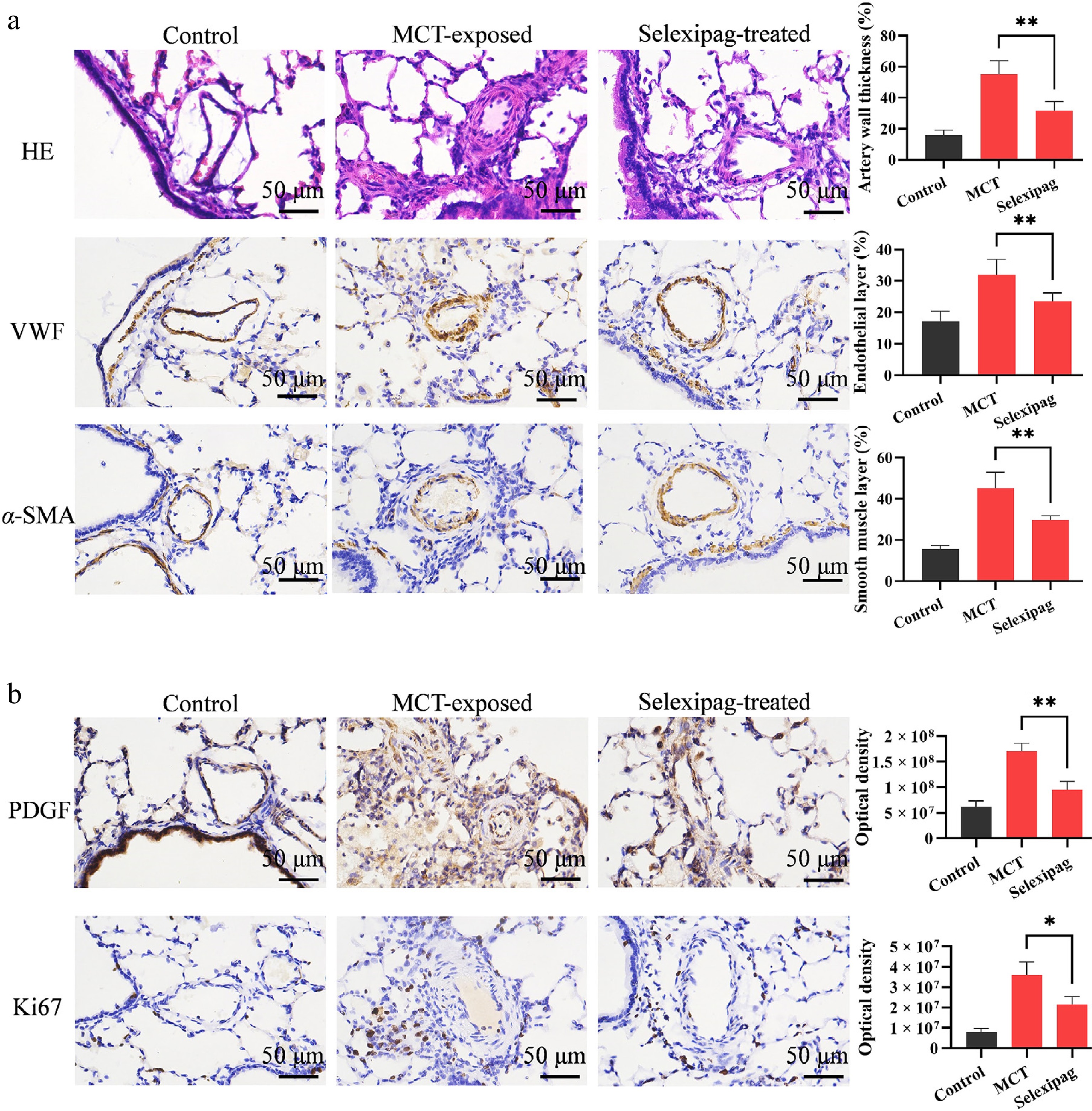
Figure 5.
Selexipag attenuates MCT-induced pulmonary vascular remodeling and reduces pulmonary PDGF-B accumulation and vascular cell proliferation. (a) Representative H&E staining and immunohistochemical staining for vWF and α-SMA in lung tissues from control, MCT-exposed, and selexipag-treated rats. MCT exposure induced marked pulmonary arterial wall thickening, endothelial hyperplasia, and medial smooth muscle hypertrophy, whereas selexipag treatment attenuated these pathological changes. Quantitative analyses of pulmonary arterial wall thickness, endothelial layer thickness, and smooth muscle layer thickness are shown on the right. (b) Representative immunohistochemical staining and quantitative analyses of PDGF-B and Ki67 in pulmonary arteries. MCT exposure markedly increased pulmonary vascular PDGF-B accumulation and the number of proliferating Ki67-positive cells, whereas selexipag treatment significantly reduced PDGF-B expression and vascular cell proliferation. Data are presented as mean ± SD (n = 3). * P < 0.05, ** P < 0.01 vs. the MCT-exposed group (unpaired Student's t-test). Scale bar = 50 μm.
-
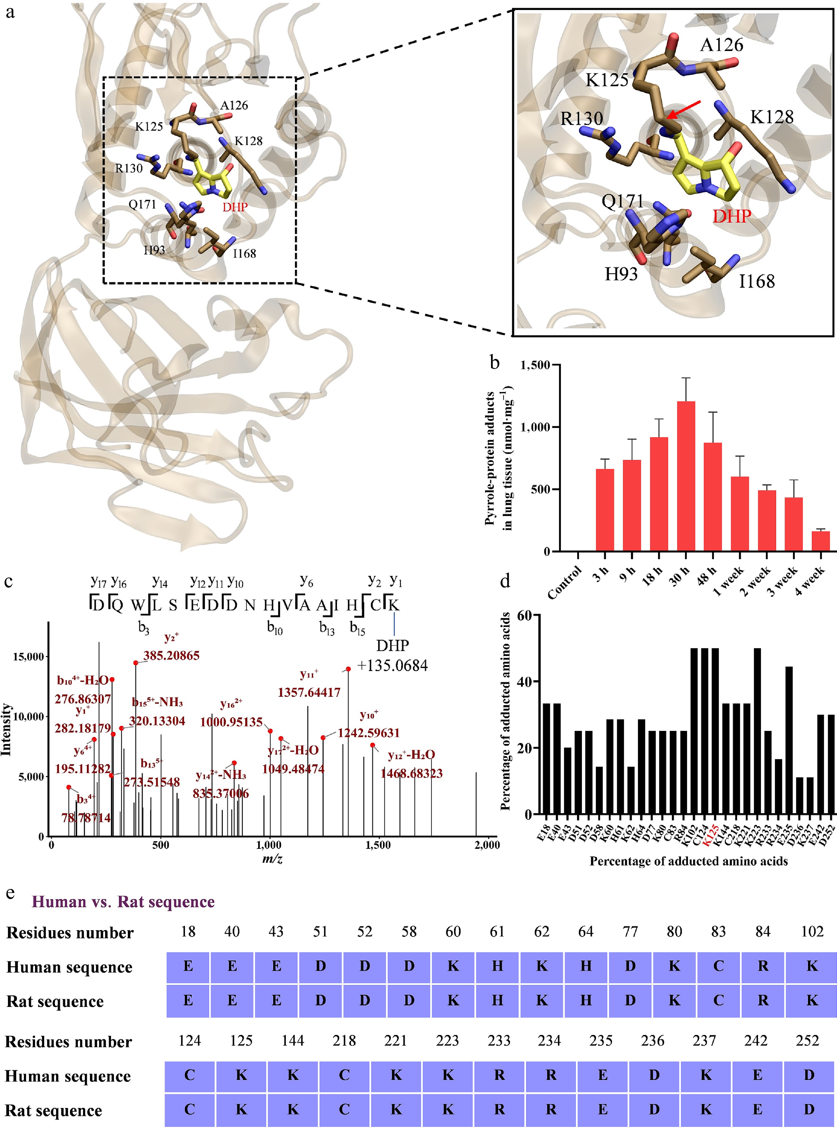
Figure 6.
Molecular docking and proteomic analysis identify PTEN as a direct target of DHP. (a) Covalent docking model illustrating covalent bond formation between DHP and the K125 residue of PTEN. (b) Concentration of pyrrole-protein adducts in rat lung tissues following MCT exposure, demonstrating the protein-binding capacity of DHP in vivo. (c) Representative MS/MS spectrum of PTEN purified from MCT-exposed rat lung tissue, confirming the covalent modification of K125 by DHP. (d) Summary of DHP-modified residues identified by proteomic analysis within the PTEN protein sequence. (e) Sequence alignment of rat and human PTEN shows that all DHP-modified amino acid residues are conserved between the two species.
-
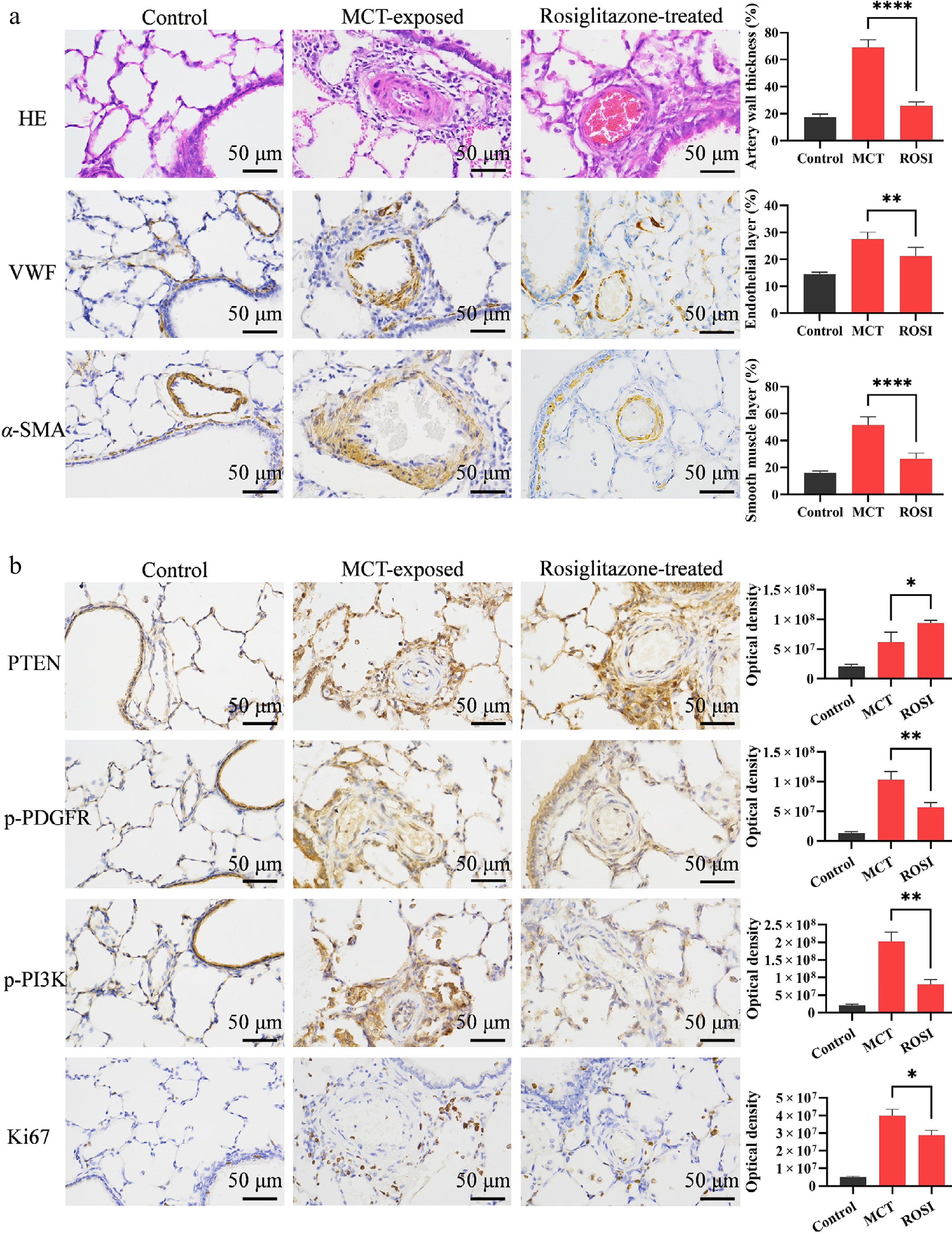
Figure 7.
Rosiglitazone alleviates pulmonary vascular remodeling by restoring PTEN expression and inhibiting the PDGFR/PI3K pathway. (a) Representative H&E and IHC (for vWF, α-SMA) images of lung tissues. ROSI treatment ameliorated MCT-induced pulmonary vascular remodeling, including reduced wall thickness, endothelial hyperplasia, and medial smooth muscle hypertrophy. (b) IHC staining and quantitative analysis of PTEN, p-PDGFR, p-PI3K, and Ki67 in lung tissues. ROSI intervention upregulated PTEN expression, suppressed p-PDGFR and p-PI3K levels, and consequently inhibited vascular cell proliferation as indicated by reduced Ki67. Data are presented as mean ± SD (n = 3). * P < 0.05, ** P < 0.01, *** P < 0.001, **** P < 0.0001 compared with the MCT-treated group (unpaired Student's t-test). Scale bar = 50 μm.
-
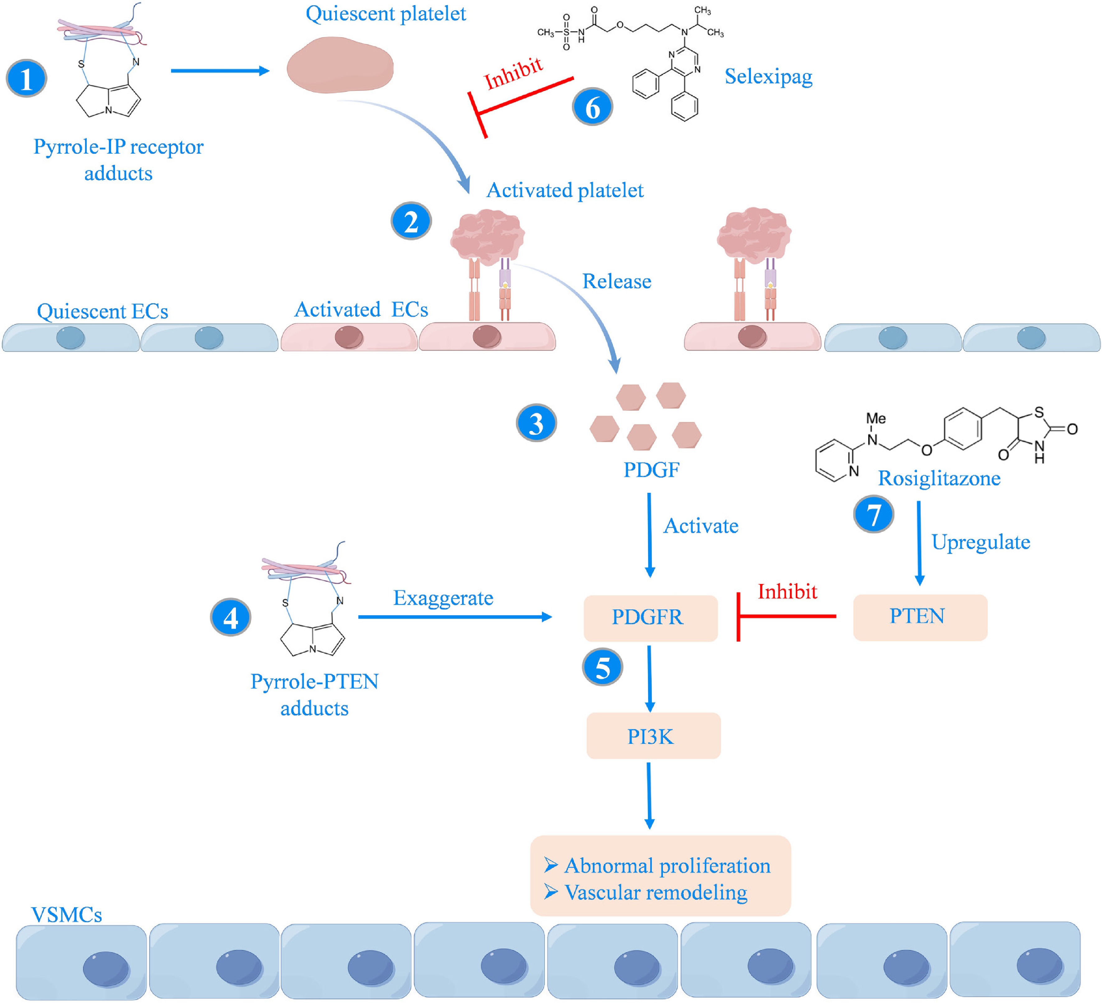
Figure 8.
Proposed mechanism of PA-induced PAH via covalent targeting of the IP receptor and PTEN and the corresponding therapeutic interventions. (1) Reactive PA metabolites (e.g., those derived from MCT) covalently modify the IP receptor through formation of pyrrole-IP receptor adducts, impairing IP receptor signaling and promoting platelet activation. (2) Activated platelets sequester in the pulmonary vasculature and release PDGF upon degranulation, leading to increased PDGF accumulation within the vascular wall. (3) Increased local PDGF levels promote activation of PDGFR signaling. (4) Simultaneously, reactive PA metabolites form pyrrole-PTEN adducts that impair PTEN function, thereby relieving its endogenous suppression of the PDGFR/PI3K axis. (5) The resulting activation of PDGFR/PI3K signaling promotes aberrant proliferation of pulmonary vascular cells and pathological vascular remodeling. (6) Pharmacological inhibition of platelet activation with selexipag decreases PDGF release, thereby attenuating PA-induced PAH. (7) Pharmacological upregulation of PTEN by rosiglitazone restores its inhibitory control over the PDGFR/PI3K pathway, thereby suppressing abnormal vascular cell proliferation and alleviating PA-induced PAH. Created with FigDraw.
Figures
(8)
Tables
(0)