-
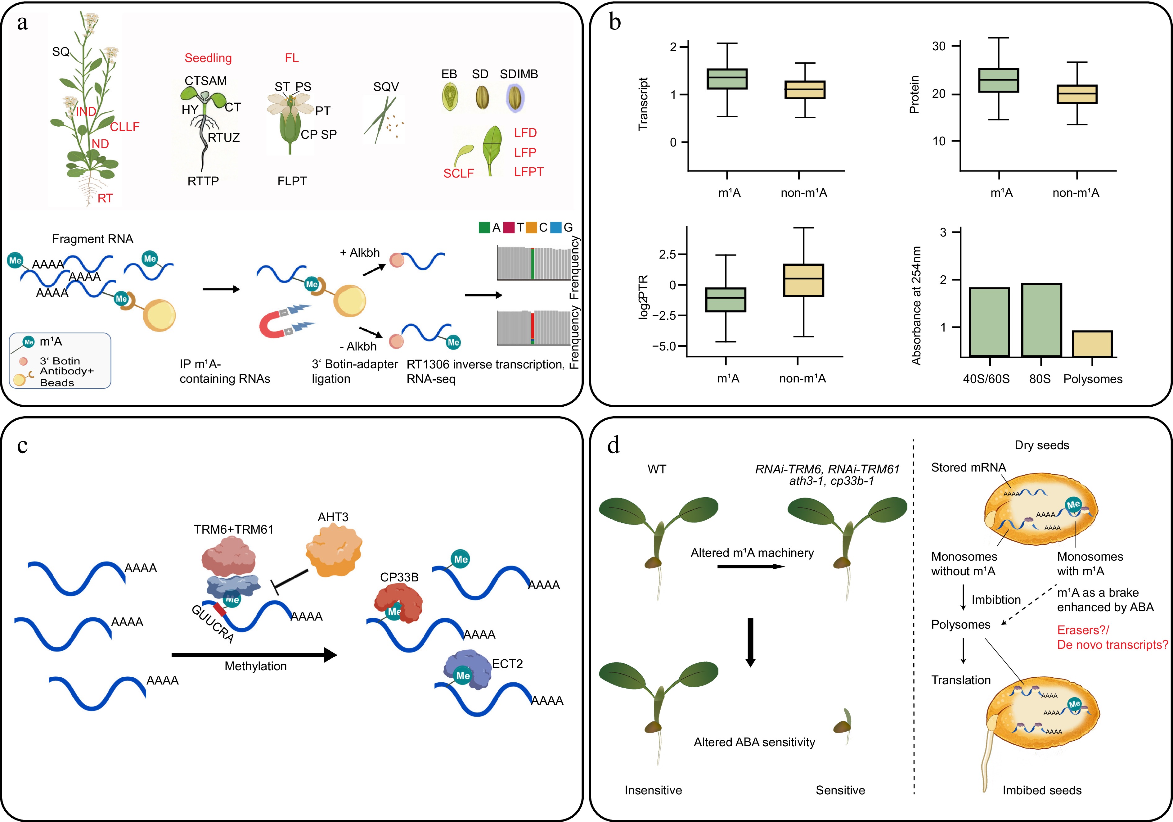
Figure 1.
Foundational m1A epitranscriptomic atlas and regulatory system in Arabidopsis. (a) Schematic of 25 Arabidopsis tissues collected in the m1A atlas and m1A-IP-seq workflow. Tissues abbreviations: EB, embryo; CP, carpel; CT, cotyledons; CTSAM, shoot tip; FL, flower; FLPT, flower petiole; HY, hypocotyl; IND, internode; LFD, leaf distal; LFP, leaf proximal; LFPT, leaf petiole; ND, node; PS, pistil; PT, petal; RT, root; RTTP, root tip; RTUZ, root upper zone; SCLF, senescent leaf; SD, seed; SDIMB, seed imbibed; SP, sepal; SQ, silique; SQV, silique valves; ST, stamen. All these 25 tissues were used in the m1A level detection, while the 10 tissues used in m1A-IP-seq were labelled in red abbreviations. For m1A detection at single-nucleotide resolution, m1A-modified mRNA fragments are immunoprecipitated, and one aliquot is treated with AlkB to remove m1A. Both aliquots were reverse-transcribed using RT1306. The enzyme's elevated mismatch rate at m1A sites in the untreated sample, relative to the AlkB-treated control, enables single-nucleotide detection of m1A. (b) m1A regulates events from transcription to translation. m1A-modified transcripts show higher mRNA and protein abundance but lower protein-to-RNA ratios (PTR). m1A is enriched in monosome fractions and depleted from polysomes. (c) m1A writer–reader network. TRM6–TRM61 catalyses m1A deposition on substrates bearing a tRNA-like stem-loop with the GUUCRA motif. ATH3 inhibits this complex by impairing the RNA-binding of the TRM6–TRM61 complex. CP33B and ECT2 read m1A; ECT2 additionally binds m6A. (d) Genetic disruption of any m1A pathway component results in ABA hypersensitivity (left panel) and m1A as a translational brake on stored mRNAs at germination (right panel). The figure was created in BioRender. Guochenxi, G. (2026),
https://BioRender.com/d9bmi84 , and with BioGDP (biogdp.com ).
Figures
(1)
Tables
(0)