












-
Dietary polyphenols represent an important class of bioactive compounds with demonstrated immunomodulatory potential[1]. These natural products exert health-promoting effects through diverse mechanisms, including antioxidant activity[2], modulation of inflammatory signaling[3], and regulation of gut microbiota composition[4]. Areca nut (Areca catechu L.), a traditional palm seed widely distributed in Southeast Asia, serves as a rich source of polyphenols including catechin[5], quercetin[6], rutin[7], resveratrol, epicatechin, and luteolin[8]. Accumulating evidence indicates that areca nut polyphenol (AP) extracts possess significant immunomodulatory activity, suggesting their potential as functional food ingredients for immune health maintenance[6,9].
The immunomodulatory efficacy of APs has been attributed to their major polyphenolic constituents, particularly quercetin and luteolin[10]. Quercetin alleviates immune suppression by inhibiting the JAK1/STAT3/HIF-1α axis and reducing oxidative inflammatory responses[11], whereas luteolin activates CD8+ T cells through reactive oxygen species (ROS)/Fe2+-dependent pathways and regulates immune cell infiltration by targeting BTK and FLT3 signaling[12,13]. For chewed areca nut or crude areca nut extracts, the associated health risks are mainly attributed to alkaloids such as arecoline. Chewing areca nut can cause oral submucous fibrosis and increase the risk of oral cancer, and arecoline has also been associated with hepatotoxicity[14,15]. In our previous study, we developed a method that effectively removed the alkaloids from areca nut, yielding a polyphenol-rich extract with improved safety while retaining its functional properties[16,17]. Importantly, the present study focuses on the biological activity of the dealkaloidized polyphenol fraction rather than whole areca nut or crude extracts. Therefore, our findings should not be interpreted as supporting or recommending the consumption of whole areca nut.
Despite these advances, the molecular mechanisms underlying AP-mediated immune regulation remain incompletely understood. Previous investigations have predominantly focused on single bioactive compounds or isolated signaling pathways, failing to capture the systems-level interactions between APs and the host's physiology. Specifically, the bidirectional communication between APs and the gut microbiota – critical mediators of polyphenol bioactivity – has not been systematically characterized. Moreover, the causal chain linking alterations in the microbial community to host-related metabolic reprogramming and subsequent immune restoration remains elusive. Consequently, an integrated approach combining predictive network analysis with multi-omics experimental validation is required to elucidate the holistic mechanisms of APs' action.
This study was designed to test the hypothesis that APs ameliorate immunosuppression through a gut microbiota–metabolite–immune axis. We used an integrative strategy combining network pharmacology with 16S rRNA sequencing, untargeted metabolomics, and label-free proteomics to investigate AP-mediated immune restoration in cyclophosphamide (CTX)-induced immunosuppressed mice. By establishing correlations between microbial community shifts, serum amino acid profiles, and splenic protein expression, we aimed to construct a mechanistic framework for AP-based functional food development targeting immune dysfunction.
-
Immunoglobulin (Ig)A and IgM test kits were purchased from Nanjing Jiancheng Co., Ltd. Methanol, acetonitrile, isopropyl alcohol, and formic acid were high-performance liquid chromatography (HPLC) grade, supplied by Thermo Fisher Technologies (MA, USA). Trypsin was purchased from Promega, USA. Hainan areca nut pits were provided by Hunan Kouweiwang Group Co., Ltd. (Yiyang, China).
Preparation of APs
-
After being dried in a vacuum freeze-dryer for 72 hours, the cores of areca nuts were pulverized. In a 10% (w/v) methanol solution, the powder was dissolved. The upper liquid phase containing the AP extract was collected after ultrasonic extraction at 50 °C for 60 min and centrifugation at 10,000 rpm for 15 min. To obtain the fat-soluble polyphenol phase, the extract was concentrated under a vacuum, redissolved in hydrochloric acid, and extracted with ethyl acetate. After treating the water phase with an acid cation exchange resin, impurities were removed and the polyphenol phase was eluted. An extract of polyphenol was made by combining the fat-soluble and water-soluble phases.
Network pharmacology analysis
-
Using the identified components in the AP extract[17] and combining them with the known constituents of APs[18,19], a compound database for the APs was constructed. The SwissADME (
www.swissadme.ch ) and traditional Chinese medicine systems pharmacology (TCMSP,https://tcmsp-e.com/tcmsp.php ) databases were used to identify the active ingredients of APs. The screening criteria were set as follows: Gastrointestinal absorption = 'high' with a druglikeness (DL) rule compliance count of ≥ 2 for SwissADME; oral bioavailability (OB) ≥ 30% with DL ≥ 0.18 for TCMSP. Potential targets of the AP components were retrieved from the TCMSP and SwissTargetPrediction (https://swisstargetprediction.ch/ ) databases, and their official gene symbols were validated using UniProt (www.uniprot.org/ ). 'Immunosuppression' was used as a keyword to search the online mendelian inheritance in man (OMIM,https://omim.org/ ) and GeneCards (www.genecards.org/ ) databases to identify target genes associated with immunosuppression. Subsequently, the overlapping targets between APs and the disease were identified through Venn diagram analysis. Cytoscape software, version 3.9.1 (https://cytoscape.org ), was used to construct and visualize the compound–target interaction network. STRING (https://string-db.org/ ) was used to construct a protein–protein interaction (PPI) network, with a minimum interaction confidence score > 0.900. Subsequently, the identified overlapping targets were subjected to Gene Ontology (GO) functional and Kyoto Encyclopedia of Genes and Genomes (KEGG) pathway enrichment analyses using the Metascape platform (https://metascape.org/ ). Data visualization was performed using an online bioinformatics plotting tool (www.bioinformatics.com.cn/ ).Animal treatments
-
Six groups (eight mice per group) were randomly formed from 48 six-week-old male institute of cancer research (ICR) mice The experimental groups included: normal control (NC), model control (MC), positive control (PC), low-dose AP extract (L-AP), medium-dose AP extract (M-AP), and high-dose AP extract (H-AP). Food and water were provided ad libitum to all mice. During the initial phase, they were provided with an irradiated basal diet for 7 consecutive days. Following this, all mice except the NC were injected intraperitoneally with CTX (80 mg/kg body weight [BW], diluted in normal saline; 100 μL injection volume) for 3 d. After establishment of the CTX model, the NC and MC groups were given daily intraperitoneal injections of normal saline, whereas the AP groups received AP extract at three different doses (20, 50, and 100 mg/kg BW) via intragastric administration for seven consecutive days. The doses of AP were determined by our previous experiments, in which the same dose range demonstrated a favorable dose–response relationship in the treatment of a fatigue mouse model[17]. The PC group received levamisole (40 mg/kg BW) via gavage (200 μL) for 7 d consecutively. Mice were weighed every 3 d. After 7 d of gavage administration, blood samples, spleen, and thymus tissue were collected for further analysis. The animal test approval number is JN.No20220330b0720515[100].
Serum biochemical indicators
-
The test kit instructions were strictly followed for the measurement of IgA and IgM. The changes in serum IgA and IgM levels were recorded according to the kit's guidelines.
16S rRNA analysis
-
The cecal contents were harvested from mice and kept at –80 °C for storage. Following DNA extraction and purification, the microbial community's structure and diversity were assessed. The total genomic DNA of cecal contents was obtained using the HiPure fecal DNA kit (Nanjing, China). Amplification of the V3–V4 region of the bacterial 16S rRNA gene was used using 338 forward/806 reverse primers. Sequencing library construction and Illumina sequencing were performed by Weizhi Jin (Nanjing, China). Paired-end sequencing reads were first merged according to the overlapping regions, with a minimum overlap of 20 bp between the forward and reverse reads. Merged sequences containing ambiguous bases ('N') were removed. Adapter, barcode, and primer sequences were trimmed, and bases with quality scores below Q20 at both ends were removed. Sequences shorter than 200 bp were discarded. Chimeric sequences were subsequently identified and removed by comparison with a reference database. Data processing was performed using Cutadapt v1.9.1, VSEARCH v1.9.6, and QIIME v1.9.1. The generated sequences were clustered using VSEARCH (with 97% sequence similarity). Alpha diversity was assessed by randomly selecting sequences from the samples.
Metabolome extraction
-
Approximately 25 mg of spleen tissue was placed into a precooled 80% methanol solution, followed by swirling and grinding with a tissue grinder for 5 min. Ultrasonic treatment was then performed in ice water for 30 min. Following centrifugation at 12,000 rpm for 10 min, the supernatant was passed through a 0.22-μm filter and then transferred into an injection vial. For serum preparation, 50 µL of the serum was blended with 200 µL of prechilled methanol, followed by vortexing. Ultrasonic extraction, centrifugation, and filtration were conducted under the same conditions as for spleen preparation. Detailed methods are provided in Supplementary Text S1 and Supplementary Table S1.
Proteomics analysis
-
Following previous reports, trypsin digestion and protein extraction were implemented[20]. Spleen tissues from the mice were placed in a 8 M urea buffer solution (containing 100 mmol/L NH4CO3), to which protease and phosphatase inhibitors were added. The supernatant was recovered after centrifuging the lysate at 4 °C and 12,000 rpm for 15 min. Protein quantification was conducted using the Bradford assay. Next, 5 mmol/L dithiothreitol (DTT) was incorporated into the solution and incubated at 56 °C for 30 min. Afterward, 15 mmol/L iodoacetamide (IAA) was added to the reaction solution, and the mixture was incubated at 25 °C for 30 min. Trypsin digestion (with a protein-to-enzyme ratio of 50:1) was carried out overnight, and the reaction was quenched with 1% formic acid. Oasis hydrophilic-lipophilic balance (HLB) columns (Waters, USA) were used for desalting, followed by vacuum concentration and lyophilization of the purified peptide fractions. The peptides were then reconstituted in 0.1% formic acid and 2% acetonitrile. The ultrahigh-performance liquid chromatography–tandem mass spectrometry (UHPLC-MS/MS) platform was used for label-free proteomic analysis. Detailed methods are provided in Supplementary Text S2.
Statistical analysis
-
Data analysis was conducted using SCIEX OS 2.1.1, Cytoscape 3.9.1, MS-DIAL 4.92, and Simca 14.1 software; the Unistream Biocloud platform;and the MetaboAnalyst web page. Graphing was performed with GraphPad Prism 8 and Origin 2018. Significance analysis was carried out using IBM SPSS Statistics, with p < 0.05 demonstrating a significant difference.
-
Network pharmacology was used to predict the immunomodulatory mechanisms of APs. With the SwissADME and TCMSP databases, 35 active compounds were identified on the basis of the gastrointestinal absorption and DL criteria (Supplementary Tables S2 and S3), which yielded 496 potential targets. Intersection with 2,515 immunosuppression-associated genes from OMIM and GeneCards databases revealed 247 overlapping targets (Fig. 1a). Construction of a compound–target network (Fig. 1b) and topology analysis identified quercetin and luteolin as the hub compounds (node degree ≥ 100), representing the primary polyphenolic constituents of APs (Supplementary Table S4). These 247 targets were subjected to PPI network analysis using STRING and Cytoscape (Fig. 1c, d), where node size and color intensity indicate the interaction strength. GO enrichment revealed that the overlapping targets were significantly associated with biological processes related to cellular stress responses, lipid metabolism, and hypoxia; cellular components including transcriptional regulator complexes and membrane rafts; and molecular functions such as kinase binding (Fig. 1e). KEGG pathway analysis further demonstrated significant enrichment in immune-related signaling pathways, notably phosphoinositide 3-kinase (PI3K)-protein kinase B (Akt), mitogen-activated protein kinase (MAPK), and hypoxia-inducible factor 1 (HIF-1) (Fig. 1f). These computational predictions were subsequently validated in a CTX-induced immunosuppression mouse model to confirm the immunomodulatory efficacy of APs and elucidate the underlying mechanisms. High-dose APs restored immune function and lymphoid tissue architecture.
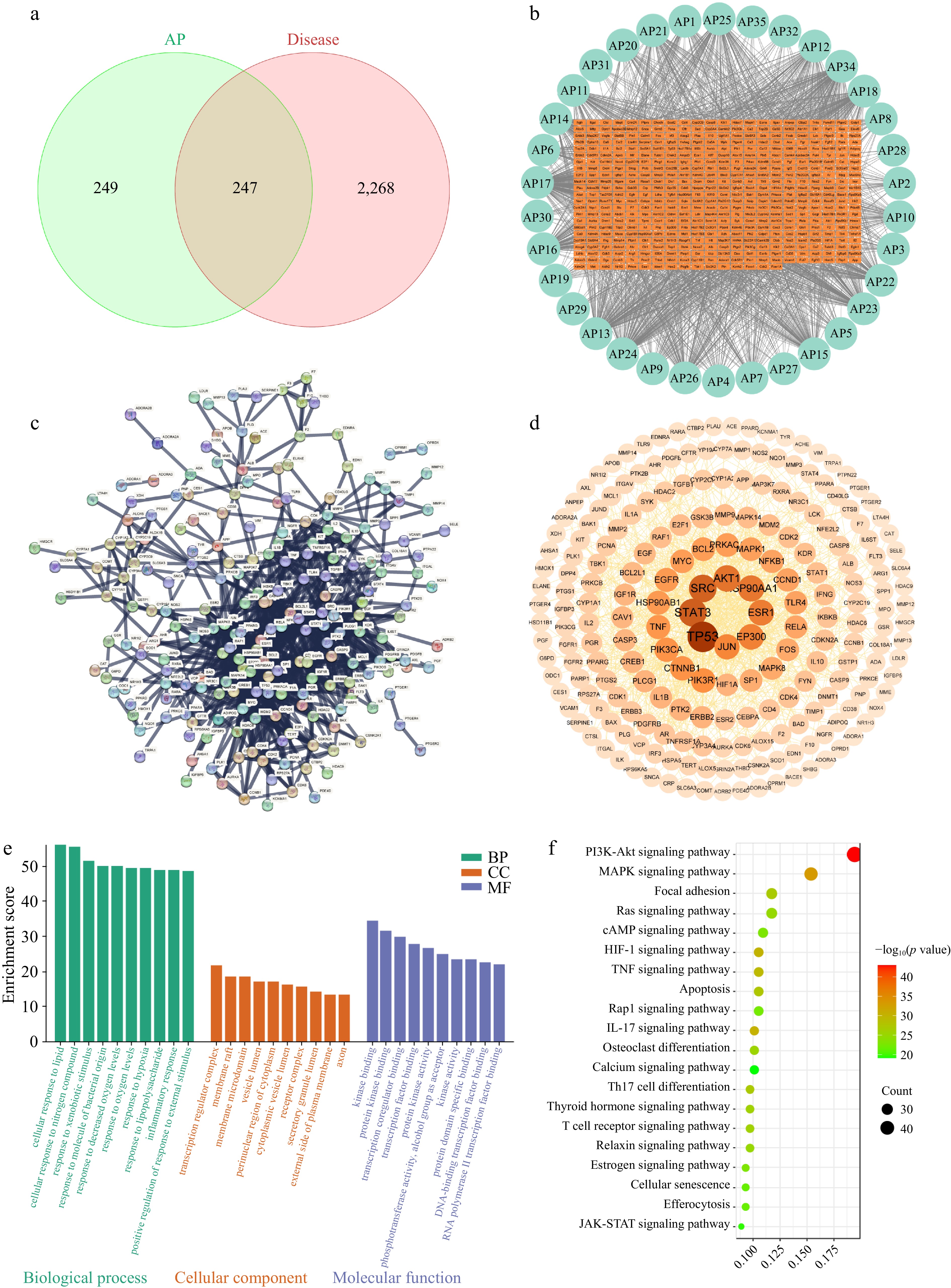
Figure 1.
Network pharmacology analysis of APs in the treatment of immunosuppression. (a) Component–disease intersection targets. (b) APs' component–target network diagram. (c) PPI network of the intersection targets of APs and immunosuppression. (d) The PPI network diagram after further analysis by Cytoscape, protein nodes with stronger interactions have larger sizes and darker colors. (e) GO enrichment analysis of the intersection targets. (f) KEGG enrichment analysis of intersection targets.
To establish the immunosuppression model, 6-week-old mice received CTX (80 mg/kg, intraperitoneal) for 3 d, followed by oral administration of APs (20, 50, or 100 mg/kg) or levamisole (40 mg/kg, positive control) for the next 7 d (Fig. 2a). The CTX injection induced significant weight loss in all treated groups by Day 3. However, the AP intervention attenuated this decline, with the H-AP group (100 mg/kg) showing the most pronounced recovery from Days 7 to 10 (Fig. 2b).
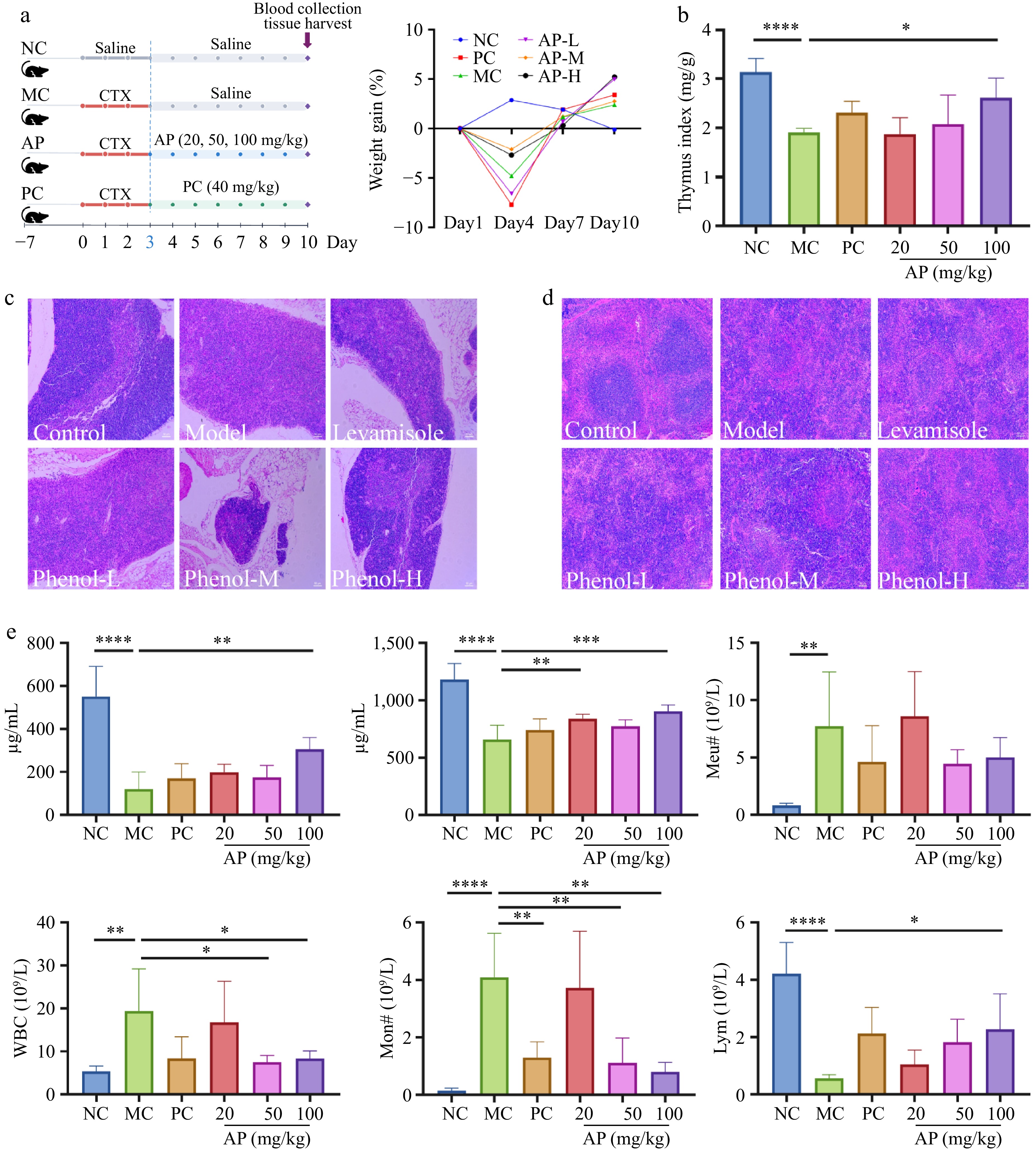
Figure 2.
Changes in the animal experiment's procedures and basic indexes of the mice. (a) Experimental protocol and changes in the BW of mice. (b) Changes in the thymus index. (c) Morphological changes in the thymus. (d) Morphological changes in the spleen. (e) Changes in biochemical indicators: IgA, IgM content changes and neutrophils, white blood cells, monocytes, and lymphocyte number changes. Note: *, p < 0.05, **, p < 0.01. ***, p < 0.001. ****, p < 0.0001.
Histological examination revealed profound alterations in the lymphoid tissue's architecture following CTX treatment. In the thymus, CTX disrupted the corticomedullary junction, reduced cortical lymphocyte density, and expanded the medullary region (Fig. 2c). Both the M-AP (50 mg/kg) and H-AP treatments restored thymic organization, with an increased cortical proportion and distinct corticomedullary boundaries. Similarly, CTX administration obscured splenic red and white pulp boundaries and reduced the white pulp area, confirming successful immunosuppression (Fig. 2d). AP treatment reversed these pathological changes in a dose-dependent manner, with H-AP restoring a distinct pulp architecture and white pulp abundance similar to that of the normal controls.
Biochemical and hematological analyses further demonstrated the dose-dependent immunomodulatory efficacy of APs. High-dose APs (100 mg/kg) significantly reversed CTX-induced reductions in serum IgA and IgM levels (Fig. 2e). The CTX treatment elevated white blood cell, neutrophil, and monocyte counts while decreasing lymphocyte populations. CTX exerts time-dependent effects on circulating leukocytes. Early after administration, CTX induces bone marrow suppression and decreases peripheral leukocytes, whereas subsequent hematopoietic recovery may cause a transient rebound of neutrophils and monocytes[21]. In this study, hematological parameters were measured during the recovery phase; thus, elevated myeloid cell counts together with persistently reduced lymphocytes may reflect asynchronous hematopoietic recovery. H-AP administration normalized these hematological parameters, whereas L-AP (20 mg/kg) and M-AP produced minimal effects. Given these findings, subsequent multi-omics analyses focused on the NC, MC, and H-AP groups to elucidate the molecular mechanisms underlying AP-mediated immune restoration.
Beneficial bacteria enrichment underlies AP-mediated gut microbiota modulation
-
To investigate whether APs restore immune function through microbiota-dependent mechanisms, 16S rRNA sequencing was performed on the cecal contents from the NC, MC, and H-AP groups. The alpha diversity rarefaction curves indicated that the current sequencing depth sufficiently covered the major microbial diversity in the samples (Supplementary Fig. S1a and Supplementary Table S5). CTX-induced immunosuppression significantly reduced microbial diversity (Shannon index) and richness (Chao index), whereas the H-AP intervention reversed these alterations, restoring α-diversity indices toward normal levels (Fig. 3a–c). β-diversity analysis indicated that the experimental treatments may have altered the overall composition of the gut microbiota (Supplementary Fig. S1b).
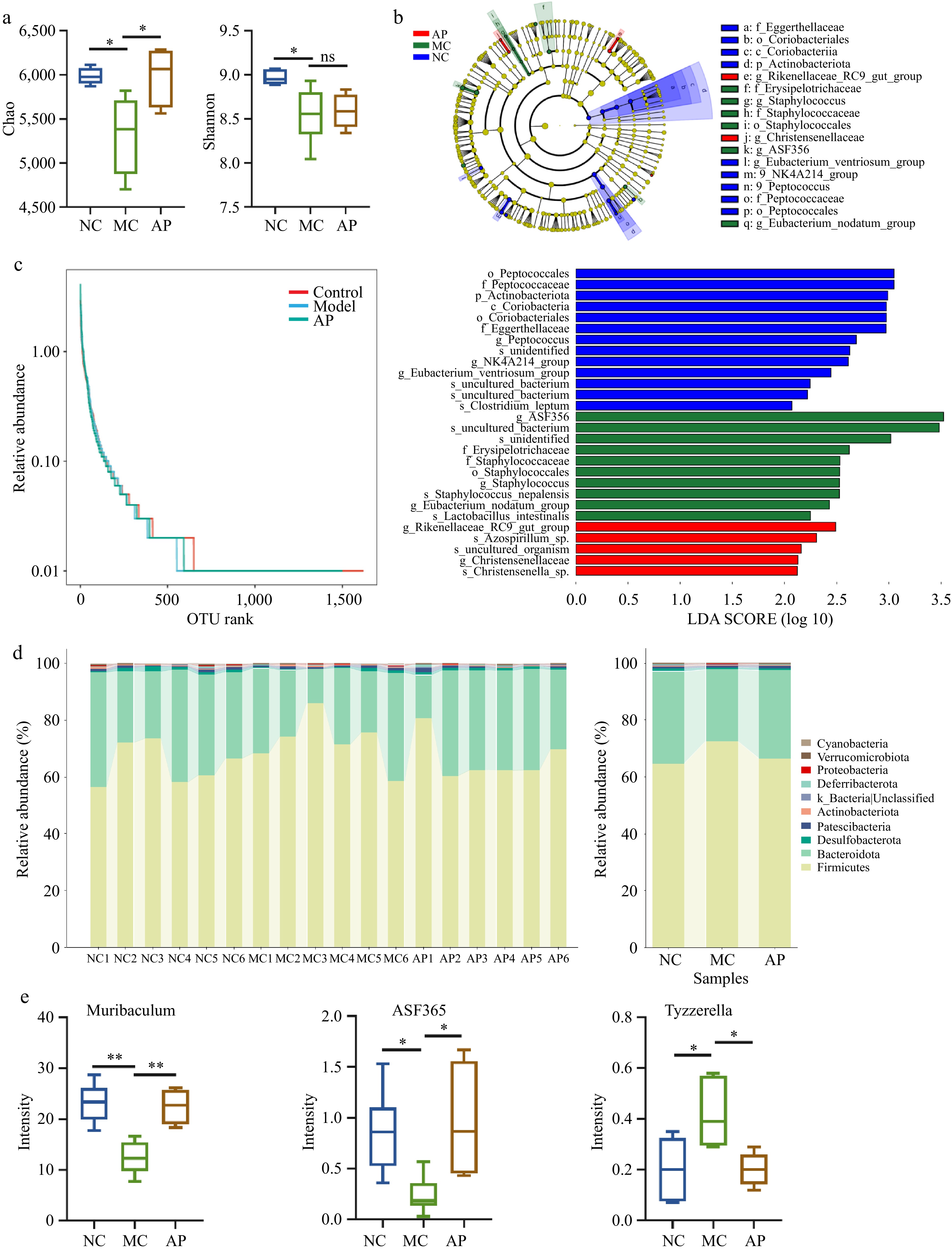
Figure 3.
APs altered the gut microbiota composition in mice. (a) Alpha diversity analysis. (b) Linear discriminant analysis effect size analysis. (c) Operational taxonomic units (OTU) rank analysis. (d) Relative abundance of differential genera. (e) Differential bacteria genera. Note: *, p < 0.05; **, p < 0.01.
At the phylum level, H-AP significantly increased the relative abundance of Bacteroidetes and Firmicutes compared with the MC group (Fig. 3d). At the genus level, analysis of compositions of microbiomes with bias correction (ANCOM-BC) identified seven and two significantly different taxa between the AP group and the NC and MC groups, respectively (Supplementary Table S6). Linear discriminant analysis effect size (LEfSe) analysis revealed more pronounced community restructuring at the genus level: Muribaculum and Clostridium ASF365 – bacterial taxa associated with immune homeostasis[22,23] – were significantly enriched following the AP intervention, whereas Tyzzerella, a marker of dysbiosis and colorectal adenoma[24], was markedly depleted (Fig. 3e). These findings indicate that AP-mediated immunomodulation is partially attributable to the selective enrichment of beneficial gut bacteria and suppression of pathobionts.
Metabolic reprogramming of amino acid and lipid pathways by APs
-
To elucidate the metabolic consequences of AP-induced microbiota modulation, untargeted metabolomics profiling was performed on serum and spleen tissues from the NC, MC, and H-AP groups. Orthogonal partial least squares discriminant analysis (OPLS-DA) revealed distinct metabolic signatures among the three groups in both sample types (Fig. 4a, b) Using the criteria of variable importance in projection (VIP) > 1 and p < 0.05, we identified 36 differential serum metabolites and 49 splenic metabolites (Fig. 4c, d).
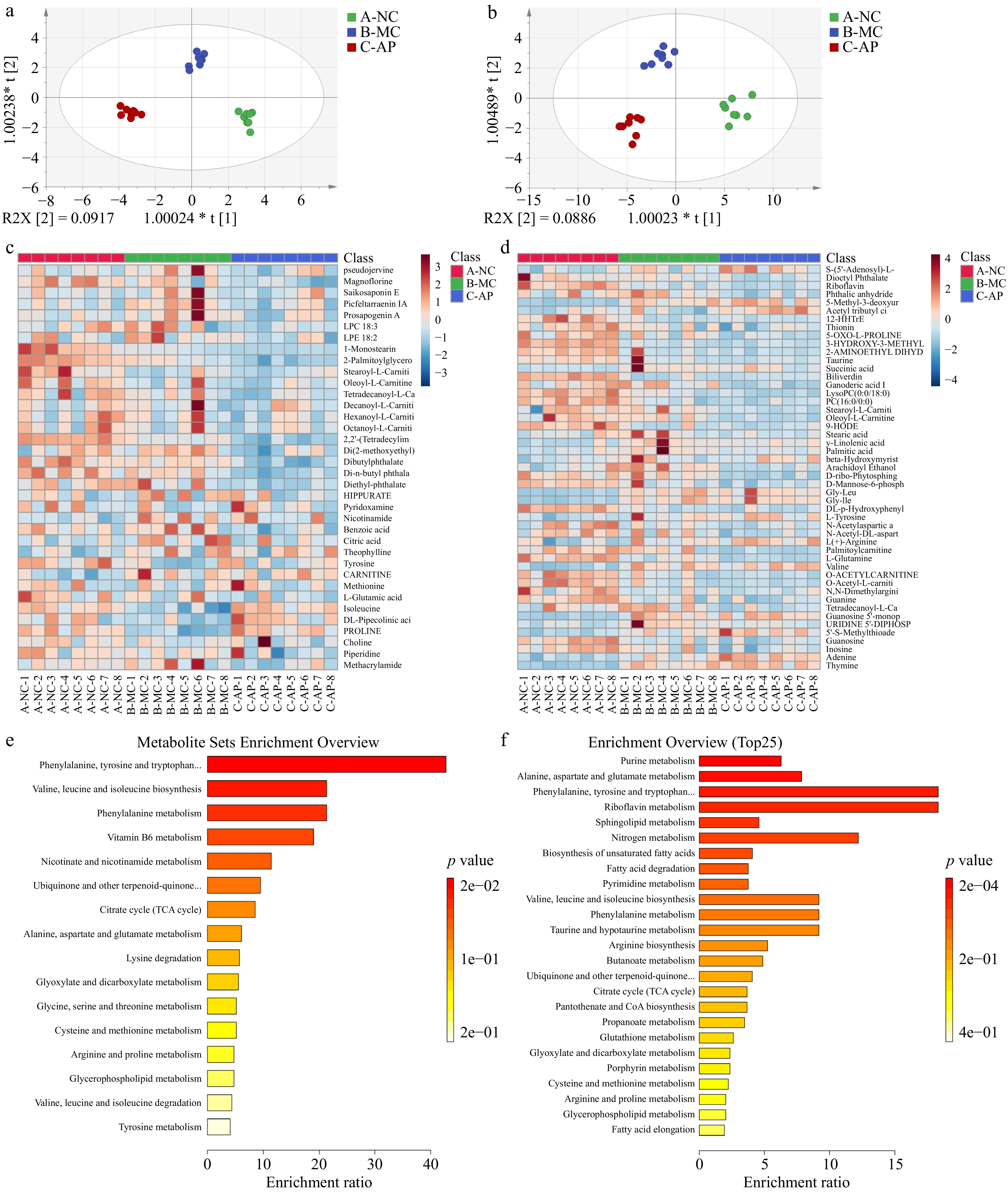
Figure 4.
Effects of the AP intervention on serum and spleen metabolites in mice. (a) Orthogonal partial least squares discriminant analysis (OPLS-DA) of serum metabolites. (b) OPLS-DA of splenic metabolites. (c) Heatmap analysis results of serum metabolites. (d) Heatmap analysis results of spleen metabolites. (e) Metabolite pathway analysis of serum differences. (f) Differential metabolite pathway analysis in the spleen.
In serum, AP treatment reversed CTX-induced depletion of amino acids, restoring proline, isoleucine, and L-glutamic acid to levels comparable with those of normal controls (Fig. 4c). KEGG pathway enrichment further identified amino acid biosynthesis as the most significantly upregulated pathway in the AP group (Fig. 4e), suggesting enhanced systemic amino acid availability.
Conversely, spleen metabolism exhibited a distinct pattern characterized by lipid downregulation. Although CTX elevated γ-linolenic acid, palmitic acid, and arachidonyl ethanolamide, the AP intervention significantly reduced these lipid mediators (Fig. 4d). Pathway analysis confirmed that amino acid metabolism and lipid metabolism were the primary affected pathways in spleen tissue (Fig. 4f). These tissue-specific metabolic alterations – systemic amino acid restoration versus splenic lipid homeostasis – suggest that APs coordinate immune recovery through compartmentalized metabolic reprogramming.
RPS23-centered proteomic network restoration by the AP intervention
-
To investigate whether AP-mediated metabolic reprogramming translates into functional protein restoration, label-free quantitative proteomics was performed on splenic tissues. Comparative analysis revealed substantial proteomic alterations in immunosuppressed mice: 999 upregulated and 66 downregulated proteins in MC versus NC (Fig. 5a, b). H-AP treatment reversed this dysregulation, normalizing 160 differentially expressed proteins (15 upregulated, 145 downregulated relative to MC), as visualized in the hierarchical clustering heatmap (Fig. 5c).
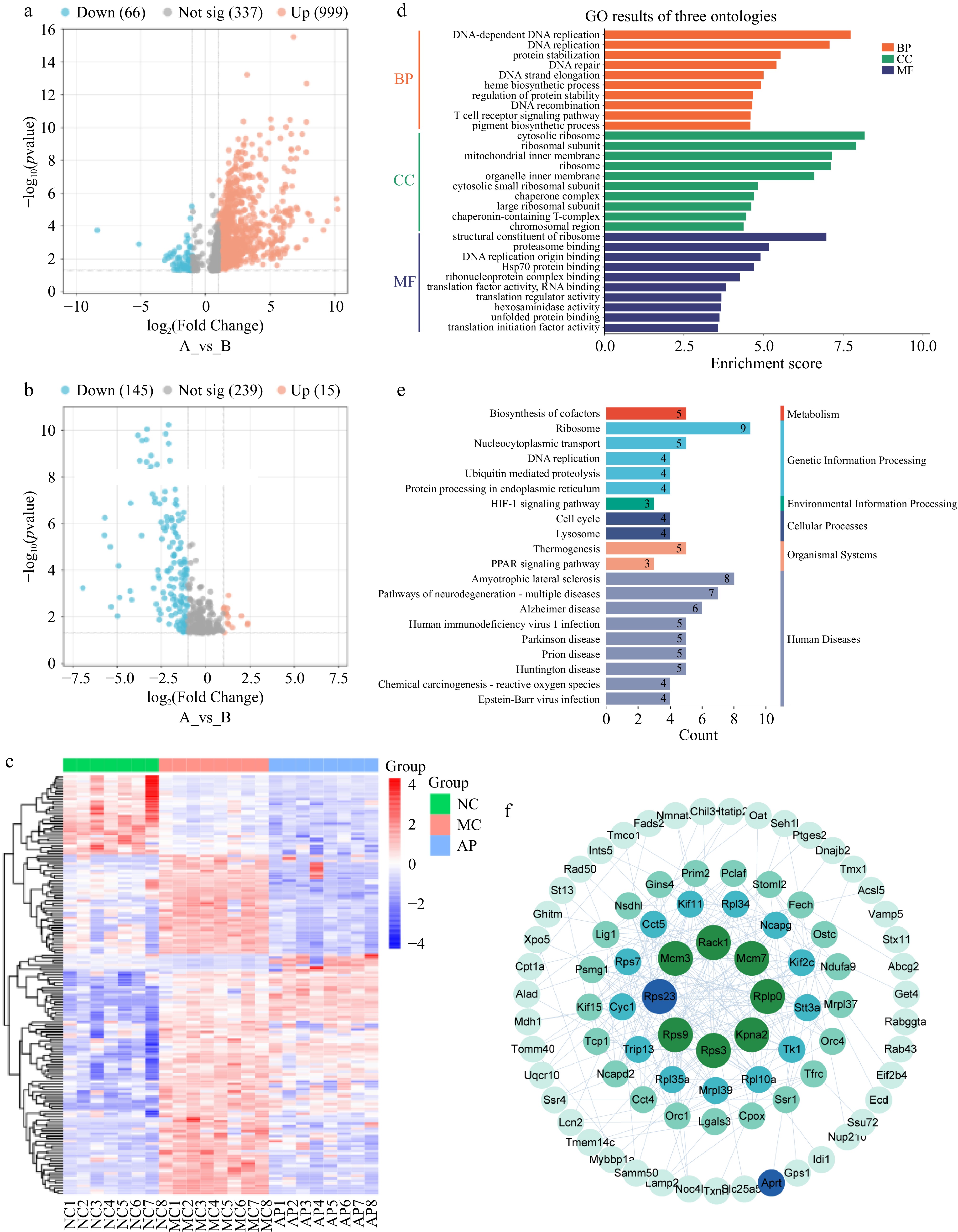
Figure 5.
Effects of the AP intervention on spleen protein in mice. (a) Volcano map analysis (NC vs MC). (b) Volcano map analysis (MC vs AP). (c) Differential protein heatmap analysis among the three groups. (d) GO enrichment analysis of spleen proteins. (e) KEGG analysis of spleen proteins. (f) Spleen protein interaction analysis.
The GO enrichment analysis indicated that these restored proteins were significantly associated with T cell receptor signaling and ribosomal protein complex binding (Fig. 5d), suggesting the recovery of immune cell activation and translational machinery. KEGG pathway analysis further identified HIF-1 and peroxisome proliferator-activated receptor (PPAR) signaling as the primary metabolic pathways modulated by APs (Fig. 5e), consistent with the observed alterations in splenic lipid metabolism.
PPI network analysis revealed ribosomal protein S23 (RPS23) as a hub node connecting immune and metabolic functions. RPS23, which was aberrantly elevated in immunosuppressed mice, was restored to normal levels following H-AP treatment (Fig. 5f and Supplementary Fig. S2). Given its established role in regulating cyclic adenosine monophosphate (cAMP)/protein kinase A (PKA) signaling and immune cell exhaustion[25], RPS23 represents a critical molecular mediator linking AP-induced metabolic changes to immune restoration.
Construction of the microbiota–isoleucine–RPS23 causal chain
-
To establish the mechanistic link between AP-induced gut microbiota alterations and immune restoration, Spearman correlation analysis was performed to integrate the microbiome, metabolome, and proteome datasets.
At the microbiota–metabolite interface, Muribaculum abundance exhibited a strong positive correlation with serum isoleucine levels (Fig. 6a), suggesting that AP-enriched Muribaculum may enhance systemic amino acids' availability. Conversely, Clostridium ASF365 was positively associated with tyrosine but negatively correlated with isoleucine and lipid metabolites (1-monostearin and 2-palmitoylglycerol), indicating distinct metabolic niches for these beneficial taxa.
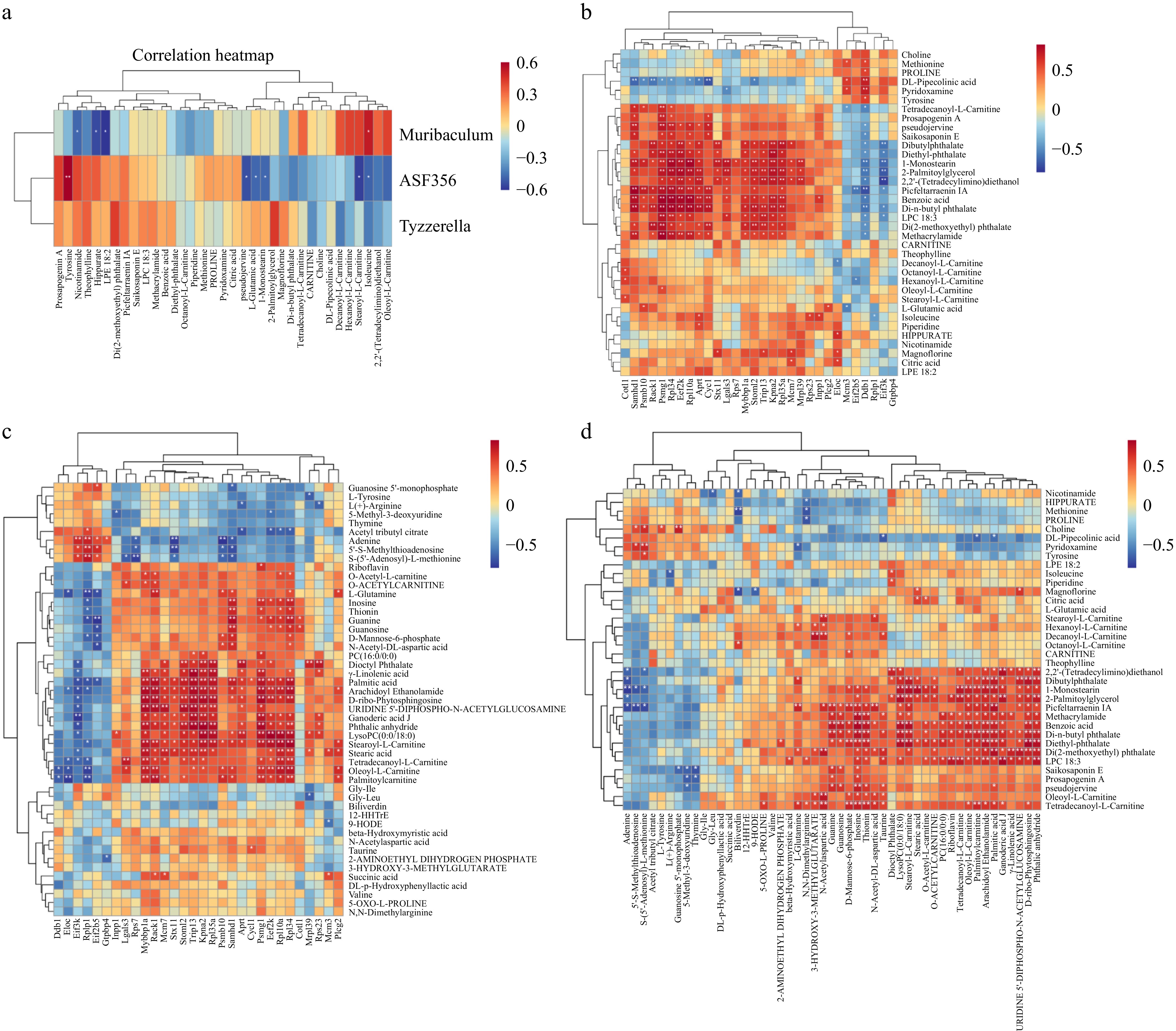
Figure 6.
Integration of multi-omics data. (a) Spearman correlation analysis of intestinal flora and serum metabolites. (b) Spearman correlation analysis between serum metabolites and spleen proteins. (c) Spearman correlation analysis of spleen proteins and spleen metabolites. (d) Spearman correlation analysis of spleen metabolites and serum metabolites. Note: *, p < 0.05; **, p < 0.01.
The metabolite–protein nexus was anchored by isoleucine, which demonstrated significant positive correlations with splenic RPS23 and adenine phosphoribosyltransferase (APRT) (Fig. 6b), which are critical proteins for ribosomal function and purine metabolism, respectively[26,27]. This finding positions isoleucine as a key metabolic mediator translating microbiota signals into functional protein networks. Additionally, splenic lipid mediators (γ-linolenic acid, palmitic acid, and arachidonyl ethanolamide) correlated with specific protein targets (Fig. 6c), whereas serum–spleen metabolite pairing revealed coordinated lipid trafficking between compartments (Fig. 6d).
Collectively, these multi-omics correlations support a mechanistic model wherein AP-driven enrichment of Muribaculum elevates systemic isoleucine, which, in turn, restores RPS23 expression and ribosomal function in splenic immune cells, thereby reversing CTX-induced immunosuppression.
-
Natural polyphenolic compounds have garnered increasing attention as functional food ingredients for immunomodulation[28]. However, their systemic mechanisms of action often defy explanation by single-target models[29,30]. This study integrates network pharmacology prediction with multi-omics validation to construct a gut microbiota–metabolism–immunity axis model underlying AP-mediated immune restoration. This finding not only elucidates the action trajectory of APs from a dietary component to systemic immune recovery, but also provides a research paradigm for understanding the multicomponent synergistic mechanisms of natural products.
The core of this axis model lies in the synergistic action of quercetin and luteolin, which were identified through network pharmacology as hub immunomodulatory constituents (node degree ≥ 100) targeting 247 overlapping genes enriched in PI3K-Akt and MAPK signaling. These polyphenols operate through distinct yet complementary mechanisms: quercetin attenuates immune suppression via Janus kinase 1 (JAK1)/signal transducer and activator of transcription 3 (STAT3)/hypoxia-inducible factor 1-alpha (HIF-1α) axis inhibition, thereby reducing oxidative inflammatory responses and splenic damage[11,31]. Luteolin induces ROS/Fe2+-dependent ferroptosis in CD8+ T cells and suppresses immune cell infiltration through Bruton's tyrosine kinase (BTK)/fms-like tyrosine kinase 3 (FLT3) phosphorylation blockade[12,13]. Notably, the coexistence of these multitarget agents within AP extracts may produce synergistic effects unattainable by single purified compounds, a pharmacological advantage inherent to botanical preparations[30,32]. In fact, the two core components identified in this study, quercetin and luteolin, have been experimentally shown to exhibit synergistic effects in another polyphenol-rich natural matrix. Li et al.[33] used a quadratic general rotary unitized design combined with microcalorimetric analysis and demonstrated a synergistic antibacterial effect between quercetin and luteolin isolated from Dracontomelon dao leaves. This finding further supports the possibility that the coexistence of quercetin and luteolin in APs may confer advantages over individual compounds through pharmacological complementarity and potential effects on bioavailability. The enrichment of these components in immune-related pathways (PI3K-Akt, MAPK, cAMP, HIF-1) thus provided a computational rationale for subsequent experimental validation using the CTX-induced immunosuppression model. Nevertheless, network pharmacology is primarily a predictive approach for identifying potential bioactive compounds, targets, and signaling pathways, and the predicted mechanisms still require further experimental validation.
APs reshape the gut microbiota composition through selective enrichment of immunomodulatory taxa rather than broad-spectrum probiotic promotion[34]. Although previous polyphenol studies emphasized modulation of the Firmicutes/Bacteroidetes ratio[35], our analysis revealed genus-level specificity as the primary driver of immune restoration. In our study, AP administration improved the gut microenvironment by increasing the Bacteroidetes/Firmicutes ratio. Muribaculum, a genus positively associated with intestinal immune activation and pathogen defense[23,36,37], emerged as the dominant responsive taxon, exhibiting strong correlation with systemic amino acid availability. This finding diverges from conventional short-chain fatty acid (SCFA)-mediated mechanisms typified by Clostridium ASF365 enrichment[22,38,39]; instead, Muribaculum appears to operate through amino acid metabolic pathways, explaining its central position in the microbiota–isoleucine–RPS23 axis. However, serum isoleucine levels are influenced by multiple factors, including dietary intake, protein catabolism, hepatic metabolism, and intestinal absorption. Therefore, the observed association between Muribaculum and serum isoleucine does not establish a direct causal relationship. Similarly, the correlations of isoleucine with splenic RPS23 and APRT should be interpreted as potential associations that require further experimental validation. The concomitant depletion of Tyzzerella, a colorectal adenoma biomarker, further underscores APs' dual action in both promoting beneficial symbionts and suppressing pathobionts, thereby reconfiguring the gut microenvironment toward immune homeostasis.
Multi-omics integration establishes the microbiota–isoleucine–RPS23 axis as the central conduit linking AP ingestion to immune restoration. This causal chain, initiated by Muribaculum enrichment, propagated through systemic isoleucine elevation, and executed via RPS23-mediated translational control, provides a mechanistic framework that transcends simple correlation. The axis operates through bidirectional metabolic signaling: gut-derived amino acids fuel immune cell proliferation, whereas splenic lipid suppression prevents inflammatory overactivation. This compartmentalized strategy ensures robust immune recovery without pathological hyperactivity. The schematic model (Fig. 7) encapsulates this sequence: AP remodeling of gut microbiota –> serum amino acid reprogramming –> splenic proteomic normalization –> B cell Ig secretion (IgA/IgM). Beyond mechanistic insight, this paradigm offers translational applications: the abundance of Muribaculum may serve as a predictive biomarker for AP responsiveness, enabling personalized nutritional interventions, whereas the isoleucine–RPS23 node presents a druggable target for adjunctive immunotherapy.
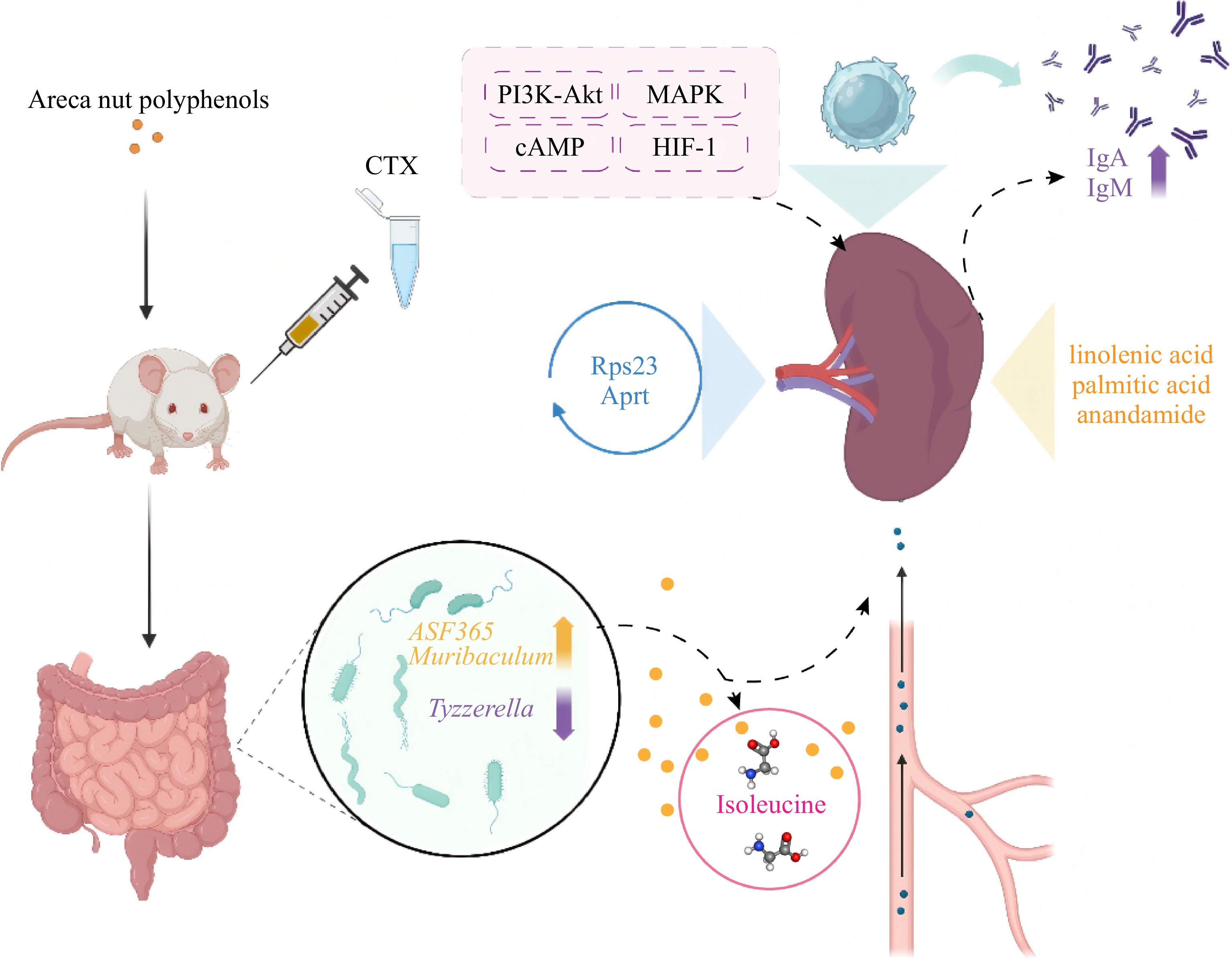
Figure 7.
Schematic diagram of potential mechanisms.
It is acknowledged that this study has several limitations. First, the exclusive use of male mice introduces a sex bias, as female hormones modulate immune and microbial responses not captured here[40]. Moreover, the CTX-induced immunosuppression model used in this study primarily reflects acute drug-induced immune injury and subsequent immune recovery. Therefore, the present findings mainly support a restorative effect of APs on acute CTX-induced immune injury and should not be directly generalized to other forms of immune dysfunction, such as those associated with chronic infection, autoimmune diseases, aging-related immune decline, or malnutrition, which involve distinct and more complex pathophysiological processes. Therefore, future research must validate this axis through fecal microbiota transplantation, isoleucine supplementation trials, and genetic RPS23 modulation to establish definitive causality and optimize therapeutic protocols.
-
This study establishes a gut microbiota–metabolite–protein axis as the mechanistic foundation for AP-mediated immune restoration. By integrating network pharmacology with multi-omics validation, we demonstrate that APs selectively enriche Muribaculum, elevate systemic isoleucine, and normalize splenic RPS23 expression, thereby reversing CTX-induced immunosuppression. This compartmentalized metabolic strategy, where systemic amino acid support is coupled with local inflammatory suppression, ensures robust immune recovery without pathological hyperactivation. The identification of Muribaculum abundance and isoleucine–RPS23 signaling as predictive biomarkers and druggable targets positions APs as a precision nutritional intervention for immune dysfunction. These findings advance our understanding of botanical polyphenol–host interactions and provide a translational framework for developing microbiome-targeted immunotherapies. However, this study still has several limitations that should be addressed in future research. Although the integrated multi-omics analysis revealed potential associations among the gut microbiota, metabolites, and proteins and provided a possible mechanistic framework for the biological effects of APs, the current findings are mainly based on correlation analyses. The proposed mechanisms therefore require further validation through targeted experiments. In addition, the AP extract is a complex polyphenol-rich extract containing multiple bioactive components. The present study mainly evaluated the overall effects of APs, but future studies should further investigate the specific roles of the major polyphenolic components identified in APs and their potential interactions. Moreover, although a mouse model was used in this study, inherent differences exist between mice and humans in terms of physiology, metabolism, and immune function. Future studies may use experimental models that more closely reflect human physiology or conduct clinical investigations to further evaluate the biological effects and translational potential of APs.
-
All procedures were reviewed and preapproved by the Animal Welfare and Ethics Committee of Jiangnan University (identification number: JN.No20220330b0720515[100]). The research followed the Replacement, Reduction, and Refinement principles to minimize harm to teh animals. This article provides details on the housing conditions, care, and pain management used for the animals, ensuring that the impact on the animals was minimized during the experiment.
-
The authors confirm their contributions to the paper as follows: contributed equally to this work: Wu B, Zhang Y; writing – original draft, conceptualization, data curation, methodology, visualization: Wu B; writing – original draft; data curation: Zhang Y; data curation, methodology, software: Zhao J; methodology, software: Shi J; methodology: Yuan L; supervision, writing – review and editing: Zhang J; supervision, writing – original draft, writing – review and editing, project administration: Xu YJ. All authors reviewed the results and approved the final version of the manuscript.
-
The raw mass spectroscopy data have been deposited in the ProteomeXchange Consortium via the PRIDE partner repository (www.ebi.ac.uk/pride/profile/reviewer_pxd061446) with project accession PXD072768.
-
The authors declare no competing interests.
-
accompanies this paper online at: https://doi.org/10.48130/fia-0026-0043.
-
Authors contributed equally: Boxiong Wu, Yu Zhang
- Supplementary Table S1 Parameter settings for MS-DIAL data processing.
- Supplementary Table S2 Active ingredients identified from SwissADME.
- Supplementary Table S3 Active ingredients identified from TCMSP.
- Supplementary Table S4 Key compounds of AP.
- Supplementary Table S5 Rarefaction depth for each sample.
- Supplementary Table S6 Significant genus-level taxa identified by ANCOM-BC (q < 0.05).
- Supplementary Fig. S1 Sequencing depth and beta-diversity analysis of gut microbiota among the NC, MC, and AP groups.
- Supplementary Fig. S2 AP restored the expression level of RPS23 protein in the spleen of immunosuppressed mice.
- Supplementary Text S1 Metabolomics instrument parameters.
- Supplementary Text S2 Proteomics instrument parameters.
- Copyright: © 2026 by the author(s). Published by Maximum Academic Press on behalf of China Agricultural University, Zhejiang University and Shenyang Agricultural University. This article is an open access article distributed under Creative Commons Attribution License (CC BY 4.0), visit https://creativecommons.org/licenses/by/4.0/.
-
About this article
Cite this article
Wu B, Zhang Y, Zhao J, Shi J, Yuan L, et al. 2026. Multi-omics reveals the gut microbiota–metabolic axis-mediated immunomodulatory activity of areca nut polyphenols. Food Innovation and Advances 5(4): 532−544 doi: 10.48130/fia-0026-0043 

Multi-omics reveals the gut microbiota–metabolic axis-mediated immunomodulatory activity of areca nut polyphenols
- Received: 05 May 2026
- Revised: 13 August 2026
- Accepted: 28 August 2026
- Published online: 28 October 2026
Abstract: Areca nut polyphenols (APs) exhibit diverse health-promoting properties, yet the mechanisms underlying their immunomodulatory effects remain poorly understood. This study followed an integrative approach combining network pharmacology, microbiome analysis, metabolomics, and proteomics to investigate the immunomodulatory mechanisms of APs. Network pharmacology identified quercetin and luteolin as the hub immunomodulatory components (node degree ≥ 100). In vivo, high-dose APs (100 mg/kg) significantly reversed cyclophosphamide (CTX)-induced immunosuppression, restoring serum immunoglobulin (Ig) A and IgM levels and normalizing white blood cell, neutrophil, monocyte, and lymphocyte counts. Multi-omics analysis results revealed that APs enriche beneficial gut bacteria (Muribaculum), enhance serum amino acid metabolism (particularly isoleucine), and restore splenic RPS23 expression and lipid homeostasis, ultimately promoting immunoglobulin secretion. These findings suggest APs ameliorate CTX-induced immunosuppression through a microbiota–metabolite–protein axis mechanism, providing mechanistic insights for developing AP-based functional foods targeting immune dysfunction.
-
Key words:
- Areca nut polyphenols /
- Immunomodulatory /
- Network pharmacology /
- Gut microbiota /
- Metabolism /
- Proteomics