












-
Of the more than 100 chemical modifications catalogued on eukaryotic mRNA, only a handful are understood in functional detail. N6-methyladenosine (m6A) has anchored the plant epitranscriptomic field for over a decade: its writers, readers, and erasers have been characterized, and its roles in development and stress physiology are widely documented[1−3]. Although N1-methyladenosine (m1A) has been detected in a few plant species, it has never been systematically characterized. A recent study by Ke et al. delivers the first single-nucleotide-resolution m1A methylome across 10 Arabidopsis tissues, identifies the complete writer–reader–inhibitor toolkit, and demonstrates that m1A regulates abscisic acid (ABA) signalling[4]. It provides the most accurate m1A map available for any plant species and establishes m1A as only the second mRNA modification, after m6A, for which a full regulatory system with a defined physiological function has been described in plants.
HTML
-
The defining technical advance of this study is its detection strategy. Earlier plant m1A studies in tomato and petunia relied on antibody-based enrichment alone, yielding broad, lower-resolution profiles[5,6]. Zhou et al. coupled m1A immunoprecipitation with the engineered RT1306 reverse transcriptase. This enzyme introduces a characteristic mutation signature at m1A sites, enabling detection at single-nucleotide resolution when paired with AlkB demethylase treatment as a negative control[7]. This approach yields a standard previously achieved only in mammalian systems.
The resulting atlas comprises 4,802 high-confidence m1A sites across 2,947 genes, drawn from 10 tissues spanning the Arabidopsis lifecycle (Fig. 1a). The majority of methylated genes (83%) carry a single site, consistent with m1A functioning as a position-specific mark, not a diffuse modification. In general, m1A is ubiquitous in Arabidopsis, with m1A/A ratios of 0.028%–0.043% detected from 25 different tissues. These values match those reported in mammals, yeast, and other eukaryotes. Such cross-kingdom conservation implies fundamental functional constraints on m1A deposition, rather than species-specific idiosyncrasy. Notably, m1A is preferentially enriched in 5’-UTRs, peaking roughly 100 nucleotides upstream of the start codon. This distribution mirrors the mammalian pattern and stands in marked contrast to the 3’-biased localization of m6A, pointing to a spatial division of regulatory labor between the two modifications. Ke et al. also identified a plant-specific feature: m1A sites cluster closer to the start codon when they reside in the second exon rather than the first. This is the opposite of the trend in mammals[8], and it suggests not only divergent m1A deposition mechanisms but also different regulatory mechanisms associating m1A with transcription, splicing, or translation, at least between plants and animals.
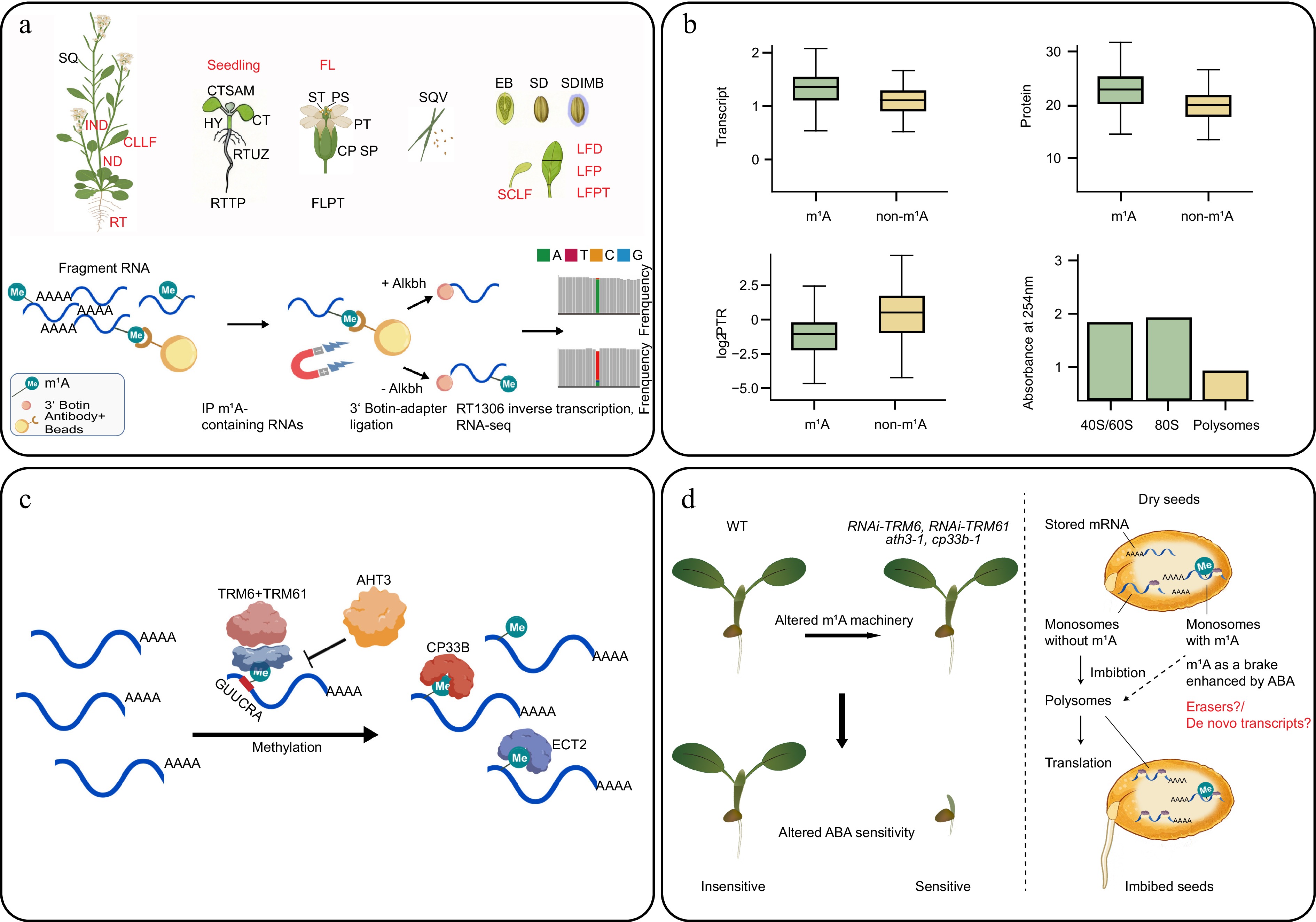
Figure 1.
Foundational m1A epitranscriptomic atlas and regulatory system in Arabidopsis. (a) Schematic of 25 Arabidopsis tissues collected in the m1A atlas and m1A-IP-seq workflow. Tissues abbreviations: EB, embryo; CP, carpel; CT, cotyledons; CTSAM, shoot tip; FL, flower; FLPT, flower petiole; HY, hypocotyl; IND, internode; LFD, leaf distal; LFP, leaf proximal; LFPT, leaf petiole; ND, node; PS, pistil; PT, petal; RT, root; RTTP, root tip; RTUZ, root upper zone; SCLF, senescent leaf; SD, seed; SDIMB, seed imbibed; SP, sepal; SQ, silique; SQV, silique valves; ST, stamen. All these 25 tissues were used in the m1A level detection, while the 10 tissues used in m1A-IP-seq were labelled in red abbreviations. For m1A detection at single-nucleotide resolution, m1A-modified mRNA fragments are immunoprecipitated, and one aliquot is treated with AlkB to remove m1A. Both aliquots were reverse-transcribed using RT1306. The enzyme's elevated mismatch rate at m1A sites in the untreated sample, relative to the AlkB-treated control, enables single-nucleotide detection of m1A. (b) m1A regulates events from transcription to translation. m1A-modified transcripts show higher mRNA and protein abundance but lower protein-to-RNA ratios (PTR). m1A is enriched in monosome fractions and depleted from polysomes. (c) m1A writer–reader network. TRM6–TRM61 catalyses m1A deposition on substrates bearing a tRNA-like stem-loop with the GUUCRA motif. ATH3 inhibits this complex by impairing the RNA-binding of the TRM6–TRM61 complex. CP33B and ECT2 read m1A; ECT2 additionally binds m6A. (d) Genetic disruption of any m1A pathway component results in ABA hypersensitivity (left panel) and m1A as a translational brake on stored mRNAs at germination (right panel). The figure was created in BioRender. Guochenxi, G. (2026),
https://BioRender.com/d9bmi84 , and with BioGDP (biogdp.com ).
-
The spatial segregation of m1A and m6A reflects fundamentally different regulatory strategies. Ke et al. demonstrated this functional distinction by integrating their methylome with publicly available proteomic data. m1A-modified transcripts consistently exhibited higher mRNA and protein levels than those of unmodified transcripts. However, their protein-to-RNA ratio (PTR), a measure of translational efficiency, was significantly lower (Fig. 1b). Polysome profiling reinforced this picture: m1A-marked mRNAs were enriched in ribosomal subunits and monosome fractions and were depleted from actively translating polysomes (Fig. 1c). The m1A-modified 5′ UTRs also exhibited higher minimum free energy than the unmodified ones. A positively charged N1-methyl group that blocks base pairing is expected to stiffen the local RNA structure, making it harder for the 43S preinitiation complex to scan through[4].
These data identify m1A as a translational brake at highly active transcripts; it permits transcript production while limiting protein output. When the restraint is lifted, protein products can be synthesized without waiting for new rounds of transcription. Much like an accomplished race car driver who skillfully balances the throttle and the brake to navigate a complex racetrack, m1A appears to fine-tune gene expression by simultaneously preserving responsiveness and preventing excessive output. In plants, constantly changing environmental conditions and the accompanying reconfiguration of phytohormone signaling pathways represent precisely such a 'complex racetrack'. Adaptive responses must be deployed rapidly, yet remain sufficiently restrained, reversible, and flexible to accommodate subsequent environmental fluctuations. The regulatory mode uncovered for m1A is remarkably well suited to these biological demands, providing an elegant mechanism for balancing rapid response with controlled gene expression.
-
TRM6 and TRM61 are known to form the tRNA m1A methyltransferase complex in Arabidopsis. Loss of this essential activity results in embryo lethality[9,10]. Ke et al.[4] now show that the same complex also methylates mRNA, but only under stringent structural conditions. Consistent with observations in humans, both a tRNA-like stem-loop and the GUUCRA consensus motif are required for substrate recognition (Fig. 1c). This structural mimicry of the tRNA T-loop represents an elegant example of evolutionary exaptation: the co-option of an ancient tRNA modification enzyme for a new regulatory function on mRNA. This stringent structural requirement also helps explain why m1A has remained difficult to characterize systematically in plants, where it is also far less abundant than m6A. Unlike m6A, whose RRACH consensus motif enables straightforward sequence-based prediction, m1A deposition depends on the three-dimensional RNA structure that cannot be inferred from the primary sequence alone.
-
The regulatory mechanism diverges further from the m6A paradigm with the discovery of ATH3 (AtTrxh3; Arabidopsis thioredoxin H-type protein)[4]. It physically associates with the TRM6–TRM61 complex and inhibits its RNA-binding activity. m1A levels are elevated when ATH3 is deleted in vivo. An inhibitor protein of an mRNA methyltransferase has not been described for any other mRNA modification writer complex. Its existence implies that m1A deposition is subject to active, tunable negative regulation.
On the reader side, CP33B and ECT2 were identified as m1A readers. ECT2 is a YTH-domain protein that was previously characterized as an m6A reader[11]. Given the distinct distributions of m6A and m1A along mRNAs, ECT2 may exert context-dependent regulatory effects depending on the modification it engages. Whether these two marks compete for ECT2 occupancy, or cooperate to produce integrated regulatory outputs, is a central open question that the tools provided by Ke et al. now make experimentally tractable. These findings further raise the possibility that additional m6A readers also recognize m1A.
However, global m6A levels did not change in m1A pathway mutants upon ABA treatment, indicating that the two modifications operate independently at the deposition level. Previous studies have shown that ECT2/3/4 function redundantly to negatively regulate ABA signaling through recognition of m6A-modified transcripts[12]. In addition, the stress-inducible reader ECT8 promotes the degradation of m6A-modified transcripts encoding negative regulators, thereby acting as a molecular sensor of stress[13]. Together, these findings suggest that functional crosstalk between m1A and m6A is more likely to occur at the level of reader-mediated decoding and downstream target regulation than through coordinated methylation deposition.
-
Ultimately, the importance of any RNA modification is determined by its physiological function. Major phytohormones, including abscisic acid (ABA), auxin, gibberellin, cytokinin, and ethylene, significantly increased the relative m1A level. A range of abiotic stresses was further examined, while only nitrogen deficiency, high pH, and light altered the relative m1A level under the experimental conditions.
ATH3 was previously reported as an ABA-binding protein[4], and m1A levels responded most strongly to ABA. Echoing these clues, knocking down the writers (RNAi-TRM6/61), deleting the inhibitor (ath3-1), or the reader (cp33b-1) all altered ABA sensitivity (Fig. 1d). The transcript of SnRK2.2, a key positive regulator of ABA signalling, contains an m1A site. Collectively, these observations suggest that m1A dynamics are physiologically important for ABA signalling, although the precise mechanism remains to be fully resolved. Since m1A may permit transcript accumulation while restraining translation, the dual role of the m1A mark may maintain ABA signaling within an optimal dynamic range, balancing responsiveness with reversibility under fluctuating environmental conditions.
-
These ABA phenotypes were evaluated in germination assays (Fig. 1d), and germination may be one of the contexts in which m1A's translational brake matters. Mature dry seeds accumulate many long-lived stored mRNAs, most of them kept as translationally silent monosomes, while the steady-state mRNA pool may not reflect the protein output[14]. On imbibition, a subset of these stored mRNAs is loaded onto polysomes and translated before the requirement of transcription[15]. De novo transcription resumes later, chiefly to sustain post-germinative growth. Newly produced transcripts face a further gate before translation: SAM8 condensation under low water potential can hold them in the nucleus[16]. Transcript accumulation and translation thus run on different schedules during germination, and it is in this window that Ke and Balarynová et al. observed altered ABA sensitivity in m1A pathway mutants. If m1A decorates stored mRNAs too, the mark could keep germination-related transcripts in reserve until water releases them, setting both how quickly and how reversibly a seed commits to germinating. Recent data point in the same direction: m1A is the most abundant RNA modification in Arabidopsis seeds, more so in a non-dormant accession than in a deeply dormant one, and its levels rise during after-ripening[17]. Whether m1A truly controls the translation of stored mRNAs, and how this intersects with ABA signalling, remains to be seen. The answer bears directly on dormancy and on uniform germination in crops.
-
Ke et al. provide the plant research community with high-quality m1A resources that it did not have before. The immediate priorities are structural and comparative studies. A cryo-EM structure of the TRM6–TRM61 holoenzyme would clarify how the GUUCRA motif and tRNA-like fold are recognized and whether tRNA and mRNA methylation activities can be experimentally uncoupled. Defining the dynamic interactome of the TRM6–TRM61 holoenzyme may help explain the differential m1A deposition in various developmental or environmental contexts. Beyond the model plant Arabidopsis thaliana, extending high-resolution m1A profiling to crop species will reveal whether translational control via m1A is a conserved strategy for tuning hormone responses under field-relevant conditions.
Another intriguing observation is that m1A and m6A may partially share the eraser and/or reader system. Identification of an m1A eraser in Arabidopsis, as they do in mammals[4], would complete the writer–reader–eraser framework. The dual-affinity reader ECT2 opens the door to a broader question that extends beyond m1A and m6A. A broader challenge now lies in defining the regulatory logic that emerges when multiple RNA modifications converge on shared effector proteins.
-
The authors confirm their contributions to the paper as follows: conceptualized the manuscript: Guo C, Li C; draft manuscript preparation: Guo C, Jiang S, Zhang S, Li C. All authors reviewed the results and approved the final version of the manuscript.
-
Data sharing is not applicable to this article as no datasets were generated or analyzed during the current study.
-
The authors declare that they have no conflict of interest.
- Copyright: © 2026 by the author(s). Published by Maximum Academic Press on behalf of Hainan Yazhou Bay Seed Laboratory. This article is an open access article distributed under Creative Commons Attribution License (CC BY 4.0), visit https://creativecommons.org/licenses/by/4.0/.
Guo C, Jiang S, Zhang S, Li C. 2026. Champion racer's maneuver—m1A mRNA modification accelerates transcription while restraining translation. Seed Biology 5: e027 doi: 10.48130/seedbio-0026-0023


|