












-
Macrofungi and myxomycetes frequently intersect in detrital habitats because both are concentrated in moist, microbe-rich substrates where organic matter is decomposed and redistributed. In forests, macrofungi shape deadwood microhabitats and produce macroscopic fruiting bodies that can persist as structured elements within decaying wood[1,2]. These fruiting bodies can retain moisture and provide sheltered surfaces and internal tissues that other organisms may exploit.
Myxomycete trophic stages exploit microbe-rich, moisture-retentive microhabitats that develop on decaying wood and litter. They are also frequently recorded in bryophyte-rich microhabitats on woody substrates, creating opportunities for spatial overlap with wood-inhabiting macrofungi[2−4]. Stable isotope patterns indicate that, in saproxylic settings, myxomycete sporocarps can occupy a consumer position relative to litter-decomposing fungi; however, bulk δ13C/δ15N data cannot identify prey and therefore do not demonstrate obligate mycophagy[5,6].
The term 'slime mould' is applied to multiple lineages; within Amoebozoa, the Eumycetozoa comprise Myxogastria, Dictyostelia and the protostelids[7−9]. In this article, 'myxomycetes' refers to Myxogastria (myxomycetes sensu stricto), because published reports of myxomycetes on macrofungal fruiting bodies and fungus-structured detritus largely concern Myxogastria[10].
This article synthesises evidence on the contexts in which myxomycetes occur on macrofungal fruiting bodies, dead basidiomata, and substrates structurally or chemically shaped by macrofungal activity, including rot-type-defined wood, rhizomorph-rich interfaces, and cultivation blocks across forest systems and mushroom cultivation environments[2,11−15]. The focus is on macrofungi as habitat-forming and resource-concentrating structures within which myxomycetes develop, whereas the reverse direction of interaction (fungi developing on myxomycete sporocarps) is outside the scope of this synthesis.
Myxomycete–macrofungal associations can reflect several non-exclusive mechanisms. Many observations represent habitat sharing, where both groups respond to similar microclimatic and resource gradients on dead organic matter. Other associations are plausibly indirect, arising because macrofungal-driven decay modifies wood physicochemistry and microstructure, thereby altering the microbial prey field and the suitability of substrates for myxomycete trophic stages. In a smaller subset of cases, antagonistic potential is supported either experimentally or by cultivation/observational reports. Experimental evidence includes directed movement toward fungal cues, plasmodial feeding on fungal mycelia, and inhibition of selected fungi in vitro, whereas tissue deterioration or lysis has chiefly been reported from cultivation or observational studies[11,15−18].
-
This study was conducted as a structured evidence synthesis using transparent literature identification, screening, and reporting principles informed by PRISMA 2020[19]; the heterogeneous evidence base was synthesised narratively rather than meta-analytically.
Information sources
-
Web of Science Core Collection, Scopus, PubMed, and CAB Abstracts were used as the principal bibliographic databases and were searched from database inception through 31 December 2025. Google Scholar was used as a supplementary discovery tool and for forward citation tracking, and reference lists of eligible articles and relevant reviews were additionally screened (backward citation searching). These sources were used to capture forest records, cultivation reports, stable isotope studies, and experimental studies relevant to myxomycete associations with macrofungal fruiting bodies or fungus-structured detritus.
Search strategy
-
Searches combined concept blocks for Myxogastria/myxomycetes/slime moulds, macrofungal fruiting bodies, fungus-structured detritus or cultivation systems, and association descriptors using Boolean operators AND/OR. The association block was designed to retrieve both descriptive and mechanistic evidence, including records of co-occurrence, colonisation, decay context, attraction, feeding, inhibition, deterioration, and cultivation impacts. A summary of the information sources, conceptual search blocks, eligible source types, screening rules, and evidence-entry counting rules is provided in Supplementary Table S1.
Eligibility criteria
-
Eligible evidence types included field records of myxomycete sporocarps or plasmodia occurring on macrofungal fruiting bodies (basidiomata and ascomata/stromata) or within fungus-structured detritus; studies linking myxomycete occurrence to fungus-defined decay contexts, including rot type and decay stage; experimental studies demonstrating attraction to fungal cues, digestion or assimilation of fungal mycelia, tissue lysis, or inhibition of fungal growth; and cultivation reports documenting colonisation and impacts on mushroom production. Eligible source types included journal articles, books and checklists, theses, and unpublished reports when they contained verifiable taxonomic, substrate, or experimental information relevant to the synthesis. English and non-English literature were eligible, provided that the relevant association data could be extracted reliably. Field records were included only when both the myxomycete and the macrofungal partner or fungus-structured substrate context were identifiable from the source; cultivation reports were included only when the crop system, myxomycete taxon, and reported outcome could be extracted.
Records focusing exclusively on Dictyostelia or protostelids were excluded. For field-record synthesis, inclusion required an identifiable myxomycete at least to genus, and records lacking a clearly described substrate context involving macrofungal fruiting bodies or fungus-structured detritus were excluded. Reports dealing only with fungi colonising myxomycete sporocarps were also excluded because they fell outside the scope of the synthesis.
Study selection
-
All database records were exported to a reference manager and deduplicated. Screening proceeded in two stages: title and abstract screening followed by full-text assessment against the eligibility criteria. Reasons for exclusion at the full-text stage were recorded. Title/abstract screening retained records that potentially involved Myxogastria, macrofungal fruiting bodies, fungus-defined decay substrates, or cultivation settings; full-text exclusion categories therefore included taxonomic scope mismatch, missing or ambiguous substrate context, and absence of extractable association evidence.
Data extraction and evidence framework
-
From each eligible record, data were extracted on the myxomycete taxon, reported macrofungal taxon or fungus-structured substrate descriptor, substrate or microhabitat context, study setting (forest vs cultivation or managed environment), and the type of evidence supporting the association. Taxon names in the tables were standardised for consistency across the manuscript; currently accepted combinations were used where readily verifiable, and historical names from the source literature were retained in parentheses only when needed for traceability (for example, Trichamphora pezizoidea reported in cultivation literature as Physarum pezizoides or P. pezizoideum[10]). An evidence-bearing source was defined as any source contributing at least one extractable field record, cultivation observation, stable isotope study, or experiment included in the synthesis. An evidence entry was the reporting unit used in the tables: for forest and cultivation material, one entry represented a distinct combination of myxomycete taxon, macrofungal partner or cultivation context, and association substrate/context, with multiple sources combined in a single row when they supported the same association; for experimental studies, one entry represented a distinct mechanistic finding relevant to a focal myxomycete–fungus system; and each stable isotope study was counted separately as indirect trophic evidence.
No formal risk-of-bias scoring or statistical weighting was applied because the evidence base was heterogeneous and largely descriptive. Instead, interpretation was guided qualitatively by evidential proximity to direct interaction: direct experimental demonstrations of attraction, feeding, or inhibition were treated as the strongest support for direct effects; cultivation reports with documented tissue deterioration were interpreted as context-specific observational evidence; stable isotope studies were treated as indirect trophic evidence; and field co-occurrence records were interpreted as descriptive habitat-association evidence only. Evidence was synthesised narratively and supported by stratified tables separating forest field records, cultivation records, and experimental or mechanistic evidence, with stable isotope studies discussed separately as indirect trophic evidence.
-
The final synthesis compiled 45 evidence entries from 38 distinct evidence-bearing sources. Because multiple sources sometimes supported the same association context, the number of evidence entries did not equal the number of sources. These comprised 34 forest field association entries from 27 sources (Table 1), five cultivation or managed-system entries from four sources (Table 2), four direct experimental/mechanistic evidence entries from five sources summarised in Table 3, and two stable isotope studies providing indirect trophic evidence[5,6].
Forest detritus systems: habitat overlap and decay-mediated coupling
-
In forest detritus, macrofungi act as principal agents of deadwood decomposition, while myxomycete trophic stages graze on microbial communities that proliferate within the same moist substrates. Shared reliance on decay-generated, microbe-rich microhabitats results in frequent spatial overlap between plasmodia and wood-inhabiting basidiomata, particularly within coarse woody debris, bark, and litter[2−4,15].
Deadwood-unit surveys and related syntheses report repeated co-occurrence of myxomycete and fungal taxa within the same decomposing wood units, consistent with shared habitat use on coarse woody debris[2]. In one survey, co-occurrence involved dozens of myxomycete and fungal species (for example, 69 myxomycete species and 66 fungal species), but such presence data alone do not demonstrate direct biotic interactions[2]. Habitat structure likely strengthens this signal, because larger trunks can buffer moisture and increase microsite heterogeneity, expanding the number of locations suitable for both fungal development and myxomycete trophic activity.
Rot type and decay gradients provide a mechanistic route for indirect linkage mediated by fungal-driven substrate transformation. White-rot and brown-rot woody debris differ in physicochemical conditions such as pH and hardness, and myxomycete diversity has been reported to differ between these decay contexts, with higher diversity on white-rot than brown-rot wood in a subalpine system[14]. Independent evidence indicates that fungal decomposer activity structures substrate conditions (including pH, water content, and decay type) that correlate with myxomycete community composition[1,6]. Decay-stage frameworks also support temporal coupling, with myxomycete taxa associated with early, intermediate, and late decomposition stages, consistent with myxomycetes tracking progressive changes in wood texture, porosity, and moisture retention that emerge during fungal decay[20,21].
Myceticolous fruiting, defined here as myxomycete sporocarps forming directly on macrofungal fruiting bodies, is documented in situ. In a microhabitat-explicit survey that included fungal fruiting bodies among sampled substrates, myceticolous records were present but less frequent than lignicolous records[22]. Species-level forest records of Myxogastria reported on fungal fruiting bodies, on macrofungal structures such as rhizomorphs, or in adjacent fungus-structured deadwood contexts are summarised in Table 1 and Fig. 1.
Table 1. Forest field records of myxomycetes associated with macrofungal fruiting bodies or fungus-structured detrital microhabitats.
Macrofungus partner Myxomycetes reported Association class Association substrate/context Ref. Aphyllophoroid basidiomycetes Arcyria denudata; Hemitrichia calyculata Adjacent substrate Wood near polyporoid basidiomata [22] Armillaria mellea Arcyria denudata Adjacent substrate Wood near rhizomorphs [23] Bjerkandera adusta Diachea leucopodia Fruiting body; adjacent substrate On basidiomata; adjacent wood near basidiomata [24,25] Calocera cornea Hemitrichia clavata; Hemitrichia decipiens Adjacent substrate Woody twigs near basidiomata [2] Cantharellus cibarius Diderma rufostriatum Fruiting body On basidiomata [26] Clavicorona pyxidata Fuligo septica Adjacent substrate Co-occurring on the same decaying wood substrate; myxomycete sporocarps not reported on basidiomata [27] Corticium pubescens Enteridium variabile Adjacent substrate Co-occurring on the same deadwood substrate; myxomycete sporocarps not reported on basidiomata [28] Corticium sp. Physarum album Fruiting body; adjacent substrate On basidiomata; adjacent wood in the same corticioid context [24,25] Cortinarius caperatus Leocarpus fragilis Fruiting body On fruiting bodies [29] Cytidia salicina Badhamia utricularis Fruiting body On living basidiomata [27] Daedaleopsis confragosa Badhamia macrocarpos; Fuligo cinerea Fruiting body Hymenophores of old basidiomata [30] Discina sp. Polyschismium carestianum Fruiting body On apothecial surface/snow-edge context (as reported) [31] Fomes fomentarius Arcyria denudata; Comatricha laxa; Fuligo cinerea; Hemitrichia leiotricha; Oligonema favogineum; Hemitrichia decipiens; Trichia scabra; Trichia varia Fruiting body; adjacent substrate On senescent basidiomata and on adjacent decayed wood in the same deadwood units (species-level reports) [2,30,32−34] Fomitopsis betulina Licea minima; Nannengaella globulifera; Oligonema affine Fruiting body Pore layer/fruiting bodies of decaying basidiomata [27,35,36] Ganoderma applanatum Arcyria denudata; Comatricha pulchella; Lycogala epidendrum; Licea glomerulifera; Nannengaella globulifera Fruiting body; adjacent substrate On basidiomata and on adjacent decayed wood (species-level reports) [2,24,25,37] Gloeophyllum abietinum Diderma umbilicatum Fruiting body On basidiomata [38] Hypholoma fasciculare Physarum psittacinum Fruiting body; adjacent substrate On living fruiting bodies; woody debris near basidiomata [27] Hypoxylon sp. Craterium minutum Fruiting body On stromata [39] Lenzites abietina Stemonitis splendens Fruiting body On basidiomata [28] Mycena renati Stemonitis fusca Fruiting body On basidiomata [40,41] Phaeolus schweinitzii Didymium nigripes Adjacent substrate On root-associated substrates where Phaeolus fruiting bodies occur (as reported) [27] Phellinus igniarius Arcyria denudata Fruiting body On old/decaying basidiomata [24,25] Phellinus sp. Stemonitopsis typhina Fruiting body On fruiting bodies [42] Phlebia radiata Badhamia utricularis Fruiting body On living basidiomata [27] Piptoporus populinus Collaria arcyrionema Fruiting body On basidiomata [43] Pleurotus sp. Didymium clavus Fruiting body Hymenophores of old basidiomata [30] Pluteus cervinus Trichia varia Fruiting body On basidiomata [40,41] Rigidoporus crocatus Stemonitopsis aequalis Fruiting body; adjacent substrate On basidiomata on a decaying log; coarse woody debris near basidiomata [26] Scutellinia sp. Stemonitis fusca Fruiting body On apothecial surface [42] Trametes betulina Arcyria denudata Fruiting body On basidiomata [44] Trametes hirsuta Badhamia utricularis Fruiting body On basidiomata [45] Trametes sp. Trichia persimilis Adjacent substrate Wood near polyporoid basidiomata [46] Tremella sp. Badhamia macrocarpos Fruiting body Living and dead basidiomata [46] Xylaria polymorpha; Xylaria longipes Lycogala epidendrum; Physarum melleum; Stemonitis fusca; Trichia varia Adjacent substrate Coarse woody debris with stromata (as reported) [2,47] 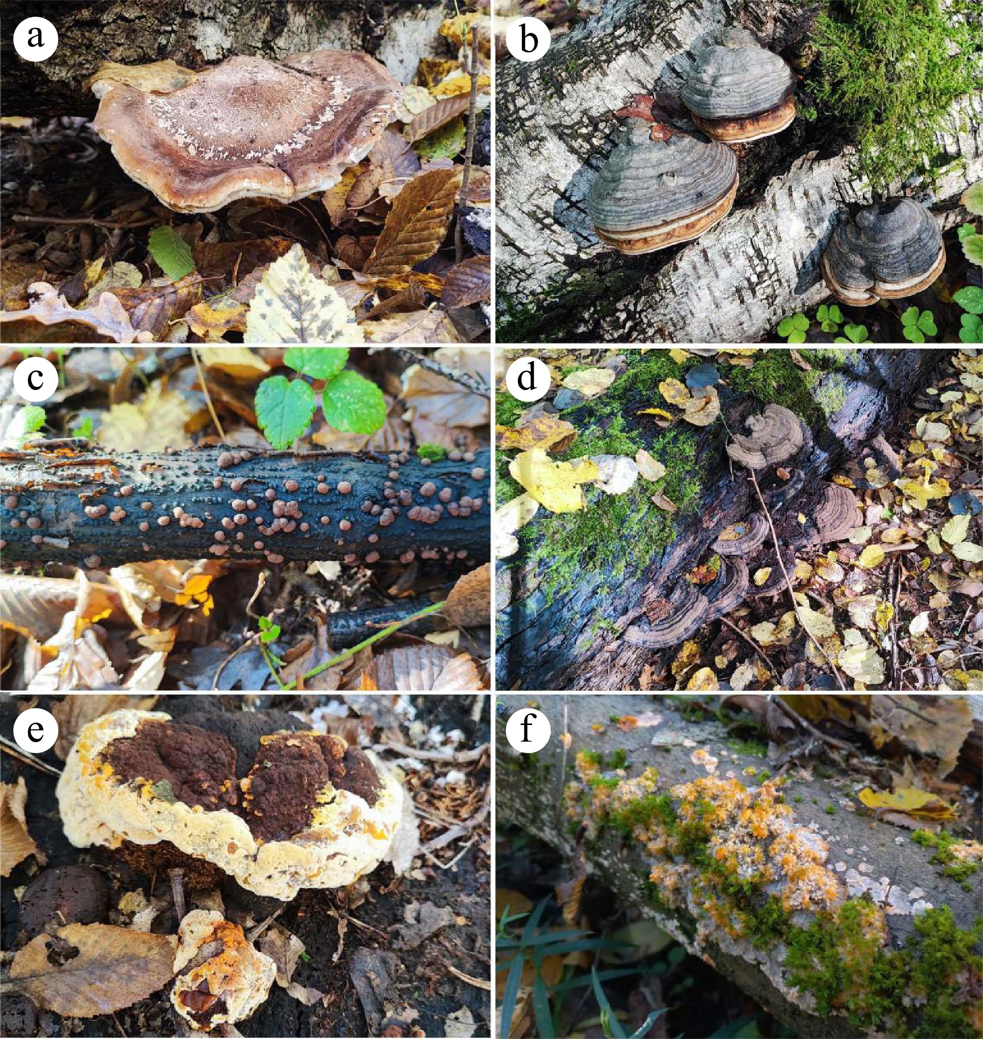
Figure 1.
Collage of macrofungal fruiting bodies that commonly serve as substrates for myxomycetes in forest detritus. (a) Fomitopsis betulina. (b) Fomitopsis pinicola. (c) Hypoxylon sp. (d) Ganoderma applanatum. (e) Phaeolus schweinitzii. (f) Phlebia radiata.
Managed and anthropogenic environments
-
Managed mushroom cultivation and other human-modified habitats can increase opportunities for myxomycete–macrofungal contact by maintaining moist microhabitats and organic-rich substrates over extended periods. In cultivation rooms and farms, repeated inoculation pressure may be sustained by dispersal via air currents and transfer via water, insects or other animals, and human activities, increasing the likelihood of recurring colonisation events[15].
The cultivation synthesis comprised five managed-system entries from four sources (Table 2). In mushroom production systems, both tissue deterioration and non-lytic competitive impacts have been reported. Badhamia utricularis has been associated with wilting, rotting, and partial lysis of fruiting bodies and primordia of cultivated Pholiota nameko and Pleurotus ostreatus, with completion of the myxomycete life cycle on mushrooms; Koch's postulates were reported as fulfilled in the underlying cultivation studies[12]. Comparable lytic outcomes have been reported for Trichamphora pezizoidea (reported in the cultivation literature as Physarum pezizoides/Physarum pezizoideum) on cultivated wood ear mushrooms (Auricularia spp.), and extracellular enzymes have been proposed as contributors to tissue deterioration[10,12]. Cultivation records also highlight impact pathways that do not require obvious tissue lysis: dense plasmodial growth and sporulation can function as a competitive 'weed' layer that occupies cultivation surfaces and suppresses fruiting, and spore-inoculation experiments have been reported to reduce mushroom production in logs[12]. Additional indoor observations show that myxomycetes can develop and sporulate on cultivated mushrooms under controlled conditions, even when damage is not emphasised[13]. Species-level cultivation records are consolidated in Table 2.
Table 2. Cultivation/managed systems: records of myxomycetes associated with cultivated macrofungi.
Cultivated macrofungus (crop) Myxomycetes reported Where observed Reported outcome Ref. Pholiota nameko Badhamia utricularis Fruiting bodies and primordia Wilting/rotting/partial lysis; life cycle completed on mushrooms; Koch's postulates reported as fulfilled in the underlying cultivation studies [12] Pleurotus ostreatus Badhamia utricularis Fruiting bodies and primordia Wilting/rotting/partial lysis; life cycle completed on mushrooms; Koch's postulates reported as fulfilled in the underlying cultivation studies [12] Auricularia spp. Trichamphora pezizoidea (reported as Physarum pezizoides/pezizoideum in cultivation literature) Fruiting bodies Lysis; extracellular enzymes proposed as contributors to tissue deterioration (as reported) [10,12] Pleurotus (controlled indoor cultures) Stemonitis herbatica; Physarum compressum Substrate and fruiting bodies Sporocarps formed on Pleurotus; development recorded in a controlled indoor setting [13] Multiple cultivated mushrooms (reported) Multiple myxomycete taxa Fruiting bodies and/or cultivation substrate Production impacts reported; includes damage and competitive surface 'weed' effects [15] Trophic and antagonistic evidence
-
This section distinguishes between indirect trophic indications and direct evidence of antagonistic potential. The synthesis drew on seven sources: four direct experimental or mechanistic evidence entries from five sources summarised in Table 3, together with two stable isotope studies that provided indirect trophic evidence[5,6,11,16−18,48].
Table 3. Direct experimental and mechanistic evidence relevant to myxomycete interactions with fungi in macrofungal systems.
Myxomycete taxon Fungal taxon/substrate Evidence type Main finding Ref. Badhamia utricularis Stereum hirsutum (basidioma pieces/extracts; mycelium) Attraction/chemotaxis Attraction to basidioma material/extracts and mycelium extracts demonstrated [17] Badhamia utricularis Stereum hirsutum (basidioma tissue) Observational lysis report Lysis/liquefaction of fungal tissue reported [16,48] Unspecified myxomycete plasmodia Multiple fungal mycelia (including some macrofungus-relevant taxa) Feeding/digestion assays Digestion/assimilation reported, with differential susceptibility among fungi [11] Enteridium variabile (reported as Licea flexuosa in source literature; mucous secretions/aqueous extracts) Cladosporium herbarum; Penicillium sp. (in vitro) Growth inhibition assays (in vitro) Secretions/extracts reported to inhibit growth of filamentous fungi in vitro; ecological relevance remains uncertain [18] Bulk stable isotope data are consistent with myxomycete sporocarps occupying a consumer position relative to saprotrophic microorganisms, including litter-decomposing fungi, in some systems[5,6]. However, bulk δ13C/δ15N data alone cannot discriminate direct consumption of fungal tissues from feeding on bacteria and other heterotrophic microbes embedded in fungal decay matrices, and δ13C overlap primarily indicates shared carbon pools rather than proof of direct mycophagy[5,6].
Experimental evidence demonstrates the biological feasibility of fungal feeding. In classical feeding assays, myxomycete plasmodia digested and assimilated fungal mycelia, and outcomes differed among fungal taxa, indicating differential susceptibility to plasmodial feeding[11]. Mechanistic studies also provide plausible pathways linking encounter to direct antagonism. Plasmodia can show directed movement toward macrofungal tissues and extracts, providing a behavioural route for targeted contact; for example, attraction of Badhamia utricularis plasmodia to extracts and tissues of Stereum hirsutum has been demonstrated experimentally[17]. Observational reports also describe lysis of fungal tissues as plasmodia traverse or colonise fungal substrates[16,48]. In parallel, in vitro assays indicate that myxomycete secretions or crude extracts can inhibit growth of selected fungi[18], but the ecological relevance for interactions with wood-decay macrofungi remains uncertain. The direct experimental and mechanistic evidence relevant to macrofungal systems is summarised in Table 3.
Claims of obligate parasitism of macrofungi by myxomycetes are uncommon in the available literature, and most records are consistent with a continuum from substrate use to facultative antagonism under conducive conditions. Reports involving taxa such as Licea parasitica are primarily linked to bark and epiphytic lichens and are not used here to infer obligate parasitism of macrofungal fruiting bodies[15,49−51].
-
Across forests and managed systems, the compiled evidence indicates that macrofungi repeatedly generate moist, structured, microbe-rich habitats that overlap with the environmental requirements of myxomycete trophic stages. Most reported associations are therefore best interpreted as substrate-mediated co-occurrence within fungus-structured detritus, together with occasional myceticolous fruiting on fruiting bodies as moisture-retentive platforms. In managed systems, artificially maintained humidity, concentrated organic substrates, and repeated handling may further alter local microhabitat conditions and increase encounter opportunities[12,13,15].
Within this shared arena, a smaller subset of studies provides direct evidence of antagonistic potential, including plasmodial digestion of fungal mycelia under experimental conditions[11], experimental attraction to fungal materials[17], and inhibition of selected fungi in vitro by myxomycete secretions or extracts[18]. By contrast, deterioration or lysis of fungal tissues has mainly been described in observational and cultivation contexts[12,16,48]. Stable isotope evidence supports consumer trophic positions for myxomycetes in saproxylic settings, but does not resolve prey identity and therefore cannot by itself establish obligate mycophagy[5,6].
In cultivation systems, persistent humidity and repeated inoculation pressure may increase encounter frequency and are associated in some reports with fruiting body deterioration and competitive surface 'weed' effects that suppress cropping[12,15]. Overall, myxomycete–macrofungal relationships are best described as habitat-coupled associations with context-dependent antagonistic potential rather than uniform parasitism.
-
Available evidence is consistent with a continuum of myxomycete–macrofungal associations, dominated by shared-habitat and substrate-mediated coupling in forest detritus, with direct antagonistic effects documented only in a smaller and heterogeneous subset of studies. In managed cultivation systems, sustained moisture and repeated inoculation pressure may increase opportunities for contact and, in some reports, coincide with production losses. A key limitation is that most available evidence is descriptive and that few studies directly test interaction mechanisms.
The authors have no acknowledgments to declare. This research received no external funding.
-
Not applicable. This article is a literature-based synthesis and did not involve new experiments with humans or animals. The authors declare that no AI tools or generative AI technologies were used during the preparation of this manuscript.
-
The authors confirm their contributions to the paper as follows: study conception and design, draft manuscript preparation: Pawłowicz T; data collection: Pawłowicz T, Bachura E, Malej GK, Kudrycka O, Kieczka A, Żebrowski I; analysis and interpretation of results: Pawłowicz T, Bachura E. All authors reviewed the results and approved the final version of the manuscript.
-
Data sharing is not applicable to this article as no new datasets were generated. All extracted evidence entries, category counts, and search-summary details are presented within the article and its supplementary information.
-
The authors declare that they have no conflict of interest.
-
accompanies this paper online at: https://doi.org/10.48130/sif-0026-0019.
- Supplementary Table S1 Summary of information sources, search framework, eligibility criteria, screening procedure, and included evidence categories used in the structured evidence synthesis.
- Copyright: © 2026 by the author(s). Published by Maximum Academic Press, Fayetteville, GA. This article is an open access article distributed under Creative Commons Attribution License (CC BY 4.0), visit https://creativecommons.org/licenses/by/4.0/.
-
About this article
Cite this article
Pawłowicz T, Bachura E, Malej GK, Kudrycka O, Kieczka A, et al. 2026. Associations between myxomycetes, macrofungal fruiting bodies, and fungus-structured detritus in forests and mushroom cultivation. Studies in Fungi 11: e020 doi: 10.48130/sif-0026-0019 

Associations between myxomycetes, macrofungal fruiting bodies, and fungus-structured detritus in forests and mushroom cultivation
- Received: 02 March 2026
- Revised: 08 April 2026
- Accepted: 20 April 2026
- Published online: 02 July 2026
Abstract: Myxomycetes (Myxogastria) commonly fruit on decaying organic matter, where they exploit moist, microbe-rich substrates. Macrofungal fruiting bodies and fungus-structured detritus can function as persistent microhabitats that retain moisture and concentrate microbial prey, and may increase opportunities for encounter between myxomycetes and macrofungi. We conducted a structured evidence synthesis of published field records, cultivation reports, and experimental studies to characterise associations between myxomycetes, macrofungal fruiting bodies, and fungus-structured detritus across forest systems and mushroom production environments. The synthesis compiled 45 evidence entries from 38 distinct evidence-bearing sources. Forest reports indicate repeated co-occurrence of myxomycetes with wood-inhabiting macrofungi, including sporocarp formation directly on basidiomata as well as on adjacent, fungus-structured deadwood. Cultivation environments can increase opportunities for contact, and cultivation reports describe fruiting body deterioration or competitive surface colonisation associated with reduced cropping, whereas experimental studies demonstrate attraction of plasmodia to fungal cues, digestion of fungal mycelia in feeding assays, and inhibition of selected fungi by myxomycete secretions in vitro. Overall, the available evidence supports a gradient from shared-habitat associations to context-dependent antagonism. Most records remain descriptive and are better explained by indirect, substrate-mediated coupling than by obligate parasitism.
-
Key words:
- Myxomycetes /
- Macrofungi /
- Mycophagy /
- Deadwood microhabitats /
- Forest detritus /
- Antagonistic interactions