













-
Cancer remains a leading cause of morbidity and mortality worldwide. In the most recent global estimates, approximately 20.6 million new cancer cases and 9.7 million cancer deaths occurred worldwide in 2024, and the annual number of new cases is projected to rise by approximately two-thirds, to about 35 million, by 2050 on the basis of population growth and ageing alone[1−4]. T-cell-based adoptive cell therapy (ACT) has gained clinical traction as an important component of multimodal cancer treatment[5,6]. ACT principally includes tumor-infiltrating lymphocyte (TIL), chimeric antigen receptor (CAR)-T, and T cell receptor-engineered (TCR-T) therapies. TIL therapy harnesses a patient's natural, polyclonal antitumor repertoire without receptor engineering, and the 2024 accelerated approval of lifileucel for advanced melanoma, the first FDA-approved TIL therapy, marked an important milestone for ACT in solid tumors[7,8]. CAR-T and TCR-T cells, by contrast, are equipped with defined antigen-specific receptors. These two receptor-engineered, target-driven modalities, and the antigens they recognize, are the focus of this review.
CAR-T and TCR-T cells differ in how they engage their targets. A CAR uses an antibody-derived binding domain to recognize an extracellularly accessible antigen independently of the major histocompatibility complex (MHC), whereas a TCR-T cell expresses a natural or engineered TCR that recognizes a short peptide presented by an MHC molecule[6,9,10]. CAR-T therapy has reshaped the treatment of hematologic malignancies: seven FDA-approved products target the B-lineage antigens CD19 or B cell maturation antigen (BCMA) in leukemia, lymphoma, or multiple myeloma[11−13]. Nevertheless, antigen heterogeneity, antigen loss or downregulation, drive escape and motivate multi-antigen strategies, while the scarcity of tumor-restricted surface antigens and an immunosuppressive microenvironment have made translation to solid tumors substantially more difficult[14−19].
Despite these barriers, both target spaces have recently yielded major clinical milestones. In 2024, afamitresgene autoleucel (afami-cel), which recognizes a melanoma-associated antigen A4 (MAGE-A4) peptide, became the first FDA-approved TCR-T therapy and the first receptor-engineered T-cell therapy approved for a solid tumor, synovial sarcoma[20,21]. For surface-antigen targeting, a 2025 randomized trial showed that satricabtagene autoleucel (satri-cel), a CLDN18.2-directed CAR-T therapy, significantly prolonged progression-free survival in patients with advanced gastric or gastroesophageal junction cancer. It was subsequently approved in China in June 2026, becoming the first CAR-T therapy approved for a solid tumor[22−24].
These advances highlight target selection as one of the central determinants of engineered T-cell efficacy and safety. The target-receptor combination helps determine which tumors can be treated, how completely they can be covered, and how much normal tissue may be affected. CARs and TCRs access different antigen spaces and generally operate at different ligand-density thresholds: CARs primarily recognize extracellularly accessible targets and often require relatively high antigen density, whereas TCRs recognize peptides derived from proteins throughout the cell only after processing and MHC presentation and can respond at substantially lower ligand densities[25−28].
To evaluate a target rather than merely describe it, each of these properties must be expressed as a measurable quantity. Tumor specificity is quantified as the tumor-to-normal ratio across a comprehensive normal-tissue reference, transcript abundance in GTEx or the Human Protein Atlas[25,29], staining intensity and H-score by immunohistochemistry, and, for peptide-MHC (pMHC) targets, detection or non-detection in multi-tissue benign immunopeptidome references such as the HLA Ligand Atlas[30], with particular attention to vital organs rather than to tumor-adjacent tissue alone. Target abundance is measurable as antigen density in molecules per cell by quantitative flow cytometry (antibody-binding capacity) for surface antigens[26], and as peptide copies per cell by targeted mass spectrometry for pMHC targets[31,32]; these values typically differ by two to three orders of magnitude between the two target classes and should be interpreted against the functional activation threshold of the specific receptor rather than as binary positivity[26,27,33]. Intratumoral uniformity is measurable as the percentage of antigen-positive tumor cells by quantitative immunohistochemistry, multiplexed imaging, or single-cell profiling. Shareability is calculable as the product of antigen prevalence and, for pMHC targets, the frequency of the restricting HLA allele in the intended population, which together define the addressable patient fraction[34]. Fitness dependence can be approximated from clonality (cancer cell fraction), driver status, and CRISPR gene-dependency scores in resources such as DepMap[35−37]. Framing target selection in these quantitative terms, rather than as a categorical definition of a 'tumor antigen', is what allows safety and efficacy to be anticipated before a receptor is built.
Although CAR-T and TCR-T therapies are usually developed, reviewed, and regulated as separate technologies, both ultimately solve the same biological problem: identifying and safely engaging actionable tumor targets. This review therefore reframes engineered T-cell therapy around the tumor targetome, the subset of tumor antigens that engineered receptors can safely and effectively engage. It extends beyond classical tumor-associated and cancer-testis antigens to shared public neoantigens derived from recurrent drivers such as KRAS and TP53[38], as well as cryptic and non-canonical antigens arising from non-coding regions, aberrant splicing, and unconventional translation[39−41]. As this target space expands, selecting the appropriate antigen becomes increasingly consequential. Accordingly, this review organizes the field around the targetome: mapping targetable antigen classes; examining how targets are discovered, validated, and engaged; and recasting barriers to efficacy and safety as target-centric failure modes together with the strategies designed to overcome them. Rather than comparing CAR-T and TCR-T as competing technologies, it treats them as complementary solutions to a common problem, the precise, safe, and durable engagement of the tumor targetome through its surface and intracellular faces.
-
The surfaceome comprises plasma-membrane proteins with extracellularly exposed regions accessible to antibodies or CARs[42], whereas the broader CAR-accessible surface target space also includes selected glycoepitopes and glycolipids[15,43]. The immunopeptidome comprises peptides displayed by MHC molecules and available for TCR recognition[31]. The tumor targetome is the subset of these surface and peptide-MHC (pMHC)-presented targets that engineered receptors can safely, effectively, and tractably engage. It is therefore determined not simply by what a tumor expresses or presents, but by tumor specificity, target abundance and accessibility, antigen presentation, and human leukocyte antigen (HLA) context (Fig. 1a).
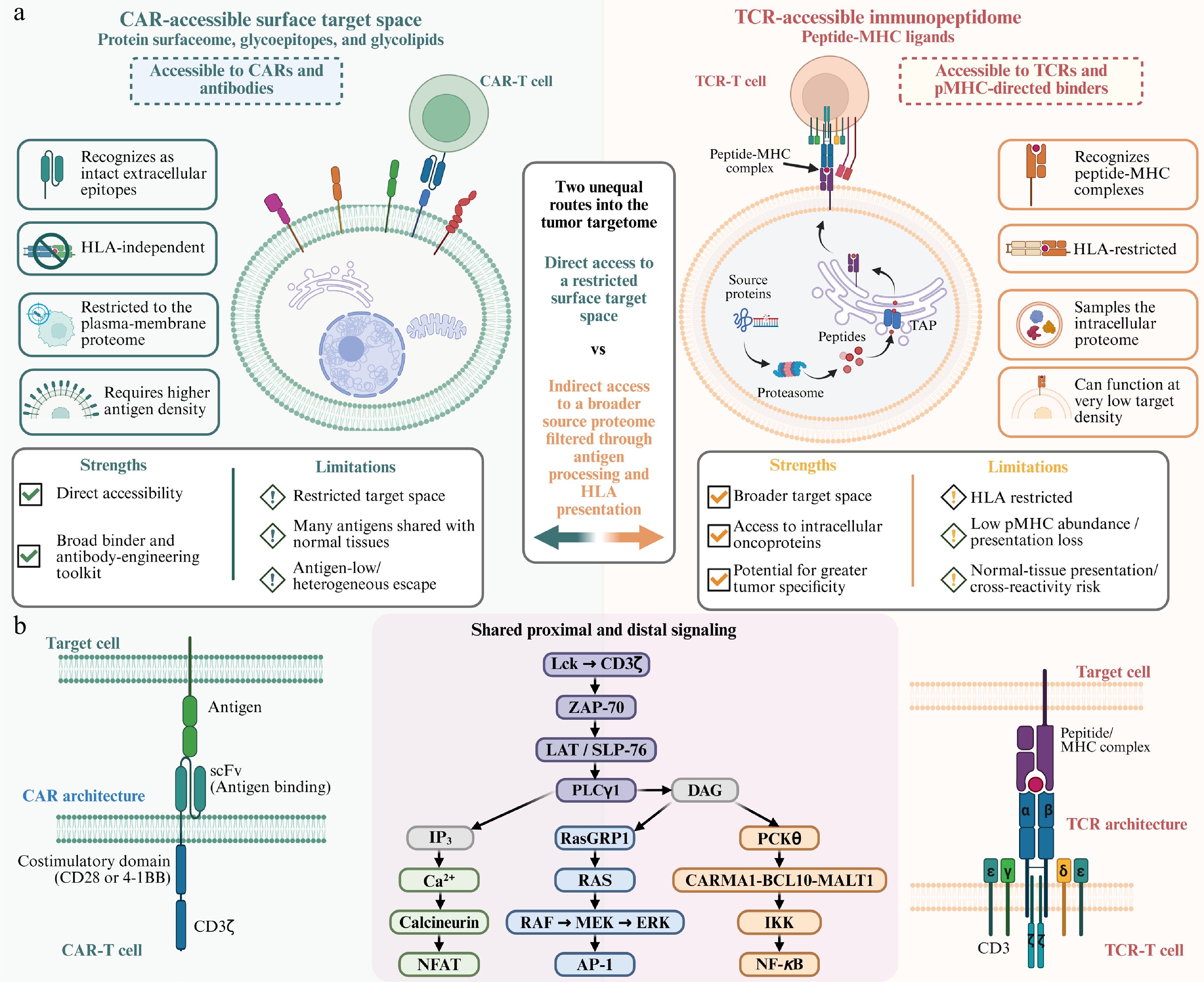
Figure 1.
Complementary target spaces and receptor architectures of CAR-T and TCR-T cells. (a) Conventional CARs directly recognize intact extracellular epitopes, surface proteins, glycoepitopes, and glycolipids, independently of HLA, but are confined to the plasma-membrane proteome, generally require relatively high antigen density, and face escape through antigen loss or low antigen density. TCRs recognize peptide-MHC complexes generated from a much broader source proteome through antigen processing and MHC presentation, reaching intracellularly derived oncoproteins at very low ligand density, at the cost of HLA restriction, low pMHC abundance, presentation loss, and cross-reactivity risk. (b) A CAR fuses recognition and signaling into a single chain (scFv, hinge and transmembrane segment, one costimulatory domain, and CD3ζ), whereas the TCR distributes these functions across the αβ heterodimer and the associated CD3γε, CD3δε, and CD3ζζ modules. Both nonetheless converge on the same signaling machinery: Lck-mediated phosphorylation of CD3ζ ITAMs, ZAP-70, the LAT/SLP-76 signalosome, and PLCγ1, whose products IP3 and DAG drive the Ca2+-calcineurin-NFAT, RasGRP1-RAS-ERK-AP-1, and PKCθ-CBM-IKK-NF-κB pathways.
Receptor architecture: synthetic CAR vs native TCR
-
CAR-T and TCR-T cells differ in receptor architecture. A CAR is a synthetic, MHC-independent receptor that integrates recognition and signaling into a single polypeptide containing an antigen-binding domain, a hinge or spacer, a transmembrane segment, and an intracellular signaling region. First-generation CARs contained CD3ζ alone and generally showed limited expansion and persistence[9,14,44]. Second-generation CARs add one costimulatory domain, most commonly CD28 or 4-1BB, and form the dominant architecture among approved products[6,44]. CD28-based CARs often produce faster activation and expansion, whereas 4-1BB-based CARs are frequently associated with slower kinetics and greater persistence, although these properties depend on the complete receptor and cellular context[44]. Third-generation CARs combine two costimulatory domains but have not shown a consistent advantage. Later designs add inducible payloads or cytokine-receptor signaling modules to remodel the microenvironment or supplement T-cell signaling[6,14,45,46]. Despite this modularity, a conventional CAR remains limited to extracellularly accessible targets displayed on the target-cell surface.
TCR-T cells instead introduce an αβ T-cell receptor and use the cell's native recognition and signaling machinery. The αβ heterodimer binds peptide-MHC through its variable domains but has short cytoplasmic tails without intrinsic signaling motifs. It associates through charged transmembrane residues with CD3γε, CD3δε, and CD3ζζ to form an octameric TCR-CD3 complex containing ten immunoreceptor tyrosine-based activation motifs[47]. Most clinically developed TCR-T therapies recognize peptides of approximately 8−11 amino acids presented by MHC class I, making recognition HLA-restricted and tying each receptor to one or a narrow group of alleles. Introduced α and β chains may also mispair with endogenous TCR chains, a liability addressed through pairing engineering or disruption and replacement of the endogenous receptor. TCR recognition is therefore structurally more complex and presentation-dependent, but it can access peptides derived from proteins throughout the cell.
Despite their different assembly, CARs and TCRs converge on overlapping proximal signaling modules. Antigen engagement promotes Lck-dependent ITAM phosphorylation, ZAP-70 recruitment, and LAT/SLP-76 signalosome formation, which activate PLCγ1; its products inositol trisphosphate (IP3) and diacylglycerol (DAG) then drive the Ca2+-calcineurin-NFAT, RasGRP1-RAS-ERK-AP-1, and PKCθ-CARMA1-BCL10-MALT1-NF-κB pathways[48,49]; CAR costimulatory domains add architecture-specific inputs such as PI3K-AKT or TRAF signaling (Fig. 1b).
These receptor architectures provide two unequal routes into the tumor targetome (Fig. 1a). A machine-learning census predicted approximately 2,886 proteins in the high-confidence human surfaceome[42], only a subset of which is likely to meet the accessibility, density, and normal-tissue criteria required for therapeutic targeting. TCRs are not confined to this surface compartment. Through antigen processing and MHC presentation, they can recognize peptides derived from intracellular oncoproteins, transcription factors, mutated drivers, and other proteins inaccessible to conventional antibodies[50,51]. Bulk immunopeptidomic analyses can identify thousands of distinct MHC-bound peptides from broad sets of source proteins, although such population-level catalogs do not establish which peptides coexist on an individual cell or at what copy number[32,52].
Antigen sensitivity, affinity, and cross-reactivity risk
-
Affinity alone does not determine antigen sensitivity. Natural TCRs commonly bind cognate pMHC with micromolar affinity[53,54], yet T cells can respond to very small numbers of agonist ligands. A few pMHC complexes can serially engage multiple TCRs[55], and, in a highly sensitive experimental system, a single agonist pMHC was sufficient to trigger cytokine secretion by a CD4+ T cell[28]. This sensitivity reflects the integrated TCR-CD3 architecture, including serial engagement, kinetic proofreading, co-receptor-mediated Lck recruitment, and amplification through the ten CD3 ITAMs. Force-dependent interactions can further sharpen ligand discrimination[56]. Many conventional CARs, by contrast, require hundreds to thousands of target molecules per cell for robust activation, although the threshold varies substantially with the antigen, epitope, receptor architecture, and functional readout[26,57]. CAR sensitivity can be tuned through affinity engineering, receptor design, and synthetic circuits[26,27,33], but partial antigen downregulation may still push antigen-positive tumors below the functional threshold and contribute to relapse after CD19-, CD22-, and BCMA-directed therapies[57−60]. On the other hand, TCRs can remain responsive at lower ligand abundance, provided that the relevant peptide continues to be processed and presented by the restricting HLA molecule.
Affinity enhancement creates a distinct safety challenge for TCRs directed against self-derived tumor antigens. Thymic selection removes many high-affinity self-reactive clones, leaving naturally available TCRs against tumor-associated self-antigens with relatively modest affinity. Artificial affinity enhancement may improve potency but can broaden peptide recognition and breach this natural specificity, as illustrated by fatal titin cross-reactivity associated with an affinity-enhanced MAGE-A3 TCR[61,62]. This constraint is less uniform for TCRs recognizing foreign viral antigens or genuinely mutation-derived neoantigens, which may be less affected by central tolerance.
Antigen presentation as a determinant of TCR target availability
-
The availability of a pMHC target is also only partly predicted by transcript or protein abundance. Presentation depends on source-protein expression and turnover, proteasomal processing, peptide transport and trimming, HLA binding, and complex stability[32,52]. Tumors can therefore escape TCR recognition without losing the source antigen by disrupting HLA expression or antigen-processing machinery. Neither face of the targetome is therefore unconstrained: the surfaceome is directly accessible but limited in scope and frequently shared with normal tissues, whereas the immunopeptidome provides a broader and potentially more tumor-selective target space but is HLA-restricted, dependent on antigen processing, and often characterized by very low abundance of each target pMHC. The receptor platforms used to engage these two target spaces are summarized in Table 1.
Table 1. The receptor spectrum for engaging the tumor targetome.
Platform Recognition module Signaling route Targetome face/
HLA contextLigand-density profile Representative example Principal limitation Second-generation
CARAntibody-derived binder, usually an scFv or VHH[6,44] Synthetic endodomain with one costimulatory domain plus CD3ζ
(3 ITAMs)[6,44]Surface proteins, glycoepitopes, or glycolipids; HLA-independent Often hundreds to thousands of molecules per cell; construct-dependent[26,27] Seven FDA-approved CD19/BCMA products; satri-cel (CLDN18.2) approved in China in 2026[11−13,22−24] Antigen-low escape; limited safe surface targets; on-target, off-tumor toxicity TCR-T (native or engineered) Native or engineered αβ TCR variable domains Native TCR-CD3 complex (10 ITAMs); co-receptor support when applicable[47,55] Intracellularly derived peptides displayed as pMHC; usually restricted to one HLA allele or narrow allele family Generally lower than for conventional CARs; highly context-dependent[28,55] Afami-cel (MAGE-A4) approved; anzu-cel (PRAME) in phase 3[20,21,23] HLA or presentation loss; low pMHC abundance;
cross-reactivity; introduced-chain mispairing[61,62,94,114,115]TCR-mimic CAR/pMHC-directed CAR Antibody-derived or designed binder to a defined peptide-HLA complex Synthetic CAR endodomain Intracellularly derived peptides displayed as pMHC; HLA-restricted Low pMHC density is a major challenge; binder- and architecture-dependent NY-ESO-1/HLA-A*02 TCR-like CAR, preclinical[160] Combines HLA restriction and pMHC cross-reactivity risk with CAR density thresholds pMHC-directed bispecific (TCRm/
MANAbody)pHLA-directed binder linked to an anti-CD3 arm Crosslinks CD3 on endogenous polyclonal T cells Intracellularly derived peptides displayed as pMHC; HLA-restricted Can redirect killing at very low pMHC abundance in preclinical systems TP53 R175H/HLA-A*02 and mutant RAS/HLA-A*03 bispecifics, preclinical[95,96] HLA restriction; repeated dosing; pMHC specificity and cross-reactivity require rigorous validation Surface-recognizing TCR-CD3 receptors (TRuC, STAR, HIT) TRuC: binder fused to TCR/CD3; STAR/HIT: antibody variable domains paired with TCR constant regions[98−100] Native TCR-CD3 machinery Surface antigens; HLA-independent Lower thresholds than matched CARs in selected studies; not a universal class property[100] Gavo-cel (mesothelin TRuC), phase 1/2[101] Still surface-restricted; limited clinical validation; target- and construct-specific therapeutic window ImmTAC (soluble TCR x anti-CD3) Affinity-enhanced soluble TCR linked to anti-CD3[102] Crosslinks CD3 on endogenous polyclonal T cells Intracellularly derived peptides displayed as pMHC; HLA-restricted High-affinity pHLA binding stabilizes engagement of low-density targets Tebentafusp (gp100/HLA-A*02:01), FDA approved with overall-survival benefit[103,104] HLA restriction; affinity-related specificity risk; repeated dosing; no living-cell persistence or memory Surface-directed CD3 bispecific (BiTE/TCE) Binder to a surface antigen linked to anti-CD3 Crosslinks CD3 on endogenous polyclonal T cells[161−163] Surface antigens; HLA-independent Depends on antigen density, geometry, avidity, and dosing Blinatumomab (CD19), teclistamab (BCMA), and talquetamab (GPRC5D)[161−163] Repeated dosing; CRS; surface restriction; no self-renewing cellular persistence Soluble T-cell engagers are included as mechanistic comparators rather than adoptive cell therapies. "HLA-independent" refers to receptor recognition, not to the source or manufacture of the effector cell. -
Tumor antigens are traditionally grouped by molecular origin[50], but for engineered cell therapy, each target is more usefully evaluated along four translational axes: tumor specificity, which shapes the therapeutic window; shareability across patients, which enables reusable receptor designs; target abundance and uniformity, which influence tumor coverage; and fitness dependence, which can increase the cost of antigen loss. Because no target is optimal across all four dimensions, target selection requires balancing these competing attributes.
Tumor-infiltrating lymphocytes: the unselected targetome
-
Before considering individual antigen classes, it is useful to define the boundary of target-directed therapy. TIL therapy engages the targetome without selecting a target: the infused product is polyclonal, its specificities are shaped in vivo by the patient's own tumor, and they are not defined, enriched, or validated in advance. TIL therapy therefore addresses an unselected targetome, drawing simultaneously on private neoantigens, shared antigens, and specificities that remain unidentified, and it is not tied to a single HLA allele or to a predetermined antigen. Clinically, a randomized phase 3 trial in advanced melanoma reported longer progression-free survival with TIL therapy than with ipilimumab (7.2 vs 3.1 months) and an objective response rate of 49%[63], and lifileucel received accelerated approval in 2024[7,8]. The trade-offs are the mirror image of those faced by engineered receptors: no companion diagnostic, HLA matching, or receptor validation is required, but the product cannot be reused across patients, its potency cannot be engineered, the active specificities are usually unknown, and manufacturing requires a resected lesion containing tumor-reactive lymphocytes. TIL therapy also remains dependent on intact antigen processing and HLA presentation and therefore shares the presentation-related failure modes discussed in Section When targeting fails: resistance and toxicity as target-centric failure modes. It thus serves as both a clinical benchmark and a conceptual limiting case: it demonstrates that patient-specific antigens are therapeutically actionable, whereas receptor-engineered products convert selected members of that space into reproducible, transferable, and quantitatively defined therapeutics. Where a defined target does not yet exist, the unselected targetome remains the practical alternative.
Tumor-associated antigens
-
The most clinically established CAR targets are tumor-associated antigens (TAAs), self-proteins that are overexpressed or lineage-restricted in cancer rather than truly tumor-specific. In hematologic malignancies, CD19 and BCMA show relatively consistent lineage-associated expression, and depletion of the corresponding normal B-cell or plasma-cell compartments can often be managed clinically. Nevertheless, heterogeneous expression, antigen downregulation, and antigen loss can contribute to relapse. Solid tumors offer few similarly tractable surface targets, and most CAR programs focus on overexpressed or lineage-associated TAAs such as HER2, EGFR, mesothelin, glypican-3, prostate-specific membrane antigen (PSMA), carcinoembryonic antigen (CEA), and CLDN18.2. CLDN18.2 targeting produced the first positive randomized trial of a CAR-T therapy in a solid tumor and the first regulatory approval of a solid-tumor CAR-T product[19,22,24]. A major limitation of these targets is their expression in normal tissues, which can produce on-target, off-tumor toxicity. A fatal event after infusion of a highly active HER2-directed CAR-T product was considered consistent with recognition of low-level HER2 in the lung, illustrating that therapeutic suitability depends on the target's expression window rather than its presence on tumor cells alone[64].
Cancer-germline (cancer-testis) antigens
-
Cancer-germline, also known as cancer-testis, antigens (CTAs) can provide greater tumor selectivity than conventional TAAs. They are normally expressed predominantly in germ cells and are largely silent in adult somatic tissues, but can become epigenetically reactivated in cancer[65]. Because most clinically pursued CTAs are intracellular, they are accessed mainly through TCR recognition of peptide-MHC complexes rather than conventional CARs. NY-ESO-1-specific TCR-engineered T cells have mediated durable regressions in synovial sarcoma and melanoma[66]; MAGE-A4 is targeted by afami-cel, the first FDA-approved TCR-T therapy[20,21]; and PRAME is targeted by anzutresgene autoleucel (anzu-cel; IMA203), which is being evaluated in the randomized phase 3 SUPRAME trial in previously treated, unresectable or metastatic cutaneous melanoma (NCT06743126)[23,67]. The major limitations of CTAs include heterogeneous and epigenetically reversible expression, variable antigen presentation, and HLA restriction. Because this silencing is methylation-dependent rather than genetic, expression can be pharmacologically restored; DNA methyltransferase inhibitors reactivate CTAs and render tumor cells susceptible to antigen-specific T cells[68], and epigenetic-modulating agents are therefore being investigated as priming strategies for TCR-directed therapy. The same plasticity, however, allows tumors to downregulate a CTA under therapeutic pressure without genetic loss.
Mutation-derived neoantigens
-
Mutation-derived neoantigens are peptides generated by tumor-specific somatic alterations and are among the most tumor-selective targets[69]. Because the mutant sequence is absent from the germline genome, they can reduce the risk of on-target, off-tumor recognition and may be less constrained by central tolerance. A key translational distinction is between private and public neoantigens. Most are private; in one study of gastrointestinal cancers, 123 of 124 neoantigenic determinants recognized by tumor-infiltrating lymphocytes were unique to individual patients[70]. Although highly tumor-specific, private neoantigens require individualized target discovery and receptor development and, when subclonal, remain vulnerable to clonal escape. Public neoantigens arise from recurrent alterations such as KRAS and TP53 and may combine tumor specificity, clonality, fitness dependence, and recurrence across patients, enabling reusable TCR designs. However, these properties vary among targets, and tumors may still escape through loss of antigen processing or HLA presentation. Clinical proof of concept has been reported in individual patients with metastatic colorectal or pancreatic cancer after targeting KRAS G12D[38,71], with additional clinical activity observed against recurrent TP53 neoantigens[72]. Across both categories, low or variable pMHC abundance and HLA restriction remain major constraints, and each receptor generally applies only to patients carrying both the relevant alteration and a compatible restricting allele.
KRAS G12D illustrates both the promise and the limits of public neoantigens. The clinically validated receptors against this target are restricted by HLA-C*08:02: adoptive transfer of HLA-C*08:02-restricted, G12D-reactive tumor-infiltrating lymphocytes produced regression of lung metastases in metastatic colorectal cancer, and an HLA-C*08:02-restricted TCR produced substantial tumor regression in pancreatic cancer[38,71]. Four constraints nevertheless limit this approach. First, restriction to a relatively uncommon allele confines eligibility to a small fraction of patients whose tumors carry the mutation; HLA-C*08:02 is carried by approximately 8%−11% of the US population[71]. Second, mutant RAS peptides are generally presented at low copy number, so recognition depends on receptors and formats with high antigen sensitivity. Third, presentation-level escape has been documented directly: in the index colorectal case, a lesion that progressed after an otherwise complete response had lost the chromosome 6 haplotype encoding HLA-C*08:02 while retaining the mutation[71]. Fourth, each receptor is specific for one mutation-allele pair, so a G12D-directed reagent does not cover G12V, G12C, or G12R, and separate receptors restricted by other alleles, such as HLA-A*11:01 or HLA-A*03:01, are required to broaden coverage.
These constraints explain why antigen sharing and HLA compatibility must be evaluated jointly rather than sequentially. A shared mutation defines a candidate population only in combination with a restricting allele, and the product of the two frequencies, reduced further by the requirement for demonstrable endogenous presentation, defines the addressable population. KRAS G12D is present in a large proportion of pancreatic and colorectal adenocarcinomas, yet the requirement for HLA-C*08:02 reduces the eligible fraction to a low single-digit percentage in most Western populations. Conversely, a common restricting allele is of no value without a recurrent and efficiently presented alteration. Reporting mutation prevalence and allele frequency together, as an addressable-population estimate for a defined target population, therefore provides a more realistic measure of translational reach than either parameter alone, and should be specified whenever a public neoantigen is nominated as a target.
Oncoviral antigens
-
Oncoviral antigens constitute a distinct non-mutational target class. These virally encoded proteins are absent from uninfected cells, and some are required to maintain the malignant phenotype. HPV E6 and E7 are the clearest examples, with TCR-T cells mediating tumor regressions in metastatic HPV-associated epithelial cancers[73,74]. EBV-specific or LMP-directed adoptive T cells have also produced responses in nasopharyngeal carcinoma and EBV-associated lymphomas, although these products generally consist of expanded virus-specific T cells rather than engineered TCR-T cells[75]. HBV-specific TCR-T cells have entered early clinical testing in HBV-associated hepatocellular carcinoma, but recognition of infected nonmalignant hepatocytes can narrow the therapeutic window[76]. MCPyV T antigens are potential targets restricted to virus-positive Merkel cell carcinoma[77].
Cryptic and non-canonical antigens
-
An expanding class of targets consists of cryptic and non-canonical antigens. Proteogenomic studies have shown that non-coding regions, alternative reading frames, retained introns, aberrant splicing, and unconventional translation can generate an underappreciated repertoire of MHC-presented peptides in selected tumors[39−41]. These sources may yield tumor-restricted and, in some cases, shared epitopes, particularly in cancers with low coding-mutation burdens. Their therapeutic potential nevertheless depends on confirmation of endogenous presentation, prevalence across tumor cells and patients, functional receptor recognition, and absent or negligible presentation in relevant normal tissues.
Post-translationally modified antigens
-
A further class of targets arises not from the genome but from aberrant post-translational modification, and it spans both faces of the targetome. Dysregulated kinase signaling generates phosphorylated peptides that are processed and presented by MHC class I and class II. MHC-associated phosphopeptides have been identified directly by mass spectrometry on primary tumors, a substantial proportion of them tumor-restricted, and are recognized by CD8+ T-cell lines that kill HLA-matched primary leukemia cells ex vivo[78,79]. Because phosphorylation reflects the transformed signaling state rather than the germline sequence, such epitopes can be shared across patients while remaining largely absent from normal tissue, although their availability varies with signaling activity, phosphatase activity, and proteasomal handling.
On the surface, aberrant glycosylation creates a structurally analogous opportunity. Truncated O-glycans exposed on mucins in carcinoma, most prominently the Tn glycoform of MUC1, generate composite glycopeptide epitopes that are absent from normally glycosylated MUC1; CAR-T cells directed against Tn-MUC1 controlled adenocarcinoma xenografts without evident toxicity[80], and related glycoepitopes such as sialyl-Tn and TAG-72 are being pursued on the same rationale. Citrullinated and other modified epitopes presented by MHC class II have likewise been proposed as targets for engineered CD4+ T cells. Post-translationally modified antigens therefore extend the taxonomy in a useful direction, because specificity derives from the modification rather than from the protein sequence. Their validation requirements are correspondingly specific: the modification must be demonstrated on tumor material, shown to survive antigen processing, and shown to be required for receptor binding; otherwise, the receptor risks recognizing the unmodified self-protein.
The CAR-accessible surface target space includes lineage-associated and overexpressed TAAs, as well as non-protein targets such as glycolipids[81]. Many of these targets are relatively abundant and recurrent across patients, but expression in normal tissues can narrow the therapeutic window. Public neoantigens, oncoviral antigens, and selected CTAs may offer greater tumor selectivity, but their utility is limited by HLA restriction, low pMHC abundance, and variable prevalence, clonality, expression, or presentation. Target availability is also dynamic rather than fixed. Polycomb repressive complex 2 can epigenetically silence the MHC class I antigen-processing pathway, and EZH2 or EED inhibition restores surface MHC-I and T-cell recognition[82]; surface targets may also be modulated post-translationally, as γ-secretase inhibition reduces BCMA shedding, increases BCMA density, and has been combined with BCMA-directed CAR-T cells in a first-in-human trial[83]. Specificity, shareability, target display, and fitness dependence must therefore be evaluated for each antigen-receptor combination rather than inferred solely from antigen class. The major classes and their context-dependent translational trade-offs are summarized in Table 2 and are represented schematically in Supplementary Fig. S1, in which each class is positioned by tumor specificity and shareability, with symbol size denoting target display and a dashed ring denoting fitness dependence. Classifying an antigen, however, does not establish that it is naturally accessible or presented, therapeutically actionable, or sufficiently restricted from normal tissues, questions addressed in the next section.
Table 2. Classes of targetable tumor antigens evaluated across four translational axes.
Antigen class Representative targets Face/receptor of choice Tumor specificity Shareability Target display/
availabilityFitness dependence Clinical/translational benchmark Lineage-associated TAA—
hematologicCD19, BCMA, CD20, CD22, CD30 Surface/CAR Low High High Low Seven FDA-approved products across B-ALL, lymphoma, and myeloma[11−13]; CD22 CAR-T active after CD19 failure[59]; CD20/CD19 bispecific[164] and CD30 CAR-T active in lymphoma[165] Overexpressed or lineage-associated TAA—solid tumors HER2, EGFR, mesothelin, glypican-3, PSMA, CEA, CLDN18.2 Surface/CAR Low-moderate High Variable/
moderate-highLow-moderate Satri-cel (CLDN18.2): first positive randomized solid-tumor CAR-T trial and first solid-tumor CAR-T approval[19,22−24] Tumor-associated glycoforms/post-translational epitopes Tn-MUC1, TAG-72, MHC-bound phosphopeptides Surface/CAR or TCE; pMHC/TCR Moderate-high Moderate Variable Low Tn-MUC1 CAR-T cells-controlled adenocarcinoma xenografts; clinical development remains early; tumor-restricted MHC-bound phosphopeptides identified by mass spectrometry[78−80] Glycolipid antigens GD2 Surface/CAR Moderate High High Low-moderate Clinical responses and neurologic improvement have been reported in H3K27M-mutant diffuse midline glioma[81,166,167] Emerging solid-tumor surface targets B7−H3 (CD276), TROP2, CDH17, CD70 Surface/CAR Low-moderate High Moderate-high Low-moderate B7−H3 CAR-T tolerable in DIPG; CDH17 CAR-T spared normal intestine (epitope sequestered at tight junctions); CD70 CAR-T gave a multiyear complete response in ccRCC; TROP2 preclinical[168−172] Stress-induced ligands NKG2D ligands (MICA/B, ULBP1−6) Surface/NKG2D-based CAR Moderate High Variable Low-moderate First-in-human NKG2D-CAR T cells were safe but showed limited expansion and persistence; shedding of soluble ligand limits surface display[173] Cancer-stem-cell-associated antigen CD133 Surface/CAR Low-moderate Moderate Variable/low Variable Phase 1 CART-133 achieved disease control in advanced CD133-positive tumors, with cytopenias consistent with on-target activity in normal progenitors[174] Lineage antigen presented as pMHC gp100, MART-1, tyrosinase, WT1 Intracellular/
TCR or soluble TCRLow-moderate High Moderate Low Tebentafusp (gp100) approved with overall-survival benefit; early MART-1 and gp100 TCR-T trials produced on-target melanocyte toxicity[103,104] Cancer-testis/
cancer-germline antigenNY-ESO-1, MAGE-A4, PRAME Intracellular/
TCRHigh Moderate-high Moderate/
variableLow NY-ESO-1 regressions; afami-cel (MAGE-A4) approved; anzu-cel (PRAME) in the phase 3 SUPRAME trial[20,21,23,65,66] Public neoantigen KRAS G12D, TP53 R175H, selected PIK3CA hotspots Intracellular/
TCR or TCRmHigh Moderate Low Variable; potentially high for clonal drivers Clinical regressions after targeting KRAS G12D and recurrent TP53 neoantigens[38,71,72] Private neoantigen Patient-specific somatic mutations Intracellular/
TCR or TILHigh Low Low Variable/
unknownMost neoantigens recognized in gastrointestinal cancers were patient-specific[69,70] Oncoviral antigen HPV E6/E7; EBV LMP/EBNA; HBV-derived antigens; MCPyV T antigens Intracellular/
TCR or virus-specific T cellsHigh, with exceptions Moderate-high Low-moderate Moderate-high HPV E7 TCR-T responses in metastatic epithelial cancers; EBV-specific T cells in EBV-associated lymphoma[73,175] Cryptic/
non-canonical antigenPeptides from noncoding ORFs, retained introns, alternative frames, and aberrant splicing Intracellular/
TCR or TCRmVariable; potentially high Low-moderate Low Unknown Tumor-restricted cryptic antigens and unannotated-ORF-derived HLA peptides identified by proteogenomics[39,88,90] Unselected targetome (TIL) Polyclonal, patient-specific; not defined in advance Intracellular/native TCR High (patient-specific) Not applicable Variable/
unknownVariable/
unknownLifileucel approved in melanoma; randomized phase 3 progression-free survival benefit vs ipilimumab[7,8,63] Scoring key: High = broadly favorable within the class; Moderate = context-dependent; Low = generally limiting. 'Shareability' denotes whether a receptor design can be reused across patients, not whether the cell product itself is off-the-shelf. 'Target display' combines surface abundance for CAR targets with pMHC abundance for TCR targets. Most clinical TCR programs are MHC class I-restricted, but MHC class II adds an additional target space for engineered CD4+ T cells. -
Target discovery begins with genomic, transcriptomic, proteomic, immunopeptidomic, and spatial profiling, followed by modality-specific validation. CAR-target pipelines assess surface localization, epitope accessibility, antigen density and heterogeneity, internalization, shedding, and normal-tissue expression. TCR-target pipelines assess HLA compatibility, endogenous processing and presentation, pMHC abundance, receptor recognition, and cross-reactivity. Both converge on tumor restriction, intratumoral coverage, functional activity, normal-tissue safety, and escape risk (Fig. 2).
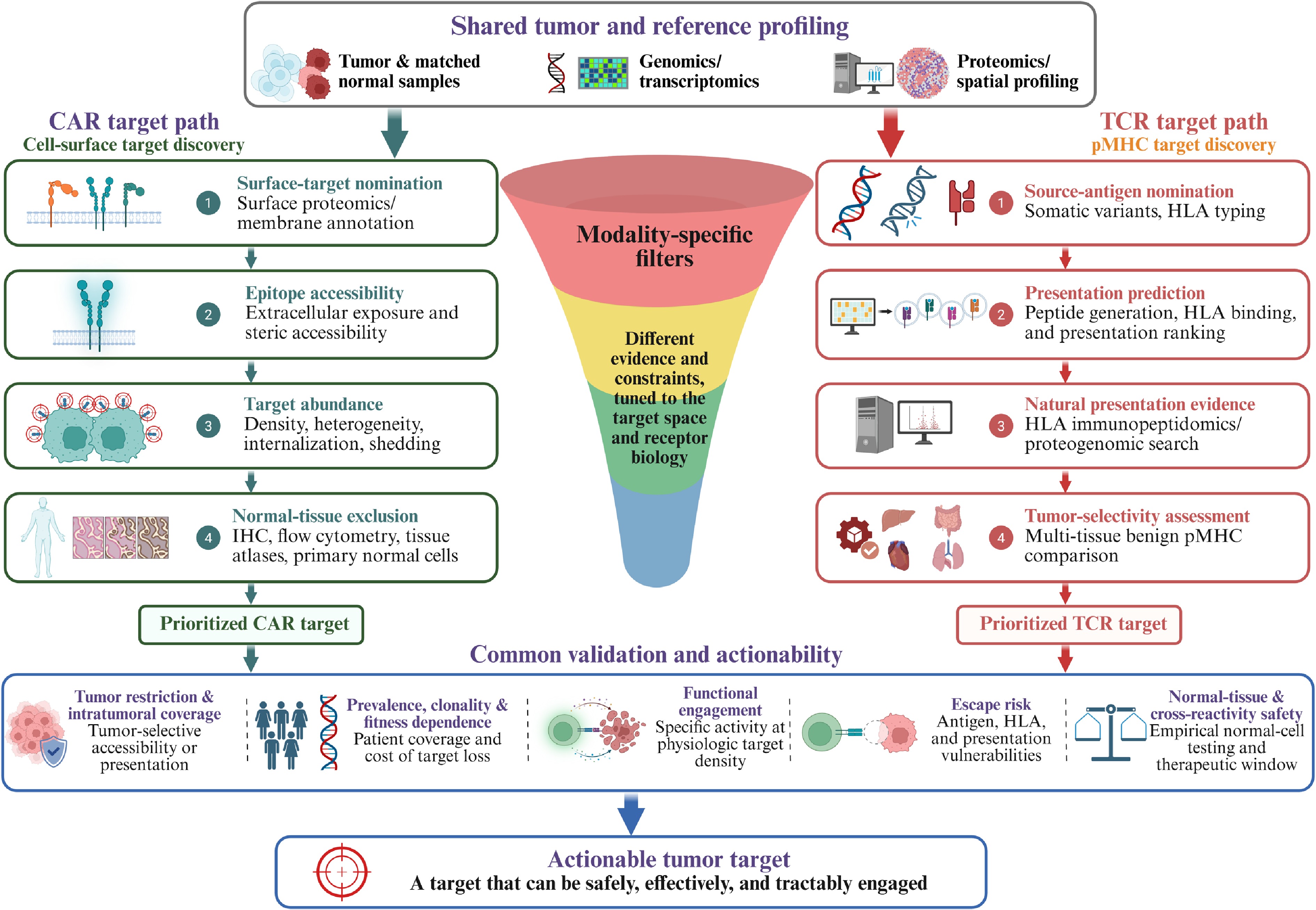
Figure 2.
Modality-specific discovery and validation of actionable tumor targets. Shared molecular profiling feeds distinct CAR and TCR target-discovery pipelines: CAR candidates are filtered for surface accessibility, density, heterogeneity, target behavior, and normal-tissue expression, whereas TCR candidates are evaluated for HLA context, predicted and natural pMHC presentation, and tumor selectivity. Both pathways converge on tumor coverage, prevalence and fitness dependence, functional activity, escape risk, and normal-tissue and cross-reactivity safety.
Converting molecular candidates into validated therapeutic targets requires increasingly stringent evidence of expression or alteration, natural accessibility or presentation, sufficient abundance and tumor restriction, and specific, functional, and safe receptor engagement. Target prevalence across patients further determines whether a receptor can be broadly reused. The steep attrition across these filters makes target discovery and validation a major bottleneck, although difficult targets may also be limited by the availability of a sufficiently specific receptor.
Validating surface targets for CAR recognition
-
For CAR targets, transcript or bulk-protein expression alone does not establish surface accessibility. Plasma-membrane localization should be confirmed by cell-surface proteomics, flow cytometry, immunohistochemistry, or spatial profiling. Quantitative assays should then define antigen density and intratumoral heterogeneity, while epitope accessibility, internalization, shedding, and expression in vulnerable normal tissues further shape efficacy and safety. Functional testing against antigen-high, antigen-low, antigen-negative, and relevant normal cells ultimately defines the therapeutic window.
Predicting HLA presentation for TCR targets
-
Mutation-derived TCR target discovery typically begins with whole-exome or whole-genome sequencing of tumor and matched normal tissue to identify somatic variants, followed by RNA sequencing to confirm expression of the altered transcript[69]. HLA genotype may be measured directly or inferred from sequencing data, but should be confirmed by validated typing before clinical application. Toolkits such as pVACtools then generate mutant peptides, predict MHC binding or presentation, and prioritize candidate neoantigens[84]. These outputs remain predictions until endogenous processing and presentation are experimentally demonstrated.
The next filter evaluates whether a candidate peptide is likely to be naturally presented by the patient's HLA molecules. NetMHCpan-4.1 and MHCflurry 2.0 use binding-affinity and mass-spectrometry-derived eluted-ligand data to predict HLA-I binding or presentation across diverse alleles[85,86]. These predictions remain imperfect because presentation also depends on source-protein turnover, proteasomal processing, peptide transport and trimming, HLA loading, and pMHC stability[52]. Candidate validation should therefore progress from computational prediction to direct evidence of endogenous presentation and, finally, specific receptor recognition and functional activity[87]. Most current pipelines focus on HLA-I, whereas HLA-II-restricted targets require distinct prediction and validation strategies.
HLA restriction and population coverage: which patients can be treated
-
HLA restriction is not only a biological property of a receptor but also an eligibility criterion, and it determines who can and cannot receive a given pMHC-directed therapy. Both approved receptor-defined pMHC-targeting products, tebentafusp and afami-cel, require HLA-A*02, and eligibility is established by validated HLA typing together with a target-expression companion diagnostic. Because HLA allele frequencies differ substantially among ancestral populations, this requirement distributes access unevenly: the HLA-A*02:01 allele frequency is approximately 27% in individuals of European descent but approximately 12% in individuals of African or African American descent, and across 263 trials of HLA-restricted therapeutics the estimated likelihood of eligibility was 53.0% for individuals of European descent vs 33.0% for African American individuals[34]. Restriction by less common alleles narrows access further; the HLA-C*08:02 allele that restricts the clinically validated KRAS G12D-reactive receptors is carried by roughly 8%−11% of the US population[71], so even a highly prevalent driver mutation reaches only a small minority of otherwise eligible patients.
Two practical consequences follow. First, the population that can benefit from a given TCR-T product is the intersection of three independent filters: the tumor harbors or expresses the target, the patient carries the restricting allele, and the epitope is genuinely processed and presented by that allele; so this addressable fraction, rather than antigen prevalence alone, is the appropriate figure to report when a target is proposed. Second, equitable access requires deliberately extending receptor development beyond HLA-A*02: identifying epitopes restricted by alleles that are common in under-served populations, such as HLA-A*11:01, HLA-A*24:02, HLA-A*03:01 and selected HLA-C alleles; developing allele-matched receptor panels against the same antigen; and, where a suitable surface target exists, favoring HLA-independent modalities such as CARs, TRuC, STAR or HIT receptors, or CAR-engineered innate cells. HLA-based population coverage is therefore best treated as a design parameter that is planned prospectively rather than as an incidental property of whichever epitope is discovered first.
Direct detection by immunopeptidomics
-
Mass-spectrometry-based immunopeptidomics provides the most direct discovery-scale evidence of naturally processed and MHC-presented peptides[31]. HLA-peptide complexes are immunoaffinity-purified, and the eluted peptides are identified by mass spectrometry. Unlike assays using exogenously loaded peptides, this approach can establish endogenous presentation. Its major limitation is sensitivity, because relevant epitopes may be present at very low abundance and deep profiling often requires substantial cell or tissue input. Thus, failure to detect a peptide does not establish its absence. Advances in sample preparation, acquisition methods, and targeted mass spectrometry are improving sensitivity and reducing input requirements.
Conventional proteomic searches are limited to peptide sequences represented in the reference database and may therefore miss peptides derived from unannotated ORFs, retained introns, alternative reading frames, and tumor-specific splice products. Detecting these non-canonical peptides requires customized proteogenomic databases generated from tumor DNA and RNA sequencing, supplemented when possible by ribosome profiling to provide evidence of active translation[88,89]. Such approaches have expanded the catalog of MHC-I ligands and identified hundreds of candidate shared or tumor-associated non-canonical HLA peptides[88−90]. Because larger search spaces increase the risk of false peptide assignments, these candidates require stringent false-discovery control, sample-specific databases, comparison with synthetic peptides, and orthogonal validation.
Establishing an acceptable therapeutic window
-
Final validation asks whether an accessible or naturally presented target provides an acceptable therapeutic window. Tumor-adjacent tissue alone is an insufficient normal comparator because it does not represent all potentially vulnerable organs. For pMHC targets, tumor immunopeptidomes should therefore be compared with multi-tissue benign references such as the HLA Ligand Atlas, while cancer atlases such as caAtlas can assess target prevalence across tumor types[30,91]. Because these resources remain incomplete, absence from an atlas does not establish tumor specificity. Sequence- and structure-based comparisons with self-peptides can prioritize potential cross-reactivities, but empirical testing against relevant normal cells remains essential.
Therapeutic usability also requires a receptor that recognizes the target with appropriate specificity and function. Functional assays should assess binding, activation, cytotoxicity, cytokine release, and responses across physiologic target densities. Machine-learning models can prioritize candidates based on predicted presentation, immunogenicity, and TCR-pMHC recognition[92,93], but reliable prediction of receptor specificity and safety remains relatively immature. For naturally derived TCRs, immunogenicity and the availability of reactive clonotypes are additional considerations; engineered TCRs, TCR-mimic binders, and de novo receptors require particularly rigorous specificity and cross-reactivity testing.
Overall, target discovery is a high-attrition funnel: candidates must be expressed or alterable, naturally accessible or presentable, sufficiently abundant and tumor-restricted, and capable of safe functional engagement. Target discovery and validation therefore remain major bottlenecks before attention shifts from what to target to how to engage it.
-
Receptor platforms can be classified by two design dimensions: the target recognized, surface antigen or pMHC, and the signaling system used, synthetic CAR modules or the native TCR-CD3 machinery. Conventional CARs combine surface recognition with synthetic signaling, whereas TCR-T cells combine pMHC recognition with native signaling. Hybrid formats occupy the other two combinations: TCR-mimic CARs direct pMHC recognition through CAR signaling, while TRuC, STAR, and HIT receptors direct antibody-based surface recognition through the TCR-CD3 complex[94−100]. Soluble T-cell engagers form a separate category because they recruit endogenous CD3 signaling without genetically modifying the responding T cell.
Why hybrid receptor formats arise
-
The rationale for hybrid receptor formats arises from fundamental differences between CAR and TCR signaling. A second-generation CAR condenses antigen recognition, costimulation, and activation into a single polypeptide, whereas the native TCR-CD3 complex distributes signaling across multiple subunits and 10 ITAMs[6,47]. This architecture contributes to distinct functional sensitivities: conventional CARs often require higher target densities, whereas TCR signaling can respond to substantially fewer ligands through coordinated triggering, kinetic proofreading, signal amplification, and co-receptor support[26,47,55]. Hybrid platforms therefore recombine recognition and signaling features across the CAR-TCR spectrum, but they do not automatically overcome the density, specificity, HLA, or presentation constraints of the systems from which they are derived (Fig. 3).
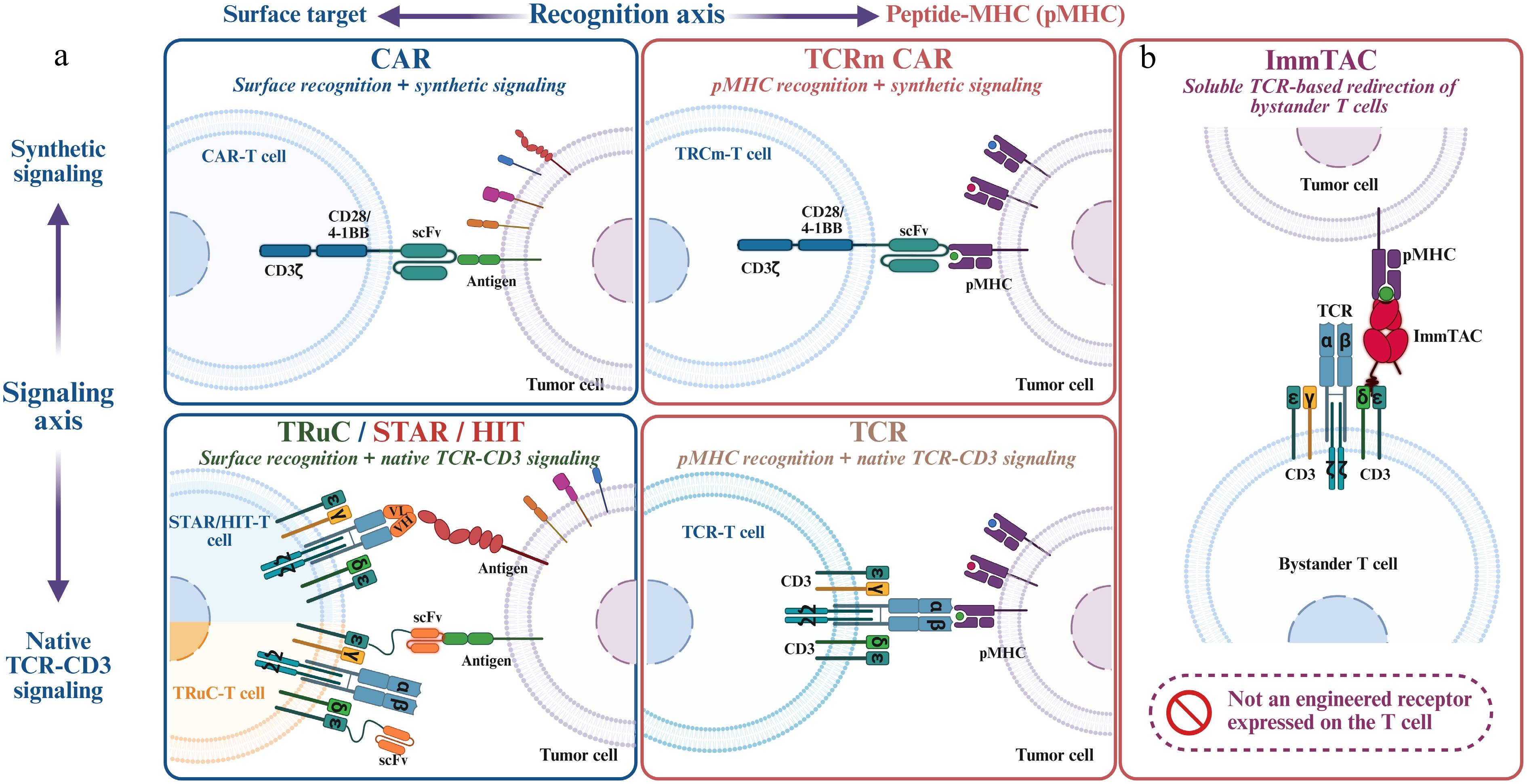
Figure 3.
Recognition-signaling design space of engineered T-cell platforms. (a) Conventional CARs combine surface-antigen recognition with synthetic signaling, whereas TCRs recognize pMHC through the native TCR-CD3 complex. TCR-mimic CARs and TRuC/STAR/HIT receptors occupy the two hybrid combinations. (b) While ImmTACs represent a soluble pMHC-directed format that recruits endogenous T cells through CD3.
pMHC recognition with synthetic signaling: TCR-mimic CARs
-
One hybrid category combines pMHC recognition with synthetic CAR signaling. Antibody-derived or designed binders against defined peptide-MHC complexes can provide TCR-like specificity and, when incorporated into a CAR, extend CAR recognition to intracellularly derived antigens. The same binders can also be configured as soluble pMHC × CD3 bispecific engagers. In preclinical studies, bispecifics targeting TP53 R175H- and mutant RAS-derived pMHCs redirected T cells against tumor cells displaying low endogenous densities of the corresponding complexes[95−97]. However, the sensitivity achieved by soluble engagers should not be assumed to translate directly to the CAR format. Across pMHC-directed platforms, major challenges include low ligand density, HLA restriction, loss of antigen presentation, and discrimination from structurally related self-peptides[94].
Surface recognition with native signaling: TRuC, STAR, and HIT
-
The reciprocal hybrid category combines antibody-based surface recognition with native TCR-CD3 signaling. In TRuCs, an antibody-derived binder is fused to a TCR or CD3 subunit and incorporated into the endogenous receptor complex; CD19-directed TRuC-T cells showed cytotoxicity comparable to second-generation CAR-T cells with lower secretion of several cytokines in preclinical studies[98]. STAR and HIT receptors instead replace the TCR variable domains with antibody-derived heavy- and light-chain variable domains, allowing assembly with endogenous CD3[99,100]. STARs generally coexist with the endogenous TCR, whereas the original HIT design used TRAC-locus insertion to achieve endogenous regulation and disrupt native TCR expression. HIT-T cells showed greater sensitivity to low-density antigens than matched CD28-based CAR-T cells in selected preclinical comparisons[100]. Mesothelin-directed gavo-cel subsequently demonstrated clinical activity, although pulmonary toxicity and CRS suggested a narrow therapeutic window for this particular target-construct combination[101].
Soluble T-cell engagers
-
Immune-mobilizing monoclonal T-cell receptors against cancer (ImmTACs) are soluble pMHC-directed T-cell engagers that link an affinity-enhanced TCR to an anti-CD3 scFv. By bridging a defined peptide-HLA complex on the target cell to CD3 on endogenous T cells, they redirect polyclonal T cells independently of native antigen specificity and provide an off-the-shelf option for HLA-matched patients[102]. Although high-affinity binding can improve engagement of low-density pHLA targets, it also heightens the risk of cross-reactivity with structurally similar self-pMHCs and therefore requires rigorous specificity screening[61,62].
Tebentafusp, which targets a gp100-derived peptide presented by HLA-A*02:01, became the first FDA-approved soluble TCR-based therapeutic in 2022. In the pivotal phase 3 trial, it improved overall survival vs investigator's-choice therapy, with a hazard ratio of 0.51 in the primary analysis and a sustained 3-year benefit (hazard ratio, 0.68; estimated survival, 27% vs 18%)[103,104]. Cytokine-mediated reactions and skin toxicities were common, consistent with T-cell redirection and recognition of the melanocytic-lineage antigen gp100. Tebentafusp illustrates both the promise and constraints of pMHC targeting: access to intracellularly derived antigens together with HLA restriction, low target density, and potential on-target effects in normal tissues.
Selected approved and late-stage therapies across these receptor and engager formats are summarized in Table 3. Receptor architecture determines how target recognition is converted into T-cell activation, whereas the target-receptor pairing strongly shapes sensitivity and the therapeutic window. Regardless of platform, efficacy requires sustained target accessibility or presentation, with pMHC-directed approaches being additionally vulnerable to loss of HLA expression or antigen processing. These failure modes are considered next.
Table 3. Selected approved and investigational T-cell therapies and mechanistically relevant T-cell-redirecting comparators.
Agent (trade name) Target Targetome face Receptor/therapeutic format Indication or development population Regulatory/development status Approved products Blinatumomab (Blincyto) CD19 Surface CD19 x CD3 bispecific engager B-cell acute lymphoblastic leukemia FDA approved, 2014[161] Tisagenlecleucel (Kymriah) CD19 Surface Second-generation CAR (4-1BB) Pediatric/young-adult B-ALL; DLBCL; follicular lymphoma FDA approved, 2017[176] Axicabtagene ciloleucel (Yescarta) CD19 Surface Second-generation CAR (CD28) Large B-cell lymphoma; follicular lymphoma FDA approved, 2017[177] Brexucabtagene autoleucel (Tecartus) CD19 Surface Second-generation CAR (CD28) Adult mantle cell lymphoma and B-cell precursor ALL FDA approved, 2020[178] Lisocabtagene maraleucel (Breyanzi) CD19 Surface Second-generation CAR (4-1BB); defined CD4:CD8 composition LBCL, CLL/SLL, FL, MCL, and MZL FDA approved, 2021; MZL added in 2025[179,180] Idecabtagene vicleucel (Abecma) BCMA Surface Second-generation CAR (4-1BB) Relapsed/refractory multiple myeloma FDA approved, 2021[88] Tebentafusp (Kimmtrak) gp100 peptide/
HLA-A*02:01Intracellular via pMHC ImmTAC: affinity-enhanced soluble TCR x anti-CD3 HLA-A*02:01-positive unresectable or metastatic uveal melanoma FDA approved, 2022—
first approved soluble TCR-based therapeutic[103,104]Ciltacabtagene autoleucel (Carvykti) BCMA Surface Second-generation CAR (4-1BB); bivalent VHH binder Relapsed/refractory multiple myeloma FDA approved, 2022[12] Teclistamab (Tecvayli) BCMA Surface BCMA x CD3 bispecific engager Relapsed/refractory multiple myeloma FDA accelerated approval, 2022[162] Talquetamab (Talvey) GPRC5D Surface GPRC5D x CD3 bispecific engager Relapsed/refractory multiple myeloma FDA accelerated approval, 2023[163] Lifileucel (Amtagvi) Polyclonal; specificities not predefined Mainly
pMHC-mediatedAutologous tumor-infiltrating lymphocytes; not receptor-engineered Previously treated unresectable or metastatic melanoma FDA accelerated approval, 2024—first approved TIL therapy[7,8] Afamitresgene autoleucel (Tecelra) MAGE-A4/
HLA-A*02
familyIntracellular via pMHC Affinity-enhanced TCR-T Patients with previously treated unresectable or metastatic synovial sarcoma meeting HLA and MAGE-A4 companion-diagnostic criteria FDA accelerated approval, 2024; pediatric label expansion in 2026[20,21] Obecabtagene autoleucel (Aucatzyl) CD19 Surface Second-generation CAR (4-1BB); fast off-rate binder Adult relapsed/refractory B-cell precursor ALL FDA approved, 2024[13] Satricabtagene autoleucel (satri-cel; CT041) CLDN18.2 Surface Second-generation CAR (CD28) CLDN18.2-positive, HER2-negative advanced gastric/GEJ adenocarcinoma after at least two prior lines China NMPA approved, June 2026—first approved CAR-T for a solid tumor[22] Selected investigational programs Anzutresgene autoleucel (anzu-cel; IMA203) PRAME/HLA-A*02:01 Intracellular via pMHC Autologous TCR-T without CD8 co-transduction Previously treated unresectable or metastatic cutaneous melanoma Recruiting randomized phase 3 SUPRAME[23] IMA203CD8 (GEN2) PRAME/HLA-A*02:01 Intracellular via pMHC Autologous TCR-T co-transduced with CD8αβ PRAME-positive recurrent/refractory solid tumors Recruiting phase 1/2; distinct from anzu-cel[181] Lifileucel is included as a regulatory benchmark for polyclonal adoptive T-cell therapy but is not a private-neoantigen-selected or receptor-engineered product. All approved receptor-defined pMHC-targeting products listed here—tebentafusp and afami-cel—are HLA-A*02-restricted; lifileucel is a polyclonal TIL product whose antigen specificities and restricting HLA alleles are not predefined. The success of afami-cel and satri-cel reflects target selection together with biomarker selection, receptor design, manufacturing, and clinical context. -
Resistance and toxicity are conventionally considered separately, but many reflect failures in the relationship among a receptor, its target, and the surrounding tissue. Efficacy can be lost when the target is downregulated, eliminated, no longer presented, or inaccessible, while safety can be compromised by target expression in vulnerable normal tissues or receptor cross-reactivity with unintended antigens. Even when recognition remains intact, inadequate trafficking, persistence, or function within an immunosuppressive microenvironment may prevent tumor control. Efficacy therefore depends in part on how reliably the target marks the tumor, and safety on how selectively recognition spares the host (Fig. 4). This target-centric framework does not fully capture systemic toxicities such as CRS and ICANS, which also depend on product characteristics, disease burden, and host responses.
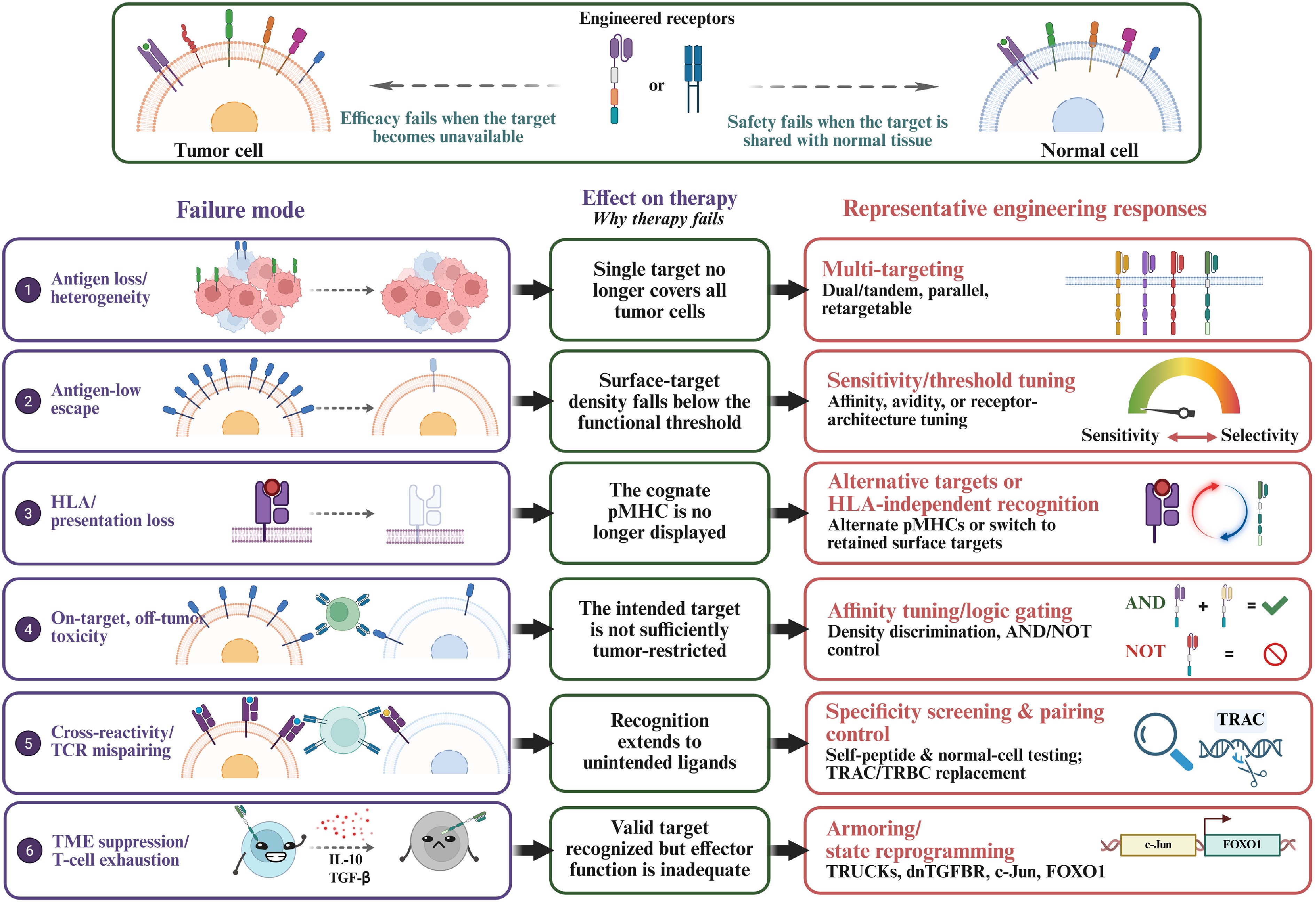
Figure 4.
Target- and effector-centered failure modes and representative engineering responses. Antigen loss or low density, impaired HLA or antigen presentation, normal-tissue target expression, receptor cross-reactivity or TCR mispairing, and tumor-microenvironment-driven dysfunction can compromise efficacy or safety. Multi-antigen targeting, threshold tuning, alternative recognition strategies, logic gating, specificity and pairing control, and armoring or cell-state reprogramming may mitigate these vulnerabilities, although no strategy is universally effective or free of trade-offs.
Antigen loss and heterogeneity
-
Antigen escape is a prominent resistance mechanism in which tumor cells lose or reduce the target recognized by the receptor[105]. In B-ALL, CD19 deletions, exon 2 mutations, and alternative splicing can generate variants that evade CAR recognition; exon 2 skipping, for example, produces an N-terminally truncated isoform that escapes killing while retaining some CD19-associated function[106]. BCMA loss has also occurred through biallelic inactivation after BCMA-directed CAR-T therapy, although its population-level frequency remains uncertain[60]. Another form of escape is lineage switching, as reported in KMT2A-rearranged B-ALL that transitioned clonally to CD19-negative myeloid leukemia after CD19-directed therapy[107].
Antigen escape may be dynamic or incomplete. In preclinical leukemia models, CAR-mediated trogocytosis transferred antigen from tumor cells to CAR-T cells, transiently lowering tumor antigen density while promoting CAR-T-cell fratricide and dysfunction[108]. Because CAR activity often depends on a minimum antigen density, partial downregulation can render antigen-positive tumors functionally resistant. Clinically, antigen-low relapse has been observed after CD19- and CD22-directed therapy; in a CD22 CAR-T trial, relapse was associated with reduced surface density rather than complete antigen loss[26,60,105].
This form of escape is particularly relevant in heterogeneous solid tumors. In recurrent glioblastoma, EGFRvIII-directed CAR-T cells trafficked to the tumor, and five of seven evaluable post-treatment specimens showed reduced EGFRvIII expression together with increased inhibitory-molecule expression and regulatory T-cell infiltration[109]. These observations are consistent with concurrent antigen selection and immunosuppressive remodeling, although causality cannot be assigned solely to CAR-T therapy. Single-target treatment may therefore enrich pre-existing or emerging antigen-low populations in heterogeneous tumors.
Loss of antigen presentation
-
A related escape mechanism disrupts pMHC presentation without requiring loss of the source protein. Because TCRs recognize peptide-MHC complexes, loss of β2-microglobulin, allele-specific HLA loss, HLA class I downregulation, or defects in IFN-γ-JAK-STAT signaling can reduce or abolish surface display of the cognate pMHC[110,111]. In an early-stage NSCLC cohort, allele-specific HLA loss was detected in approximately 40% of tumors and was more frequent in metastatic samples, consistent with selection during tumor evolution[110]. This vulnerability is particularly relevant to engineered TCRs, which are generally restricted to a single HLA allele or a narrow allele family. In one patient with breast cancer treated with a p53 R175H-specific TCR, an objective response lasted approximately six months; a progressing lesion retained mutant p53 but had lost HLA-A*02:01[72]. Thus, tumors can preserve a biologically important source protein while eliminating the restricting HLA or antigen-presentation pathway required for TCR recognition.
On-target off-tumor toxicity and cross-reactivity
-
Safety failures occur when target recognition is not sufficiently restricted to malignant tissue. On-target, off-tumor toxicity arises when the intended antigen is also expressed in vulnerable normal tissues, with severity influenced by antigen density, receptor potency, dose, tissue trafficking, and organ sensitivity. A fatal HER2 CAR-T case was considered consistent with recognition of low-level pulmonary ERBB2, although the mechanism was not directly proven[64]. CEA-directed TCR-T cells caused severe but transient colitis by recognizing normal colonic epithelium[112], whereas neurologic toxicity after BCMA-directed CAR-T therapy raised the possibility of on-target recognition in the central nervous system[113]. These cases illustrate that the therapeutic window depends on both target distribution and the intensity of receptor engagement. CD19 is comparatively tractable because normal B-cell depletion, although associated with hypogammaglobulinemia and infection risk, is often clinically manageable.
Cross-reactivity, in which a receptor recognizes an unintended peptide or antigen, is a particular concern for affinity-enhanced TCRs. In a MAGE-A3 trial, an engineered TCR cross-recognized a titin-derived cardiac peptide. Despite differing from the intended peptide at four of nine positions, the titin peptide adopted a similar HLA-A*01-bound conformation and was recognized by the same TCR. Routine cell-line screening failed to detect the risk; after two fatal cardiac events, peptide screening identified titin, and iPSC-derived cardiomyocytes confirmed functional recognition[61,62].
In a separate MAGE-A3 trial, a high-avidity TCR that cross-recognized MAGE-A9 and MAGE-A12 peptides was associated with severe neurologic toxicity in three of nine patients, including two deaths[114]. Low-level MAGE-A12 expression in the brain was considered a possible mechanism, although causality was less firmly established than for titin in the cardiac cases. The two trials therefore highlight distinct hazards: structural mimicry by an unrelated self-peptide and cross-recognition of a homologous peptide from a related protein expressed in normal tissue.
In TCR gene transfer, introduced α and β chains may mispair with endogenous chains, generating receptors with untested specificities. This mechanism caused lethal graft-vs-host-like pathology in mouse models, although analogous clinical events have not been established[115]. These findings support strategies that enforce correct chain pairing or disrupt and replace the endogenous TCR; the intended receptor must additionally undergo rigorous self-peptide and normal-cell cross-reactivity testing.
Failure within the tumor microenvironment
-
Finally, a target may remain present, tumor-restricted, and correctly recognized, yet therapy can still fail because the effector cells cannot reach the tumor or function effectively within it. Abnormal vasculature and stroma can restrict trafficking, whereas suppressive myeloid and regulatory cells, TGF-β, inhibitory checkpoints, hypoxia, acidity, and nutrient limitation impair T-cell activity[15]. Antigen-independent tonic signaling in some CAR designs can further promote premature dysfunction or exhaustion[116]. These mechanisms define the boundary of a target-centric framework: the target itself may remain therapeutically valid, but inadequate trafficking, persistence, or effector competence can still prevent durable tumor control.
Across the failure modes considered in this section, the target may be lost, downregulated, no longer presented, shared with vulnerable normal tissues, or mimicked by an unintended antigen; alternatively, the effector cells may fail to reach or function within the tumor. These mechanisms often coexist, and each requires a different countermeasure, none of which eliminates resistance or toxicity without introducing additional trade-offs.
-
Many target-centric failure modes motivate engineering strategies to improve precision, breadth, and control, although the relationship between a failure mechanism and its countermeasure is rarely one-to-one, and each intervention introduces new trade-offs (Fig. 4). One direct lever is receptor-target engagement strength. When an antigen is abundant on tumor cells but expressed at lower levels in vital tissues, reducing CAR affinity can shift the activation threshold toward antigen-high tumors while limiting recognition of antigen-low normal cells, as demonstrated preclinically for HER2 and EGFR[117,118]. This approach is particularly relevant to targets such as HER2, for which low-level normal-tissue expression may carry substantial risk[53]. Antigen density should therefore be viewed as a quantitative determinant of activation rather than a binary marker of target positivity[26].
Tuning recognition sensitivity through co-receptor engineering
-
Recognition sensitivity can also be modulated through co-receptor engineering. Co-expression of CD8αβ can enhance signaling through MHC class I-restricted TCRs and enable engineered CD4+ T cells to contribute more effectively to antitumor responses. IMA203CD8 is a second-generation, PRAME-directed TCR-T product co-expressing a PRAME-specific TCR and CD8αβ and is being evaluated in the phase 1/2 ACTengine study (NCT03686124)[119].
Logic gating for selectivity
-
Whereas affinity tuning exploits differences in antigen density, combinatorial recognition integrates multiple antigen inputs to improve selectivity. AND-like designs pair an attenuated activation receptor for one antigen with a costimulatory receptor for another, favoring robust responses against dual-positive cells[120]. Synthetic Notch receptors use a priming antigen to induce a CAR against a second target, creating spatially restricted priming rather than strict cell-by-cell AND logic because primed cells may also kill neighboring targets expressing only the induced CAR antigen[121]. Inhibitory CARs containing PD-1- or CTLA-4-derived domains provide NOT-like control by suppressing activation in the presence of a protective normal-tissue antigen[122].
A related preclinical NOT-gate strategy exploits allele-specific HLA loss: an activating receptor recognizes an antigen shared by tumor and normal cells, whereas an inhibitory receptor recognizes an HLA allele retained by normal tissues but lost by the cancer, converting HLA loss into a selectivity signal[123]. Programmable adaptor-based systems such as SUPRA CARs use interchangeable soluble adaptors to redirect specificity and implement selected AND-, OR-, or NOT-like functions[124]. Although this enables retargeting of a single cellular product without further genetic modification, activity remains dependent on adaptor affinity, dose, biodistribution, and persistence.
Multi-antigen targeting to limit escape
-
Whereas logic gating primarily improves selectivity, multi-antigen targeting broadens tumor coverage and may increase the barrier to antigen-loss escape[105]. In preclinical glioblastoma models, a HER2/IL13Rα2 tandem CAR encoded OR-like recognition within a single receptor, responding to either antigen, showing enhanced activation against dual-positive cells, and reducing antigen escape relative to monospecific controls[125]. Such designs can reduce, but not eliminate, resistance because tumors may lose both targets or exploit non-antigen mechanisms. Conceptually, they address the antigen heterogeneity and treatment-driven selection observed clinically with EGFRvIII-directed CAR-T therapy[109].
Controlling chain mispairing and receptor expression
-
TCR therapies face a structural liability because introduced α- and β-chains may mispair with endogenous chains. Murine constant regions and engineered interchain disulfide bonds can improve correct pairing, but do not eliminate mispairing and may introduce immunogenicity concerns[126,127]. TRAC disruption alone removes the endogenous α-chain while leaving the endogenous β-chain available for mispairing. By contrast, simultaneous TRAC and TRBC editing with orthotopic replacement of both introduced chains improved pairing fidelity and maintained near-physiological receptor regulation in preclinical studies[128].
Targeted CAR integration into the TRAC locus can disrupt the endogenous TCR while placing CAR expression under endogenous regulation. In the original CD19 model, this strategy produced more uniform expression, reduced tonic signaling, and delayed differentiation and exhaustion relative to conventional retroviral delivery[129]. These effects are context-dependent and may not extend to all CAR constructs or integration designs.
Armoring and cell-intrinsic fitness
-
Even when target recognition is preserved, the tumor microenvironment can limit therapeutic efficacy. Cytokine-armored CARs, including inducible IL-12 and IL-18 designs, can recruit endogenous immune cells and remodel suppressive niches in preclinical models[45,130−132]. Suppressive pathways can also be intercepted; a phase 1 trial of PSMA-directed CAR-T cells expressing a dominant-negative TGF-β receptor showed feasibility, CAR-T expansion, and preliminary biological activity[133]. However, one marked biochemical response was accompanied by grade 4 CRS, sepsis, and death, highlighting the need to balance enhanced antitumor function with inflammatory risk.
Cell-intrinsic fitness can also be engineered. Antigen-independent tonic signaling can promote CAR-T-cell dysfunction in an architecture-dependent manner[116]. By contrast, c-Jun overexpression enhanced proliferative capacity and antitumor function, while FOXO1 overexpression promoted memory-associated transcriptional and metabolic programs, persistence, and tumor control in preclinical models[134,135]. Whether such transcriptional reprogramming improves clinical outcomes remains unknown.
Genome and base editing in the clinic
-
Precise genome editing has also reached clinical evaluation. In a first-in-human study, CRISPR-Cas9 was used to disrupt TRAC, TRBC, and PDCD1 before lentiviral introduction of an NY-ESO-1-specific TCR. The edited cells expanded and persisted for several months in three patients, supporting the feasibility of multiplex-edited TCR-T-cell therapy, although the study was not designed to assess clinical efficacy[136].
Base editing has been used to generate donor-derived CD7 CAR-T cells by disrupting CD7 to prevent fratricide, CD52 to confer resistance to alemtuzumab-based lymphodepletion, and TRBC to reduce graft-vs-host risk. In an expanded phase 1 study of nine children and two adults, all 11 achieved morphologic remission by day 28, nine proceeded to allogeneic transplantation, and seven remained in remission 3−36 months after transplantation[137,138]. CRS, multilineage cytopenias, infections, and two CD7-negative relapses nevertheless remained important limitations.
In vivo generation of engineered T cells
-
All the strategies above presuppose ex vivo manufacturing. An alternative is to generate the engineered cell inside the patient, which removes leukapheresis, ex vivo culture, and product release testing, and may reduce or remove the need for lymphodepletion. Two delivery platforms dominate. Targeted lipid nanoparticles (tLNPs) carry receptor-encoding mRNA and are directed to T cells by antibodies against a lineage marker such as CD5, CD7, or CD8; CD5-targeted LNPs generated transient fibroblast activation protein-directed CAR-T cells in vivo[139], and CD8-targeted LNPs delivering anti-CD19 CAR mRNA reprogrammed CD8+ T cells from healthy donors and patients, controlled tumor growth in humanized mice, and produced deep but reversible B-cell depletion in non-human primates[140]. Expression from mRNA is transient, which limits durable antitumor activity but provides an intrinsic safety limit and permits repeat dosing. The second platform uses engineered lentiviral vectors that combine a detargeted fusogen with a cell-specific binder; because the transgene integrates, a single administration can give stable receptor expression, and CD7-targeted vectors delivering a CD20-directed CAR generated functional CAR-T and CAR-NK cells without lymphodepletion in preclinical models. Both approaches have entered first-in-human testing in B-cell malignancies and autoimmune disease (e.g., NCT06539338 and NCT06528301).
From a target-centered perspective, in vivo generation introduces a second and distinct targeting problem: in addition to the tumor target engaged by the receptor, a delivery target on the effector cell must be selected. Its requirements mirror those developed in Section Discovering and validating the targetome: from molecular profiling to actionable targets: restricted expression on the intended effector subset, sufficient surface density, efficient receptor-mediated internalization for LNPs or productive fusion for vectors, and tolerable consequences of off-target transduction, particularly of B cells or of malignant cells that express the delivery antigen. Further considerations include vector or nanoparticle immunogenicity, anti-drug antibodies against the targeting moiety that may limit redosing, insertional mutagenesis for integrating vectors, in vivo expansion that cannot be characterized by a manufacturing release assay, and regulatory expectations for biodistribution and shedding. If these issues are resolved, in vivo generation would substantially reduce cost and time to treatment and could make engineered cell therapy accessible outside specialized centers.
Viewed together, these strategies address distinct vulnerabilities: affinity tuning and logic gating may widen the therapeutic window, multi-antigen targeting may reduce escape, specificity screening may mitigate cross-reactivity, and pairing engineering or endogenous-TCR disruption can limit mispairing. Armoring, transcriptional reprogramming, and genome editing may further improve function and durability, but at the cost of greater biological, manufacturing, and regulatory complexity. The challenge is therefore to determine which added functions provide sufficient clinical value to justify this additional burden.
-
The preceding sections treat the targetome as the set of antigens displayed by malignant cells. Accumulating evidence indicates that this definition is too narrow: durable responses in solid tumors depend not only on recognition of tumor cells but also on the composition and state of the surrounding stroma, and the non-malignant compartment presents its own set of addressable targets. Tumor-associated macrophages (TAMs) are the most abundant immune population in many solid tumors and support angiogenesis, matrix remodeling, T-cell exclusion, and resistance to both checkpoint blockade and adoptive therapy. Because suppressive TAM states are marked by surface proteins that are largely absent from tumor cells. They define a microenvironmental targetome that can be engaged with the same receptor technologies described above. Three complementary strategies have emerged.
Depleting or reprogramming suppressive myeloid cells
-
The first strategy is direct depletion or conversion of suppressive myeloid cells. FRβ-directed CAR-T cells selectively eliminated FRβ+ TAMs in syngeneic models, enriched pro-inflammatory monocytes, increased infiltration of endogenous tumor-specific CD8+ T cells, and prolonged survival; notably, preconditioning with FRβ CAR-T cells before mesothelin-directed CAR-T cells improved the activity of the tumor-directed product, whereas simultaneous administration did not[141]. More recently, IL-12-armored CAR-T cells directed against FOLR2 or TREM2 produced sustained remodeling of the microenvironment in metastatic ovarian and lung cancer models, with expansion of CXCL9+ immunostimulatory macrophages and of endogenous tumor-reactive CD8+ T cells, activity at low cell dose and without lymphodepletion, and tumor control that depended in part on FAS expression by cancer cells[142]. Because these receptors are directed at a stromal state rather than at a tumor antigen, they are in principle tumor-antigen agnostic and are not defeated by tumor-cell antigen loss or by loss of HLA expression.
Engineering the myeloid compartment and remodeling the niche
-
The second strategy engineers the myeloid compartment itself. CAR macrophages combine phagocytosis, cytokine secretion, and antigen presentation with defined specificity; anti-HER2 CAR macrophages, first described preclinically[143], were subsequently manufacturable and tolerable in a first-in-human phase 1 trial in 14 patients with HER2-overexpressing solid tumors, with evidence of tumor trafficking, microenvironmental activation, and induction of T-cell responses[144]. The third strategy remodels the niche from within the tumor-directed product: inducible IL-12 and IL-18 payloads recruit and reprogram endogenous myeloid and lymphoid cells[45,130−132], and dominant-negative TGF-β receptors render T cells insensitive to a dominant suppressive signal[133]. Stromal targets other than macrophages, including fibroblast activation protein on cancer-associated fibroblasts and antigens on the tumor vasculature, follow the same logic.
-
Throughout this review, the targetome has been treated as a map of therapeutically actionable antigens. Yet this landscape is not static: under therapeutic pressure, tumors can alter surface-antigen expression, suppress antigen presentation, and enrich pre-existing or emerging escape variants. A paradigm that characterizes the target landscape once it develops a receptor, and assumes target stability, is therefore poorly suited to an evolving tumor. Longitudinal molecular profiling and computational modeling could support a more adaptive framework in which the targetome is repeatedly reassessed, emerging vulnerabilities are prioritized, and therapeutic engagement is modified to anticipate or respond to escape. Four developments support this vision (Fig. 5).
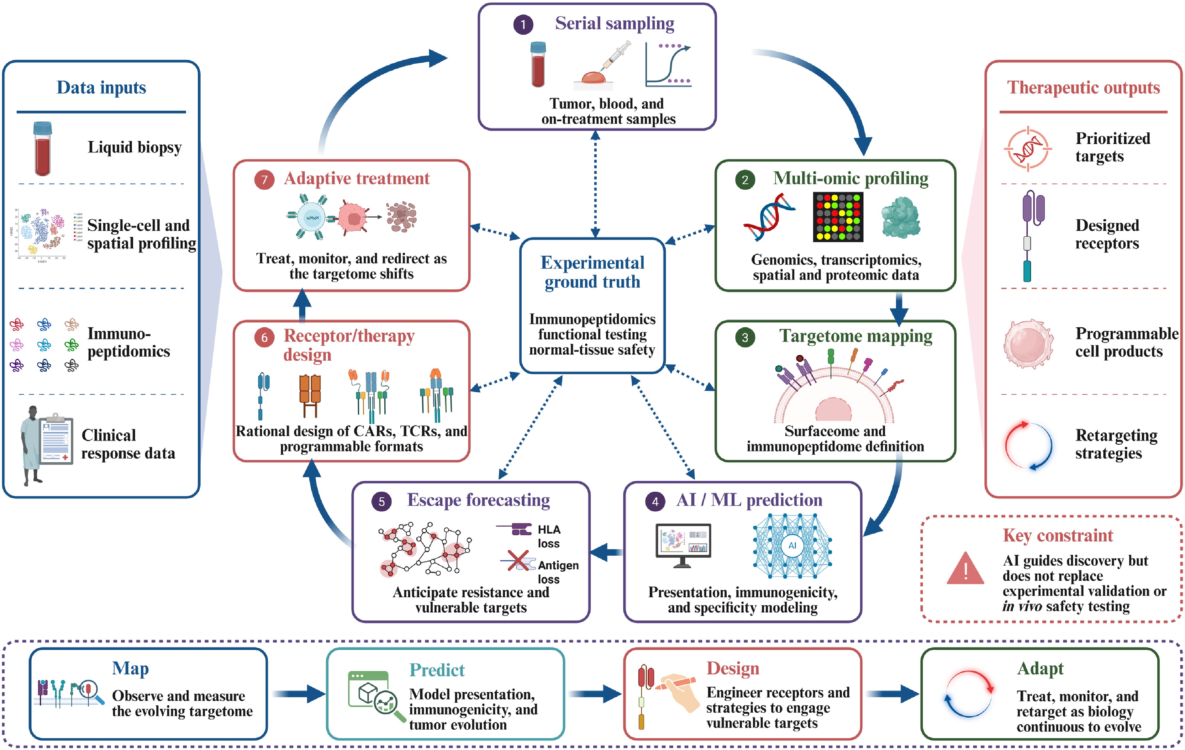
Figure 5.
AI-assisted adaptive targetome workflow. Serial sampling and multi-omic profiling enable repeated mapping of the evolving tumor targetome. Computational prediction and escape forecasting guide target and receptor prioritization, while immunopeptidomic, functional, cross-reactivity, and normal-tissue validation provide experimental ground truth. These outputs inform receptor design and adaptive treatment, but computational models do not replace experimental safety testing.
Computational target prioritization
-
The first development is improved computational target prioritization. Pan-allele models estimate the likelihood that candidate peptides will bind to or be presented by a patient's HLA molecules[85], while deep networks trained partly on mass-spectrometry-defined ligands improve prediction of MHC-I presentation and prioritize candidate immunogenic neoepitopes beyond binding-affinity prediction alone[93]. BigMHC, for example, separately models presentation and immunogenicity to improve candidate ranking[93]. These models can narrow the experimental search space but remain predictive tools and cannot establish endogenous presentation or T-cell recognition without experimental validation.
Structure-based receptor design
-
The second development applies computational methods to receptor recognition and design. AlphaFold2 and ESMFold have advanced protein-structure prediction; AlphaFold3 extends modeling to diverse biomolecular complexes, and RFdiffusion together with learned sequence-design models enables de novo generation of protein backbones and binders[145−149]. Specialized approaches are also beginning to model TCR-pMHC docking and prioritize candidate interactions[92,150]. Nevertheless, these tools do not yet reliably predict receptor specificity, cross-reactivity, signaling activity, or therapeutic safety and therefore cannot replace experimental screening.
De novo design has generated pMHC-binding modules outside conventional antibody and TCR scaffolds. In one study, RFdiffusion and learned sequence design produced an α-helical TCR-mimic binder against NY-ESO-1-HLA-A02:01 with nanomolar affinity and a peptide-focused, TCR-like docking geometry. When configured as a soluble T-cell engager, the binder redirected T cells against target-positive cells. A structure-informed screen of 14,363 immunopeptidomics-derived HLA-A02:01 peptides prioritized two cross-reactive peptides that were subsequently validated experimentally[151]. In a separate study, a de novo-designed NY-ESO-1-HLA-A02:01 minibinder was incorporated into a CAR and mediated killing of antigen-positive melanoma cells; the platform was also extended to a neoantigen-HLA-A01:01 complex for which no experimental structure was initially available[152]. These studies show that pMHC recognition modules can be designed directly from target structure and deployed in soluble or cellular formats, although their generalizability, cross-reactivity profiles, and normal-tissue safety remain to be established.
Modeling escape and evolutionary constraint
-
The third development is modeling evolutionary constraints and escape risk. Neoantigen fitness models combine predicted mutant-vs-wild-type MHC binding with sequence-based proxies for T-cell recognition and have stratified outcomes in retrospective checkpoint-blockade cohorts. Longitudinal pancreatic cancer studies further showed preferential depletion of high-quality neoantigens during tumor evolution[36,37]. Combined with clonality, driver dependence, and allele-specific HLA-loss data, these approaches may help prioritize targets whose loss would reduce tumor fitness and identify plausible presentation-mediated escape routes.
External control and retargetability
-
The fourth development is increased external control and retargetability of engineered cells. Small-molecule ON-switches enable pharmacologic control of receptor activity[153], whereas multi-input synthetic circuits allow engineered cells to integrate antigen and contextual signals and execute predefined programs[154,155]. Universal adaptor-directed CAR systems use interchangeable soluble adaptors to redirect target specificity and implement selected multi-antigen logic without further genetic modification of the cellular product[124]. Although these approaches enable pharmacologic control, programmed responses, and adaptor-mediated retargeting, most remain preclinical and still depend on external detection of tumor evolution and deliberate intervention; they do not autonomously identify emerging escape variants or select new targets[153,154].
These advances suggest a potential adaptive workflow in which serial liquid biopsy and, when feasible, tissue-based single-cell, spatial, surface-proteomic, and immunopeptidomic profiling repeatedly reassess the evolving target landscape. Presentation and evolutionary models could prioritize emerging vulnerabilities, computational tools could guide receptor selection or design, and programmable or adaptor-based cells could be redirected when clinically appropriate. Cellular foundation models such as Geneformer may support gene-network analysis and target prioritization but do not yet integrate antigen presentation, receptor specificity, normal-tissue safety, and tumor evolution into a unified framework[156].
The quality and breadth of the available data constrain this framework. Immunopeptidomic measurement, functional validation, cross-reactivity testing, and normal-tissue safety assessment remain indispensable, because predicted specificity does not guarantee in vivo safety. Model performance depends on training data, evaluation design, and experimental context. In contrast, integrated datasets linking TCR specificity, antigen presentation, normal-tissue profiles, and longitudinal tumor evolution remain scarce. Additional layers of profiling, receptor design, and cellular control also increase analytical, manufacturing, validation, and regulatory complexity. The adaptive targetome should therefore be viewed as an aspirational framework rather than an established therapeutic workflow.
These considerations point toward an adaptive strategy in which longitudinal measurement and computation guide, rather than replace, experimental validation and enable therapeutic engagement to evolve with the tumor.
-
Adoptive cell therapy comprises a diverse group of cellular immunotherapies that harness or engineer immune cells to recognize and eliminate cancer. CARs directly recognize extracellular targets independently of HLA, whereas TCRs access a broader source proteome through peptide-MHC presentation. These complementary routes differ in target availability, ligand density, normal-tissue expression, HLA coverage, and susceptibility to escape.
A target-centered framework connects antigen classification and discovery with receptor selection, resistance, safety, engineering, and manufacturing. Efficacy may be compromised by target loss, reduced display, impaired presentation, or inadequate tissue access, whereas safety may be limited by recognition of vulnerable normal tissues or unintended antigens. Engineering strategies can mitigate selected vulnerabilities, but each introduces additional biological, manufacturing, or regulatory trade-offs.
These challenges are particularly acute in solid tumors, which seldom provide targets that are simultaneously tumor-restricted, uniformly displayed, accessible, and resistant to escape. Progress will therefore require integrating target selection with receptor design, biomarker-defined patient selection, manufacturing, and clinical context. Receptor format and sensitivity must be matched to target location, abundance, heterogeneity, HLA context, and normal-tissue distribution, while longitudinal monitoring and controllable therapeutic platforms may help anticipate or respond to tumor evolution. These choices carry immediate clinical consequences: the target defines eligibility and the companion diagnostics needed to establish it, constrains how therapies are sequenced because prior exposure to the same antigen may select for antigen-low disease, shapes retreatment decisions that depend on whether the target is retained at relapse, and guides rational combinations, including agents that increase target expression or restore antigen presentation.
The framework is also not confined to T cells. A field once effectively synonymous with CAR-T cells is now better described as CAR-engineered immune cells, in which the receptor acts as a modular targeting device transferable to lineages with distinct biology, including NK cells, γδ T cells, NKT cells, and macrophages[143,157]. Each of these platforms has now generated primary clinical evidence rather than conceptual promise alone: HLA-mismatched cord-blood-derived CD19 CAR-NK cells produced responses without cytokine release syndrome, neurotoxicity, or graft-vs-host disease in CD19-positive lymphoid tumors[158]; autologous GD2-directed CAR-NKT cells co-expressing IL-15 produced objective responses in children with neuroblastoma in a phase 1 trial[159]; and anti-HER2 CAR macrophages were manufacturable and tolerable in a first-in-human phase 1 trial with evidence of tumor trafficking and microenvironmental remodeling[144]. Because these lineages differ in tissue tropism, innate recognition, persistence, and allogeneic compatibility, the question is shifting from whether a receptor can be built against a target to which effector cell is best suited to engage it in a given tumor and clinical context. Keeping the target at the center provides a coherent framework for determining which targets are actionable, how they should be engaged, how recognition may fail, and how therapy can be delivered safely, reproducibly, and at scale.
This work was supported by the Advancing a Healthier Wisconsin Endowment (GR-06816 to Fumou Sun), the American Cancer Society Institutional Research Grant IRG #22−151−37-IRG (FP-29085 to Fumou Sun), the Medical College of Wisconsin Research Affairs Committee New Faculty Pilot Grant (Fumou Sun), the William G. Schuett, Jr., Multiple Myeloma Research Fund (Fumou Sun), the National Institutes of Health (R01DE034360 to Joseph Zenga), the National Heart, Lung, and Blood Institute (K01 HL164972 to Jing Dong), the Blood Cancer United Translational Research Program (6701−25 to Siegfried Janz and Peiman Hematti), the William G. Schuett, Jr., Multiple Myeloma Research Endowment (Siegfried Janz), and the Riney Family Multiple Myeloma Research Initiative (Siegfried Janz). Figures were created with
BioRender.com .-
Not applicable.
-
The authors confirm their contributions to the paper as follows: study conception: Dhakal B, Zenga J, Sun F; study structure design and writing the manuscript, with input from all authors: Wang S, Sun F; figure and table preparation: Wang S, Sun B, Rai S, Yang X, Memon A; providing critical revision for important intellectual content: Kearl T, Hematti P, Shah N, Dong J, Malarkannan S, Janz S, D'Souza A, Dhakal B, Zenga J; all authors contributed references, reviewed the results, and approved the final version of the manuscript.
-
Data sharing is not applicable to this review as no datasets were generated or analyzed.
-
The authors declare that they have no conflict of interest.
-
accompanies this paper online at: https://doi.org/10.48130/targetome-0026-0050.
- Supplementary Fig. S1 The antigen-class landscape of the tumor targetome.
- Copyright: © 2026 by the author(s). Published by Maximum Academic Press on behalf of China Pharmaceutical University. This article is an open access article distributed under Creative Commons Attribution License (CC BY 4.0), visit https://creativecommons.org/licenses/by/4.0/.
-
About this article
Cite this article
Wang S, Sun B, Rai S, Yang X, Banday S, et al. 2026. The tumor targetome in CAR-T and TCR-T cell therapy. Targetome 2(5): e051 doi: 10.48130/targetome-0026-0050 

The tumor targetome in CAR-T and TCR-T cell therapy
- Received: 05 August 2026
- Revised: 30 August 2026
- Accepted: 10 September 2026
- Published online: 16 October 2026
Abstract: Genetically engineered T cells are reshaping cancer treatment. Chimeric antigen receptor (CAR)-T cells have produced durable and, in selected settings, potentially curative responses in hematologic malignancies, whereas the first T cell receptor-engineered T-cell (TCR-T) therapy received FDA approval in 2024 for a solid tumor. CARs recognize extracellularly accessible targets independently of the major histocompatibility complex, whereas TCRs read MHC-presented peptides, giving the platforms access to complementary target spaces. Target choice governs both efficacy and toxicity. Target prioritization therefore balances four translational dimensions: tumor specificity, population shareability, sufficient abundance and intratumoral uniformity, and fitness dependence, whereby target loss imposes a biological cost on the tumor. This review reframes both modalities around the tumor targetome: the subset of tumor antigens that engineered receptors can safely, effectively, and tractably engage. This space extends beyond the protein surfaceome to glycoepitopes and glycolipids, and through peptide-MHC presentation to the far larger intracellular proteome. We map major antigen classes and their discovery through immunopeptidomics, proteogenomics, structural biology, and machine learning, together with CAR, TCR, hybrid, and soluble formats that engage them. We then review target failure through antigen escape, loss of presentation, and normal-tissue or off-target recognition, and analyze receptor engineering, multi-antigen targeting, genome editing, manufacturing, and in vivo strategies designed to mitigate these vulnerabilities. A target-centered framework unifies CAR-T and TCR-T therapy and supports rational development of safer and more durable engineered T-cell treatments.