












-
The rapid expansion of nanotechnology has ushered in a new era of innovation across industries, from medicine and energy to consumer products[1]. However, this progress has been paralleled by growing concerns about the potential environmental health risks posed by emerging nanoscale contaminants (ENCs), especially engineered nanomaterials (NMs, 1–100 nm) and nanoplastics (NPs, 1–1,000 nm)[1]. These ENCs present unique challenges for traditional toxicological assessment due to their complex physicochemical properties, dynamic transformations in biological systems, and often elusive mechanisms of toxicity. The conventional "black box" toxicological approach, based primarily on endpoint measurements and histological observations, proves insufficient for unraveling the intricate interactions between ENCs and living systems[2]. This limitation underscores the critical need for advanced technologies that can visualize, quantify, and predict the behavior and effects of these materials in biological systems.
Bioimaging technologies have emerged as powerful tools revolutionizing the understanding of nanomaterial behavior in biological systems[2]. These approaches provide unprecedented spatial and temporal resolution for tracking the journey of ENCs from their entry points into organisms to their final destinations at the cellular and subcellular levels. The integration of multimodal imaging platforms, coupled with advances in sensor technology and computational analytics, has revolutionized the capacity to probe the fundamental interactions between ENCs and biological systems. This perspective examines the current state of bioimaging applications in nanotoxicology, with a particular focus on ENCs, and explores future directions for research and technological development.
-
Advanced bioimaging techniques provide unprecedented insight into the interactions between NECs and with biological systems, offering critical insights that bridge the gap between conventional in vitro assays and whole-organism responses (Table 1). Techniques, despite offering the highest elemental sensitivity or spatial resolution, such as laser ablation-inductively coupled plasma mass spectrometry (LA-ICP-MS) and transmission electron microscopy (TEM), are fundamentally destructive and preclude longitudinal study, limiting observations to static snapshots[3,4]. Although synchrotron-based X-ray fluorescence and nano-secondary ion mass spectrometry (NanoSIMS) enable exquisite label-free mapping[5,6], they are not suited for observing dynamic biological processes in live specimens. At the whole-organism level, non-invasive modalities like positron emission tomography (PET), magnetic resonance imaging (MRI), and Micro-CT can track bulk distribution[7−9], but lack the resolution to reveal interactions at the cellular scale. It is precisely this gap, the need to observe nanoscale interactions within a living, functioning system, that highlights the indispensable role of fluorescence imaging.
Table 1. Widely employed bioimaging techniques
Imaging technique Spatial resolution Strength Limitation Confocal microscopy ~200 nm High specificity; suitable for living cells Requires fluorescent labeling TEM < 1 nm Ultrahigh resolution; detailed morphological information Sample fixation required; limited field of view LA-ICP-MS 1–10 μm High sensitivity; multi-element analysis Destructive to samples; limited molecular information Synchrotron-based
X-ray fluorescence~50 nm Label-free; high penetration depth Limited accessibility; complex data analysis MRI ~50–100 µm Whole-body, excellent anatomy Low sensitivity for agent PET 1–2 mm Ultra-sensitive quantification Radioactivity, poor resolution NanoSIMS ~50 nm Isotopic/elemental sub-cellular map Destructive, complex STED ~30–70 nm Live-cell super-resolution High laser power required STORM ~20 nm Localizes single molecules over time Very slow; fixation Micro-CT ~1–50 µm Non-destructive 3D imaging; high-throughput
capability; quantitativeVery low soft tissue contrast without stains; low sensitivity for trace NMs Fluorescent imaging provides a unique capability for real-time, three-dimensional visualization of ENCs inside living cells and tissues[10]. By employing suitable fluorescent probes, it is possible to directly monitor critical dynamic processes, including cellular uptake, intracellular trafficking, and localization within specific organelles. Fluorescent imaging has been widely applied for the in vivo detection and visualization of NECs, owing to its unique combination of excellent selectivity, low cost, and operational simplicity. While super-resolution methods such as stimulated emission depletion microscopy (STED) and stochastic optical reconstruction microscopy (STORM) break the diffraction limit of light, achieving resolutions of 20–70 nm[11,12], they are less suited for dynamic in vivo studies. STED's high laser power poses challenges for live-cell imaging, and STORM typically requires fixed specimens and lengthy reconstruction procedures. More importantly, fluorescent imaging provides the temporal and spatial resolution essential for establishing causative links between nanoparticle presence and toxicological mechanisms. Therefore, while a correlative approach using multiple techniques is ultimately required for a comprehensive understanding, live-cell fluorescence microscopy stands as the pivotal tool for directly probing the dynamic bio-interfaces that define the toxicity of ENCs.
-
Identification and tracking of ENCs within biological systems present a significant challenge for conventional fluorescent probes. Traditional organic dyes (e.g., fluorescein, rhodamine) and fluorescent proteins (e.g., GFP) have been instrumental but are hampered by significant shortcomings[13]. The irreversible photodegradation of fluorophores under prolonged illumination severely curtails the duration of time-lapse experiments, preventing the observation of chronic, low-dose effects that are highly relevant to environmental exposure scenarios. Many conventional dyes experience fluorescence quenching at high concentrations or upon aggregation, a phenomenon known as aggregation-caused quenching (ACQ) effects—a common state for ENCs within biological environments—leading to a loss of signal precisely where it is most needed[13]. ACQ refers to the fluorescence attenuation of conventional fluorophores at high concentrations or in aggregated states due to π-π stacking or excitonic interactions, whereas AIE describes the unique property of AIEgens that emit intense fluorescence upon aggregation, resulting from restricted intramolecular motion and enhanced radiative decay[13]. This fundamental distinction underscores why AIEgens are superior for tracking ENCs in biological environments where aggregation is common. Semiconductor nanocrystals, such as quantum dots (QDs), offer exceptional brightness and remarkable photostability. Their size-tunable emission wavelengths are ideal for multiplexed imaging. However, their potential cytotoxicity, often resulting from the leaching of heavy metal ions, can confound toxicity studies, limiting their utility as benign tags for ENCs. In stark contrast to ACQ-type fluorophores, aggregation-induced emission luminogens (AIEgens) exhibit exceptional photostability and emit strong fluorescence upon aggregation, properties that have proven advantageous across diverse fields[13], especially in environmental sciences and toxicological studies[14,15]. The efficacy of confocal microscopy is intrinsically linked to the performance of the fluorescent probe. This represents a critical area of technological development, as the probe must be a reliable and non-perturbing reporter. AIEgens exhibit weak emission in solutions but become highly luminescent in their aggregated or solid state. This property, coupled with exceptional photostability[13], makes them ideally suited for labeling and tracking ENCs, which frequently aggregate within cells. AIEgens can be incorporated directly into the matrix of polymers or nanoparticles[16], creating "self-reporting" ENCs whose long-term journey and degradation can be monitored with high fidelity, directly linking their subcellular location to mechanistic toxicological events. This section delineates the application of AIE-based sensing strategies across a hierarchy of biological complexity, from subcellular interactions to whole-organism biodistribution.
-
The cellular interior represents a complex, dynamic environment where the fate of ENCs—from initial attachment and internalization to intricate trafficking and final deposition—dictates their ultimate therapeutic or toxicological impact. It is through advanced bioimaging that the cell is transformed from a "black box" into a transparent, navigable landscape. Functionalized AIEgen-labeled ENCs allow us to distinguish between non-specific adsorption and receptor-specific binding in real-time[17]. Through long-term, time-lapse confocal microscopy, the entire endocytic process can be tracked without signal decay, as shown in studies using AIEgen-labeled nanoplastics in live cells[18]. Researchers can precisely determine whether uptake occurs via clathrin-mediated endocytosis, characterized by the rapid formation of small, discrete AIEgen-positive vesicles, or via slower pathways like macropinocytosis, evidenced by the incorporation into large, irregular macropinosomes (Fig. 1)[19,20]. This ability to monitor these dynamics over hours or even days is crucial for understanding the slow, chronic effects relevant to environmental exposure.
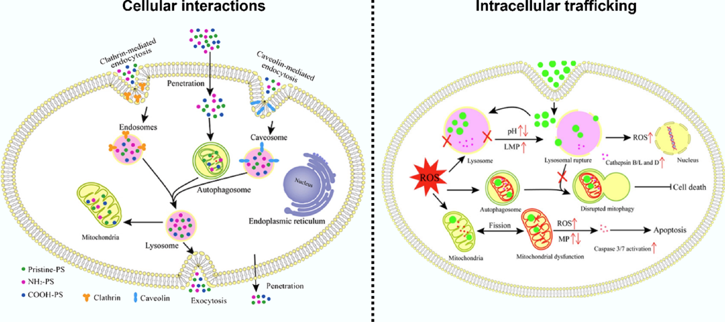
Once internalized, the journey of the AIEgen-tagged ENCs through the endo-lysosomal system is vividly illuminated. The progressive acidification and maturation of early endosomes into lysosomes trigger a corresponding increase in AIEgen fluorescence intensity, providing a direct visual and quantitative measure of trafficking kinetics[21]. This is where AIEgens reveal their true power for mechanistic toxicology. A pivotal event like lysosomal pH variation, a key mechanism of ENC-induced toxicity, can be captured with clarity (Fig. 1)[22,23]. In a dual-staining experiment, the sudden dispersion of a bright, punctate AIEgen signal from a lysosome (labeled with a pH-sensitive dye like LysoTracker) into a diffuse cytosolic pattern, concurrent with the loss of the LysoTracker signal due to neutralization of the pH, provides incontrovertible visual evidence of membrane rupture[20].
Beyond trafficking, the high signal-to-noise ratio and photostability of AIEgens make them ideal for super-resolution microscopy techniques like STED and STORM. Nanoscale visualization is critical for elucidating subtle mechanisms of organelle dysfunction, as it can determine whether ENCs are merely contained within an organelle or are directly associated with specific membrane proteins[24], such as those on the outer mitochondrial membrane[25]. This technology's potential is fully realized through its seamless integration into correlative microscopy workflows. Here, AIEgens provide the dynamic, live-cell component: a specific event, such as mitochondrial fission triggered by an AIEgen-tagged ENC, is first identified in a living cell[20]. The same cell is then rapidly fixed, and the exact location of the ENC is confirmed with the nanoscale structural resolution of TEM or its elemental signature via NanoSIMS[26]. This seamless fusion of dynamic functional data with ultra-high-resolution structural information creates a comprehensive picture that no single technique could achieve.
-
The cellular journey of ENCs represents only the first chapter in their biological narrative. To fully appreciate their systemic impact, ascension to the tissue and organ level is required, where complex architectures—comprising multiple cell types, vascular networks, and specialized functional units—create a heterogeneous landscape that dictates ultimate biodistribution, persistence, and pathological consequences. At the tissue and organ level, bioimaging provides a critical bridge between cellular observations and whole-organism pathophysiology by enabling a logical, hierarchical investigative approach that progresses from unbiased 3D mapping to mechanistic insight. The exceptional photostability of AIEgens allows for their integration with tissue clearing and light-sheet fluorescence microscopy, facilitating the complete 3D cartography of ENC distribution throughout entire organs without signal decay[27]. This spatial mapping then directs targeted investigation, as the stability of the AIEgen signal enables correlative analysis on the exact same tissue section, which can be used to identify the storage pools of ENCs and their potential targets of action[16,28]. Furthermore, the brightness and photostability of AIEgens permit real-time visualization in ex vivo models, such as observing the timed translocation of ENCs across the blood-brain barrier, thereby quantifying the kinetics of barrier penetration that underlie systemic dissemination[29]. Thus, by progressing from organ-wide distribution patterns to cellular uptake heterogeneity and finally to site-specific biological responses, AIEgen technology transforms tissue imaging from descriptive histology into a predictive, mechanistic tool that logically connects ENC biodistribution to pathological outcome.
-
The ultimate validation of the environmental health risk posed by ENCs lies in understanding their systemic journey within a living organism—from initial exposure and distribution to accumulation and final clearance. Whole-body in vivo imaging represents the critical frontier for this assessment, aiming to non-invasively quantify the pharmacokinetics of ENCs in real time. The primary challenge in this domain is the formidable scattering and absorption of light by biological tissues, which severely limit penetration depth and spatial resolution. This is where the tailored design of AIEgens, particularly those emitting in the near-infrared (NIR) windows, becomes a transformative solution, enabling a shift from endpoint tissue analysis to dynamic, longitudinal quantification. The exceptional photostability of AIEgens is their paramount advantage for in vivo studies. Unlike conventional fluorophores that rapidly photobleach, AIEgens maintain their signal integrity over days or even weeks[28,30]. This allows for repeated imaging of the same animal, which is essential for accurately calculating key pharmacokinetic parameters such as biological half-life and for distinguishing between rapid clearance pathways and long-term sequestration in organs of the mononuclear phagocyte system. The ability to use each subject as its own control significantly reduces inter-animal variability and enhances the statistical power of longitudinal studies.
Imaging in the first near-infrared window (NIR-I, 750–900 nm) has been a significant advancement, offering deeper tissue penetration compared to visible light. AIEgens engineered for NIR-I emission, when used with fluorescence molecular tomography (FMT) or highly sensitive whole-animal imaging systems, allow for the real-time tracking of ENCs in live rodents. For example, the administration of NIR-I AIEgen-tagged nanoparticles via oral gavage or intravenous injection allows researchers to quantitatively monitor their passage through the gastrointestinal tract or their systemic dissemination to secondary organs, respectively[31]. However, NIR-I imaging still encounters significant photon scattering, which can blur fine details and limit resolution at depth.
The second near-infrared window (NIR-II, 1,000–1,700 nm) represents the cutting edge, where AIEgens demonstrate their fullest potential because their longer wavelengths experience drastically reduced scattering and minimal tissue autofluorescence compared to NIR-I. AIEgens like TT3-oCB and BPN-BBT exhibit emissions in the NIR-II, resulting in a dramatic improvement in spatial resolution, typically achieving 10–50 μm in deep tissues, which is sufficient for visualizing organ-level biodistribution and vascular dynamics[32,33]. This allows for high-fidelity visualization of ENC biodistribution in deep tissues. The high resolution and superior signal-to-background ratio also enable the visualization of the dynamics of ENCs within the vasculature of deep-seated organs and the tracking of their ability to cross protective barriers like the blood–brain barrier in real time[29]. The true power of AIEgen-based in vivo imaging extends beyond aesthetically pleasing images to predictive pharmacokinetics (Fig. 2)[34]. By collecting a time series of 3D images, researchers can extract precise, organ-specific time-activity curves for the ENCs. This quantitative data forms the foundation for building computational toxicokinetic models that can predict the bioaccumulation potential of novel nanomaterials based on their physicochemical properties, moving the field away from resource-intensive, post-mortem tissue analysis. Furthermore, the ability to track multiple AIEgen-labeled ENCs with distinct emission wavelengths simultaneously within the same animal allows for direct, side-by-side comparison of their biodistribution and clearance pathways, greatly enhancing the efficiency of environmental hazard ranking[18].

Figure 2.
Application of NIR-II AIEgens labeled ENCs in studying their kinetics in adult fish.
In conclusion, AIEgen-enabled whole-body imaging transforms in vivo toxicology from a descriptive, endpoint analysis into a dynamic, quantitative, and predictive science. By providing non-invasive, longitudinal data on the absorption, distribution, metabolism, and excretion of ENCs, this technology is indispensable for constructing a comprehensive safety assessment framework that can keep pace with rapid innovation in nanotechnology.
-
The application of advanced bioimaging to the study of ENCs, while transformative, is fraught with significant technical challenges that must be addressed to ensure data accuracy and biological relevance. These hurdles can be categorized into four interconnected domains: (1) probe fidelity and the perturbation of the ENC's native identity; (2) the physical trade-offs between spatial resolution, temporal resolution, and penetration depth; (3) the analytical complexity of vast, multi-dimensional datasets; and (4) the potential for the imaging process itself to induce biological perturbation or artifacts.
A primary challenge lies in the conflict between detectability and environmental relevance. Labeling strategies are indispensable for tracking but can alter the surface properties of the ENC, while label-free techniques struggle with identification, especially for carbon-based materials. Furthermore, a fundamental physical constraint persists that high-resolution techniques like TEM are limited to static, processed samples, whereas whole-body modalities like MRI or PET lack the resolution to reveal subcellular events. The high-content data generated creates an analytical bottleneck, necessitating advanced computational tools for quantification. Finally, concerns regarding phototoxicity from high-intensity illumination, sample preparation artifacts, and the need for high exposure doses to achieve detection confound toxicological interpretations and limit environmental relevance. These challenges, however, represent active frontiers for innovation. The path forward will be defined by technological convergence, smarter probes, and data-driven analytics, moving the field from descriptive observation to predictive toxicology.
-
The foremost trajectory involves breaking the longstanding resolution-penetration trade-off through integrated correlative workflows. For example, the combination of light-sheet fluorescence microscopy for rapid, live-organism imaging with subsequent targeted analysis via electron microscopy or NanoSIMS on the same specimen will become more automated and accessible. Advancements in multimodal contrast agents are crucial. For instance, AIEgens can be engineered to contain heavy atom clusters (e.g., gold or iodine) for enhanced micro-CT contrast, or isotopic labels (e.g., 13C or 15N) for NanoSIMS mapping, as demonstrated in studies of silver nanoparticle transformations[35]. This approach allows correlative imaging without perturbing ENC behavior. The development of artificial intelligence (AI)-driven adaptive microscopy, where the system uses real-time analysis to identify critical events and automatically switches to a higher-resolution modality, represents a paradigm shift towards intelligent, hypothesis-generating instrumentation.
-
The future lies in extracting mechanistic insights, not just generating images. This will be driven by the integration of bioimaging with spatiotemporal omics technologies. Correlating the precise subcellular location of an ENC with the transcriptomic and proteomic profile of its immediate microenvironment will directly link its presence to molecular pathway activation. To manage this data deluge, the field must embrace artificial intelligence and machine learning not only for automated analysis but also for prediction. Deep learning models trained on high-content imaging libraries could eventually forecast the biodistribution and potential toxicity of a novel nanomaterial based solely on its physicochemical properties, guiding safe-by-design principles pre-synthesis[36]. Establishing centralized, open-access data repositories with standardized metadata will be essential for training these models and ensuring reproducible science. In conclusion, by consciously addressing these technical challenges through strategic innovation, bioimaging can fulfill its promise as a rigorous, predictive tool. The goal is to build a predictive toxicological framework for ENCs. By visualizing their entire journey—from initial cellular interaction to long-term sequestration—and correlating this spatiotemporal data with molecular responses, it becomes possible to move beyond observational hazard identification. This paradigm shift is essential for ensuring the sustainable and safe development of nanotechnology, effectively illuminating the path from nanomaterial innovation to environmental health protection.
-
Advanced bioimaging has fundamentally shifted the paradigm of nanotoxicology, transforming the biological system from a "black box" into a transparent, navigable landscape. By providing unprecedented spatial and temporal resolution, these technologies allow researchers to track the complete journey of ENCs—from initial cellular entry and intricate subcellular trafficking to systemic distribution and ultimate clearance. Among the available tools, fluorescence imaging, particularly when empowered by AIEgens with their exceptional photostability and aggregation-induced emission properties, has proven indispensable for dynamic, long-term, and mechanistic studies. The ability to correlate the precise location of an ENC with subsequent biological responses, from lysosomal membrane permeabilization to organ-level pathological changes, provides a causal link that is unattainable with conventional endpoint assays. However, the field must continue to address significant challenges, including the trade-offs between resolution and penetration, the potential for probes to alter ENC behavior, and the analytical complexity of large, multi-dimensional datasets. The path forward lies in technological convergence. The integration of multimodal imaging platforms, the development of multi-functional probes, and the application of artificial intelligence for image analysis and predictive toxicokinetic modeling represent the next frontier. By embracing these advancements, bioimaging will evolve beyond a descriptive tool into a predictive science. This progression is critical for building a robust, mechanistic, and efficient safety assessment framework capable of keeping pace with the rapid innovation in nanotechnology, thereby ensuring that its benefits are realized without compromising environmental and human health. Ultimately, the continued refinement of bioimaging is not merely a technical goal but a prerequisite for the sustainable and safe development of the nano-age.
We thank the reviewers for their helpful and constructive comments. The TOC graphic was created using
BioRender.com .-
Not applicable.
-
The authors confirm their contributions to the paper as follows: methodology, investigation, formal analysis, writing—original draft: Neng Yan; funding acquisition and supervision: Neng Yan, Fei Dang, Alessandro Parodi, Jianbo Shi; writing—review and editing: Neng Yan, Fei Dang, Parodi Alessandro, Jianbo Shi. All authors reviewed the results and approved the final version of the manuscript.
-
Data sharing is not applicable to this article as no datasets were generated or analyzed during the current study.
-
This study was supported by grants from the Natural Science Foundation of China (Grant Nos 42025704, 42477250, 42207319 and 42222702), the National Key Research and Development Program of China (Grant No. 2024YFC3714600), and the Natural Science Foundation of Hubei Province (Grant No. 2025AFA048). Alessandro Parodi was supported by the grant of the state program of the "Sirius" Federal Territory "Scientific and technological development of the "Sirius" Federal Territory" (Agreement No. 3-03 dated 18.02.2025).
-
The authors declare that they have no conflict of interest.
-
Full list of author information is available at the end of the article.
- Copyright: © 2025 by the author(s). Published by Maximum Academic Press, Fayetteville, GA. This article is an open access article distributed under Creative Commons Attribution License (CC BY 4.0), visit https://creativecommons.org/licenses/by/4.0/.
-
About this article
Cite this article
Yan N, Parodi A, Dang F, Shi J. 2025. Shedding light on the invisible: aggregation-induced emission-based bioimaging for assessing the environmental impact of emerging nanoscale pollutants. New Contaminants 1: e019 doi: 10.48130/newcontam-0025-0017 

Shedding light on the invisible: aggregation-induced emission-based bioimaging for assessing the environmental impact of emerging nanoscale pollutants
- Received: 06 October 2025
- Revised: 16 November 2025
- Accepted: 24 November 2025
- Published online: 17 December 2025
Abstract: The rapid proliferation of emerging nanoscale contaminants (ENCs) poses significant challenges for traditional toxicological assessment due to their unique physicochemical properties and complex interactions with biological systems. This perspective highlights the transformative role of advanced bioimaging technologies in addressing these challenges by enabling direct visualization and quantification of ENCs within living systems. The strengths and limitations of current imaging techniques are explored, ranging from electron microscopy for ultra-high resolution to non-invasive whole-body imaging modalities. Particular emphasis is placed on the advantages of aggregation-induced emission luminogens (AIEgens), which overcome critical limitations of conventional fluorophores by offering exceptional photostability and biocompatibility. AIE-based bioimaging techniques enable real-time, dynamic tracking of ENCs and facilitate multiscale imaging, from elucidating subcellular uptake mechanisms to organelle interactions. Looking ahead, prevailing technical challenges are discussed, including the trade-offs between resolution and penetration depth, along with the need for robust quantitative analysis. The integration of smarter probes like AIEgens, multimodal imaging platforms, and artificial intelligence-powered analytics is expected to drive a paradigm shift from descriptive observation to predictive toxicology. This shift will make it possible to visualize the entire journey of ENCs in biological systems, link spatial and temporal behavior to molecular responses, and develop computational models for forecasting toxicity. Such advancements are essential for evidence-based risk assessment and the safe-by-design innovation of nanomaterials, ensuring the sustainable development of nanotechnology while protecting environmental health.
-
Key words:
- Emerging contaminants /
- Nanomaterials /
- Nanoplastics /
- Bioimaging /
- Aggregation-induced emission