-
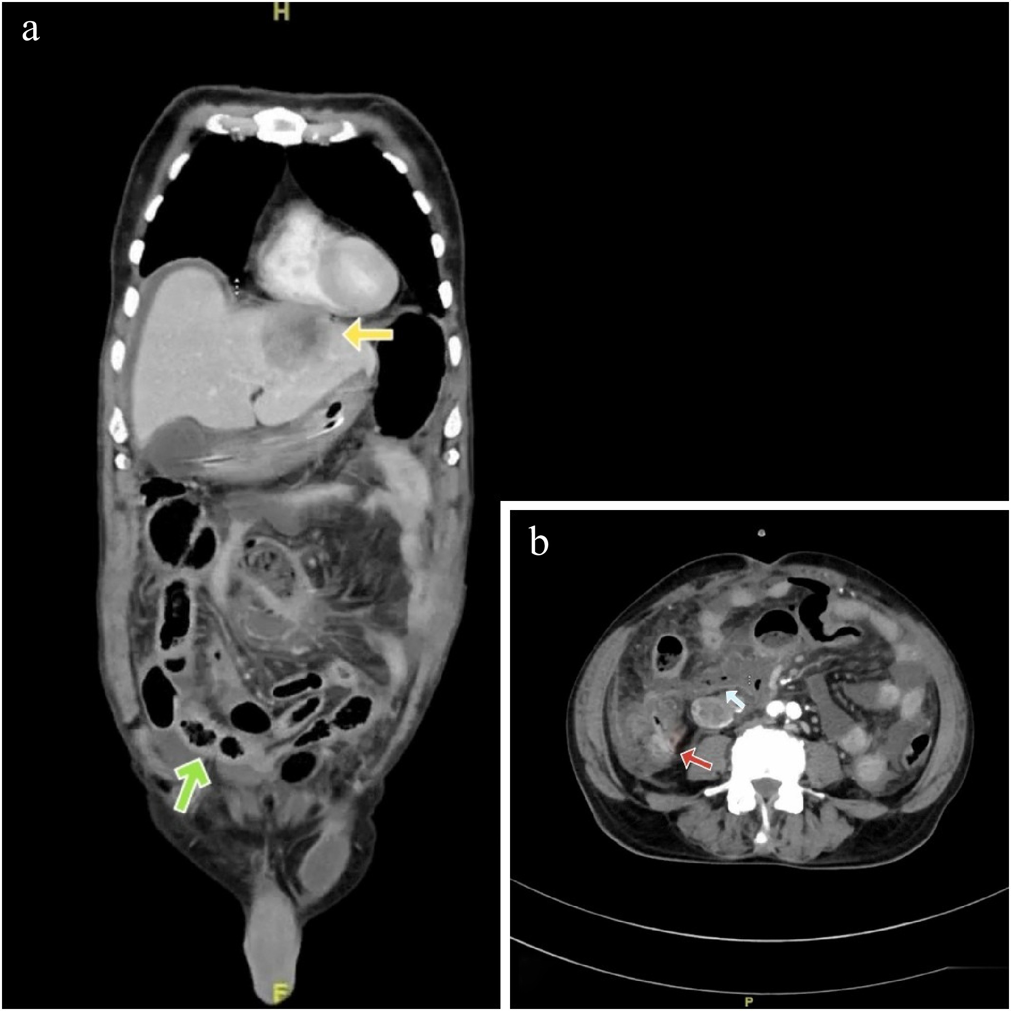
Figure 1.
(a) Yellow arrow - heterogenous, peripherally enhancing lesion in segments 2, 3 of liver (Hounsfield units: min- 10, max- 120, avg- 84). Green arrow - hyperenhancing circumferential thickening of the terminal ileum and ileo-caecal junction (Hounsfield units: min- 15, max- 100, avg- 60). (b) Red arrow showing thickened enhancing wall of ileo-caecal junction and a light blue arrow showing extraluminal gas pockets (Hounsfield units: min- 600, max- 1000, avg- 890).
-
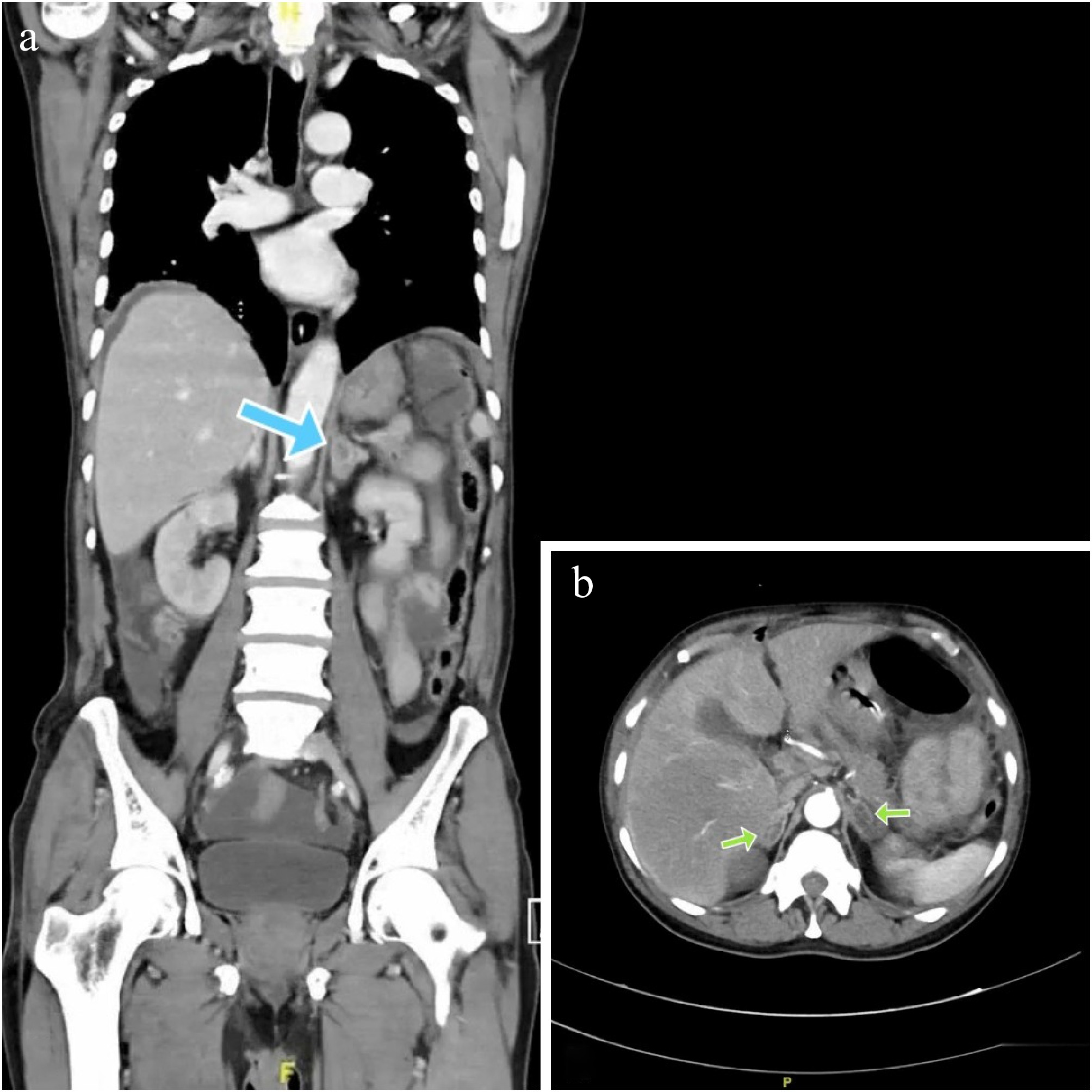
Figure 2.
(a) Blue arrow showing a hypodense lesion in the left supra-renal gland. (b) Green arrows showing bilateral hypodense adrenal lesions (Hounsfield units: min- 20, max- 104, avg- 38).
-
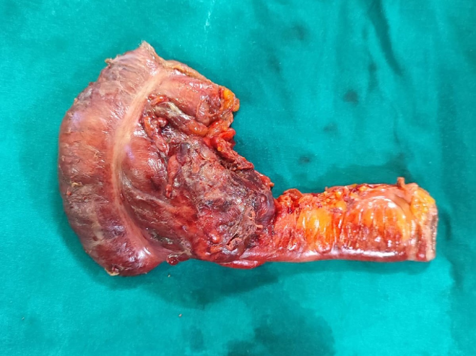
Figure 3.
Emergency limited right hemicolectomy specimen showing the growth involving the ileo-caecal junction.
-
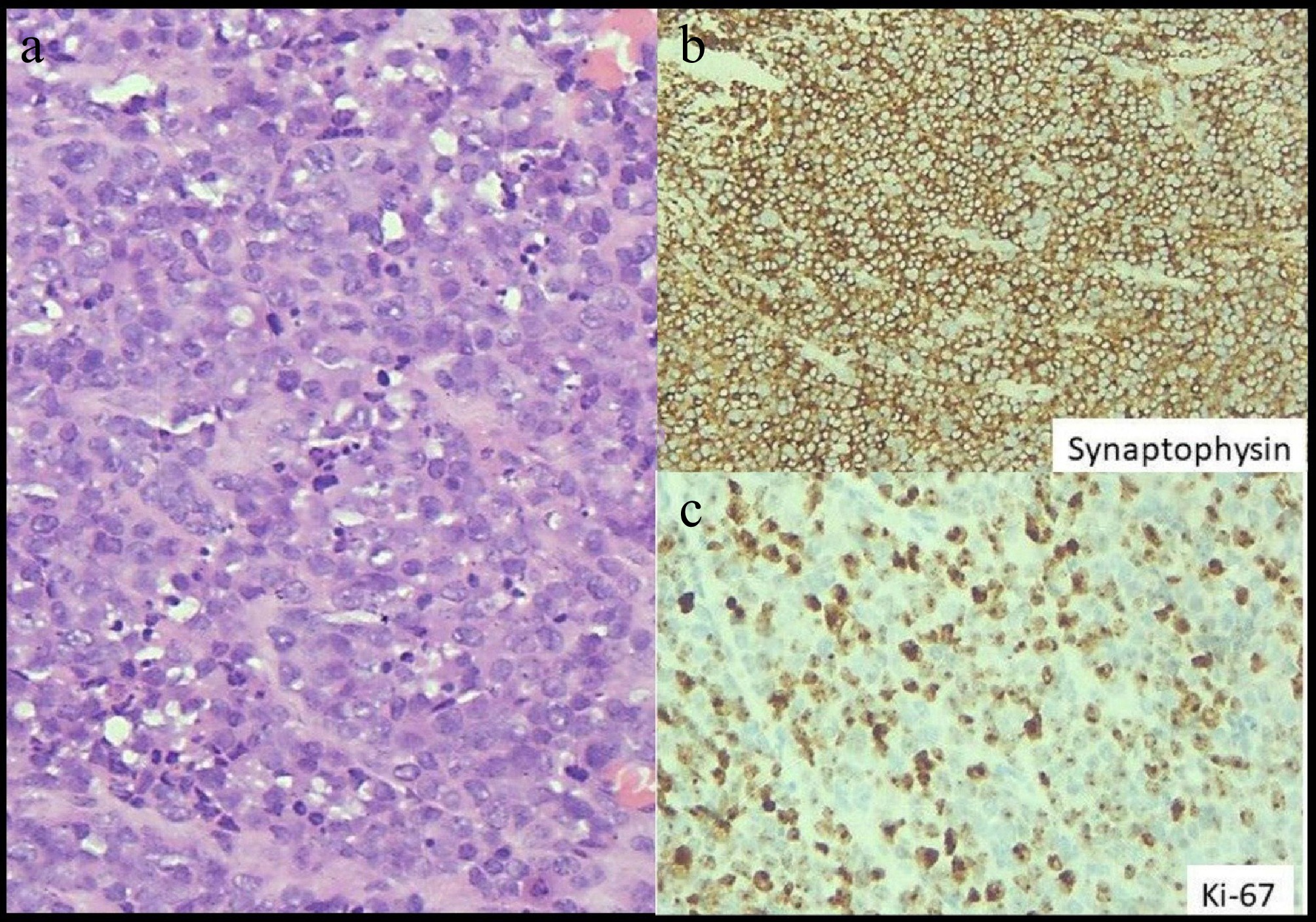
Figure 4.
(a) Highlights sheets of tumour cells with round to oval nuclei and ill-defined cell membrane. Nuclei exhibits stippled chromatin, with high mitotic and apoptotic activity. Haematoxylin and eosin stain, x 400. (b) Highlights diffuse strong cytoplasmic expression of Synaptophysin in tumour cells, immunohistochemistry with Ventana antibody. Diaminobenzidine stain, x 400. (c) Highlights high proliferation index in tumour cells as exhibited by Ki-67 immunoperoxidase stain, immunohistochemistry with Ventana antibody. Diaminobenzidine stain, x 400.
Figures
(4)
Tables
(0)