-
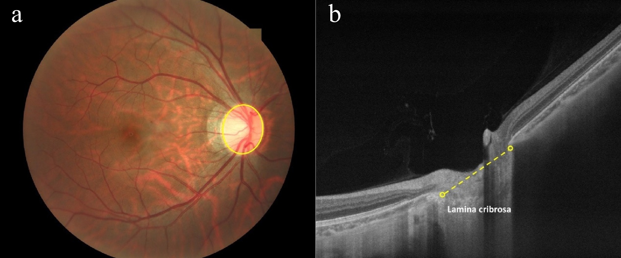
Figure 1.
Optic disc size definitions in different ophthalmic modalities. (a) Color fundus photography: The optic disc is outlined by the visible part of the neuroretinal rim and optic cup (yellow oval). (b) Optical coherence tomography: The optic disc size is measured at the end of the lamina cribrosa, specifically the peripapillary Elschnig border tissue of the peripapillary scleral flange (yellow dashed line). (Source: Personal fundus images provided by Mr. Jinze Zhang, Sun Yat-sen University).
-
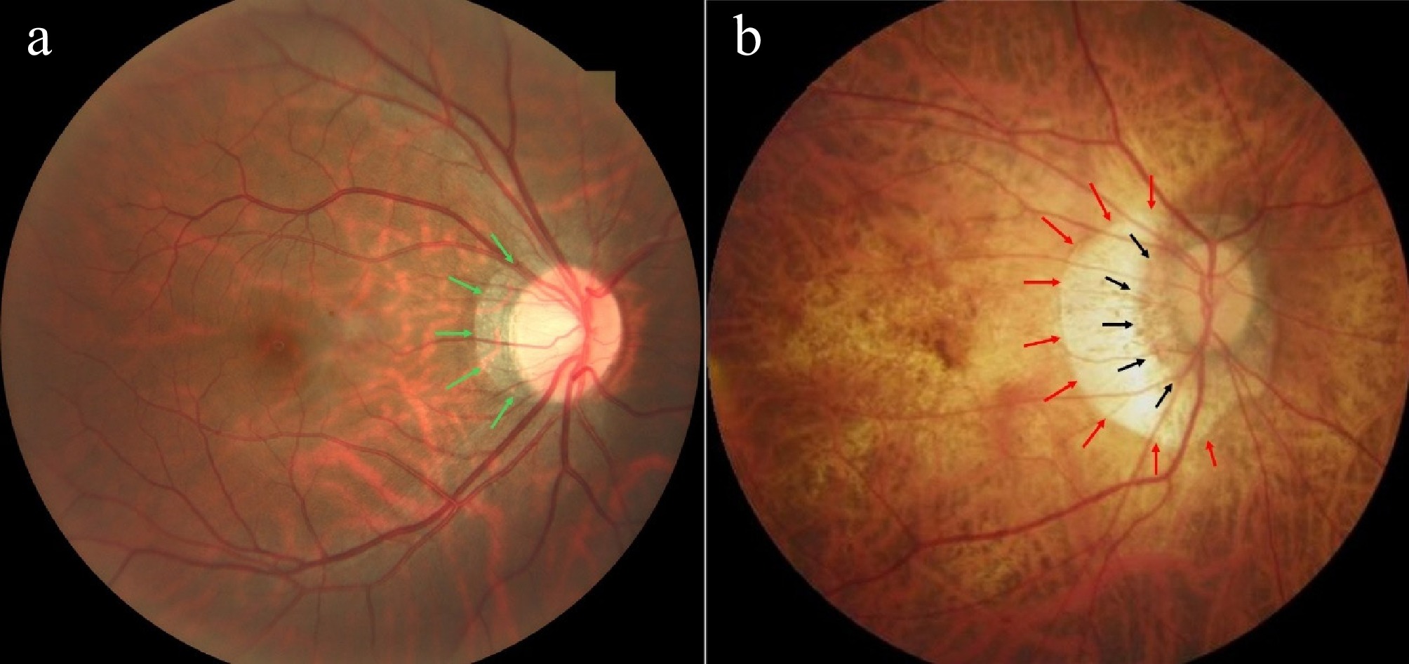
Figure 2.
Illustration of different parapapillary zones in color fundus photography. (a) Beta zone (green arrows) refers to the presence of Bruch's membrane (BM) and the absence of retinal pigment epithelium (RPE). (b) Gamma zone (red arrows) refers to the area around the optic disc where BM and RPE are absent; Delta zone (black arrows) refers to the thinning and elongation of peripapillary scleral flange. (Source: HPMI database,
https://doi.org/10.6084/m9.figshare.24800232.v1 ). -
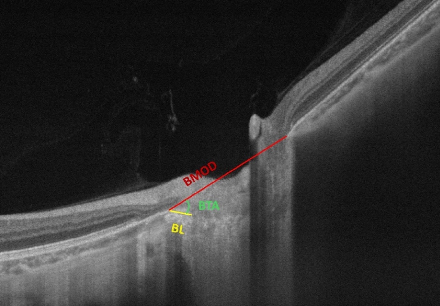
Figure 3.
Illustration of Bruch's membrane opening distance (BMOD), border length (BL), and border tissue angle (BTA) in optical coherence tomography. BMOD refers to the distance between the two points of the Bruch's membrane opening (BMO) on a horizontal B-scan; BL refers to the straight-line distance from the temporal BMO point to the border tissue and the scleral termination; BTA refers to the angle formed between the BMOD line and the BL line. (Source: Personal fundus images provided by Mr. Jinze Zhang, Sun Yat-sen University).
Figures
(3)
Tables
(0)