-
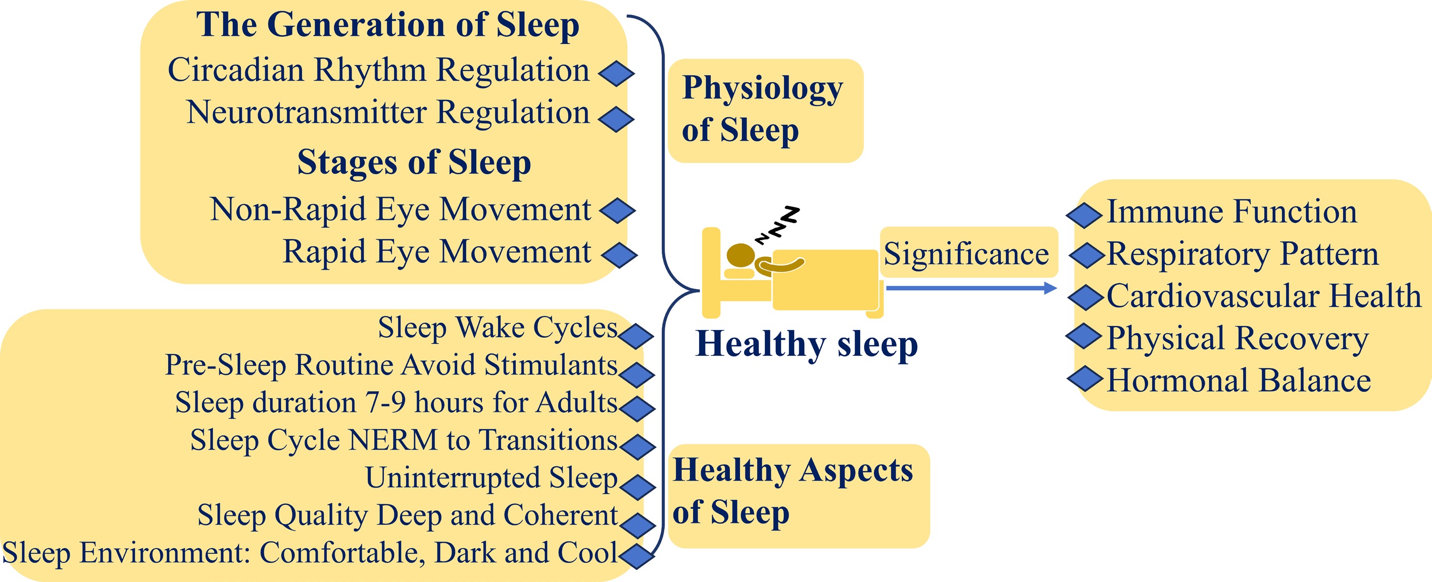
Figure 1.
The physiology of healthy sleep and its importance (created by the authors).
-
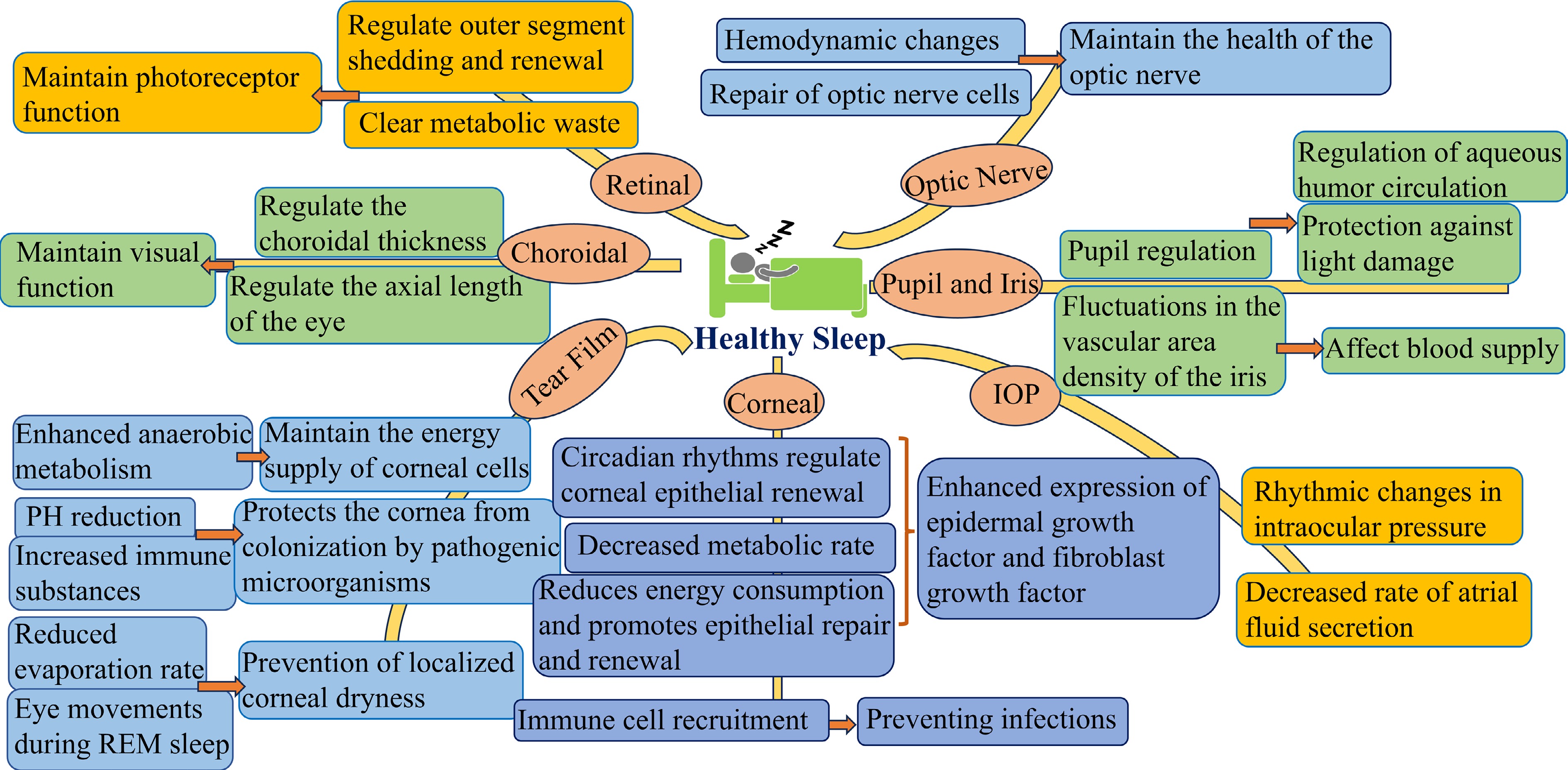
Figure 2.
The relationship between sleep and eye health (created by the authors).
-
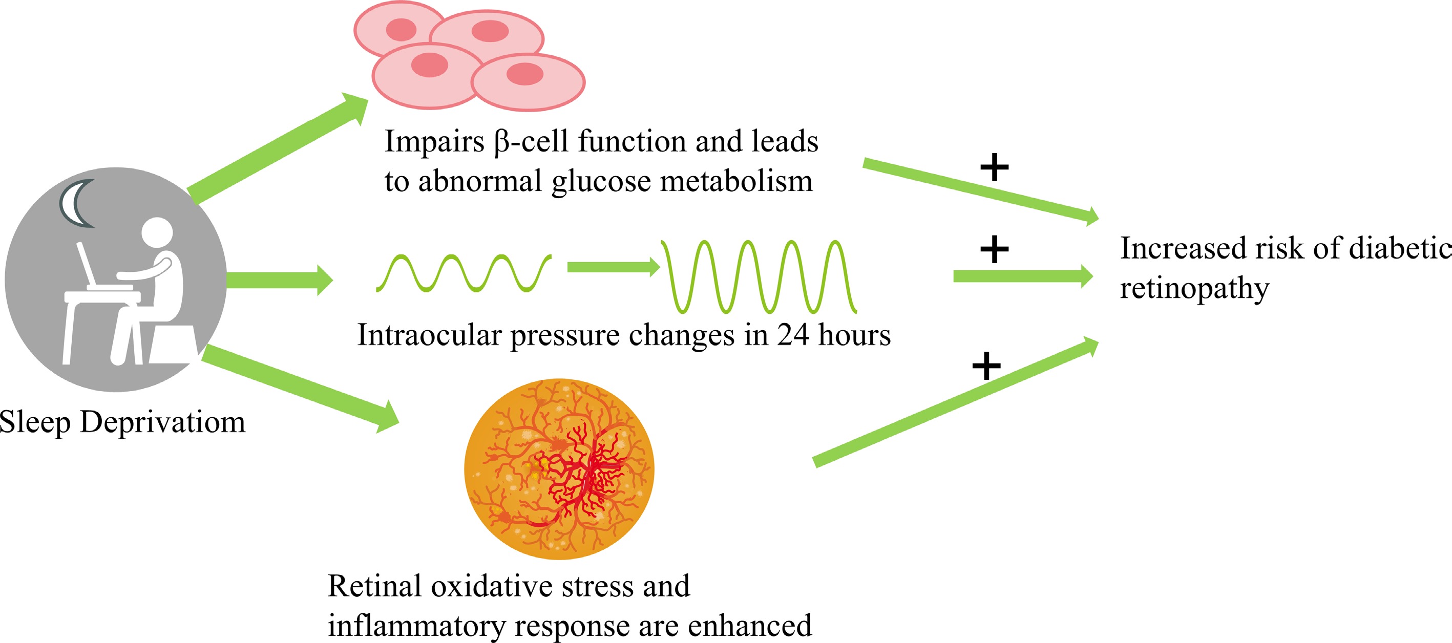
Figure 3.
Mind map diagram illustrating the mechanisms through which sleep deprivation impacts diabetic retinopathy. (Selected artwork (retina) depicted in the figure is derived from or adapted from images provided by Servier Medical Art (Servier;
https://smart.servier.com ), licensed under a Creative Commons Attribution 4.0 Unported License). -
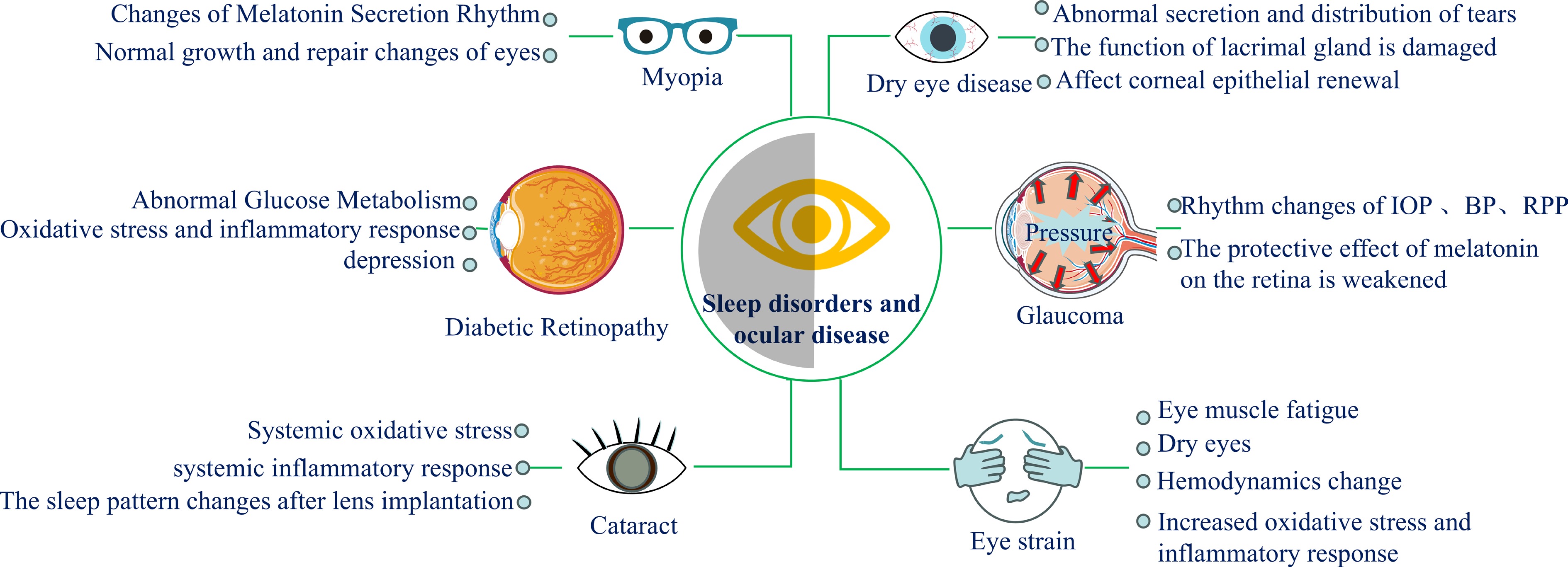
Figure 4.
Mind map diagram for sleep disorders and ocular disease. Selected artwork (the 'diabetic retinopathy' and 'glaucoma') depicted in the figure is derived from or adapted from images provided by Servier Medical Art (Servier;
https://smart.servier.com ), licensed under a Creative Commons Attribution 4.0 Unported License. -
Activity Function Impact UV protection Protects against UV ray damage Minimizes oxidative stress on the lens Antioxidant increase Boosts levels of antioxidants like GSH Protects lens cells from oxidative damage ROS neutralization Neutralizes ROS Maintains lens clarity and function Nutrient supply Supplied by aqueous humor and vitreous humor Supports overall lens health Low oxygen maintenance Maintained by low oxygen in aqueous humor and vitreous humor Regulates factors like VEGF-A, crucial for lens transparency Aqueous humor reduction Reduces flow during sleep Enhances repair mechanisms during sleep Table 1.
Protective mechanisms of the lens during sleep.
-
No. Subpart of the eye Changes in tear film and components Relevant paper 1 Tear film Disrupted tear production due to immune dysregulation. Altered tear composition due to hormonal changes. Decreased tear clearance due to metabolic changes. 2 Intraocular
pressure (IOP)Immunological response affecting aqueous humor dynamics. Impact of sleep disturbances on ocular blood flow regulation. Reduced waste clearance affecting trabecular meshwork function. Sleep-related changes in scleral compliance influencing IOP. 3 Cornea Impaired epithelial regeneration due to metabolic changes. Reduced corneal sensitivity due to hemodynamic changes. Increased risk of inflammation due to immune activation. Corneal thickness variation due to sleep stages and IOP fluctuations. 4 Lens Accumulation of metabolic by-products (e.g., advanced glycation end-products). Changes in lens hydration levels due to circadian rhythm disruption. Oxidative stress leading to cataract formation. Reduced antioxidative enzyme activity linked to poor sleep. 5 Optic nerve Immunological responses leading to optic nerve inflammation. Hemodynamic changes impacting optic nerve head blood flow. Increased intraocular pressure influencing optic nerve head integrity. Altered venous pressure and autoregulation affecting optic nerve function. 6 Retina Impaired waste clearance leading to retinal cell dysfunction. Changes in retinal blood flow due to sleep-related hemodynamic shifts. Disrupted circadian rhythm affecting retinal cell metabolism. Altered neural signaling in the retina due to sleep deprivation. Table 2.
Common and uncommon pathways impacting subparts of the eye due to poor sleep.
Figures
(4)
Tables
(2)