-
-

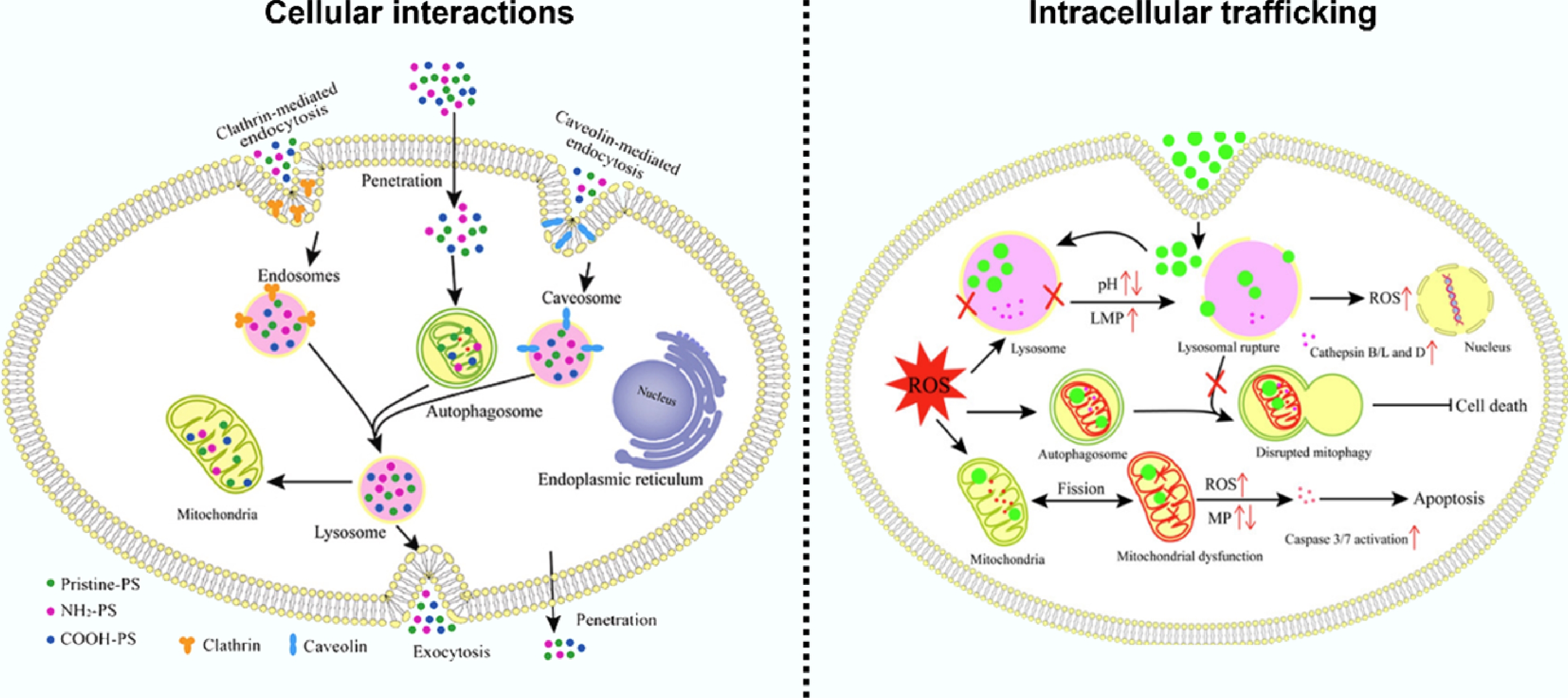
Figure 2.
Application of NIR-II AIEgens labeled ENCs in studying their kinetics in adult fish.
-
Imaging technique Spatial resolution Strength Limitation Confocal microscopy ~200 nm High specificity; suitable for living cells Requires fluorescent labeling TEM < 1 nm Ultrahigh resolution; detailed morphological information Sample fixation required; limited field of view LA-ICP-MS 1–10 μm High sensitivity; multi-element analysis Destructive to samples; limited molecular information Synchrotron-based
X-ray fluorescence~50 nm Label-free; high penetration depth Limited accessibility; complex data analysis MRI ~50–100 µm Whole-body, excellent anatomy Low sensitivity for agent PET 1–2 mm Ultra-sensitive quantification Radioactivity, poor resolution NanoSIMS ~50 nm Isotopic/elemental sub-cellular map Destructive, complex STED ~30–70 nm Live-cell super-resolution High laser power required STORM ~20 nm Localizes single molecules over time Very slow; fixation Micro-CT ~1–50 µm Non-destructive 3D imaging; high-throughput
capability; quantitativeVery low soft tissue contrast without stains; low sensitivity for trace NMs Table 1.
Widely employed bioimaging techniques
Figures
(2)
Tables
(1)