-
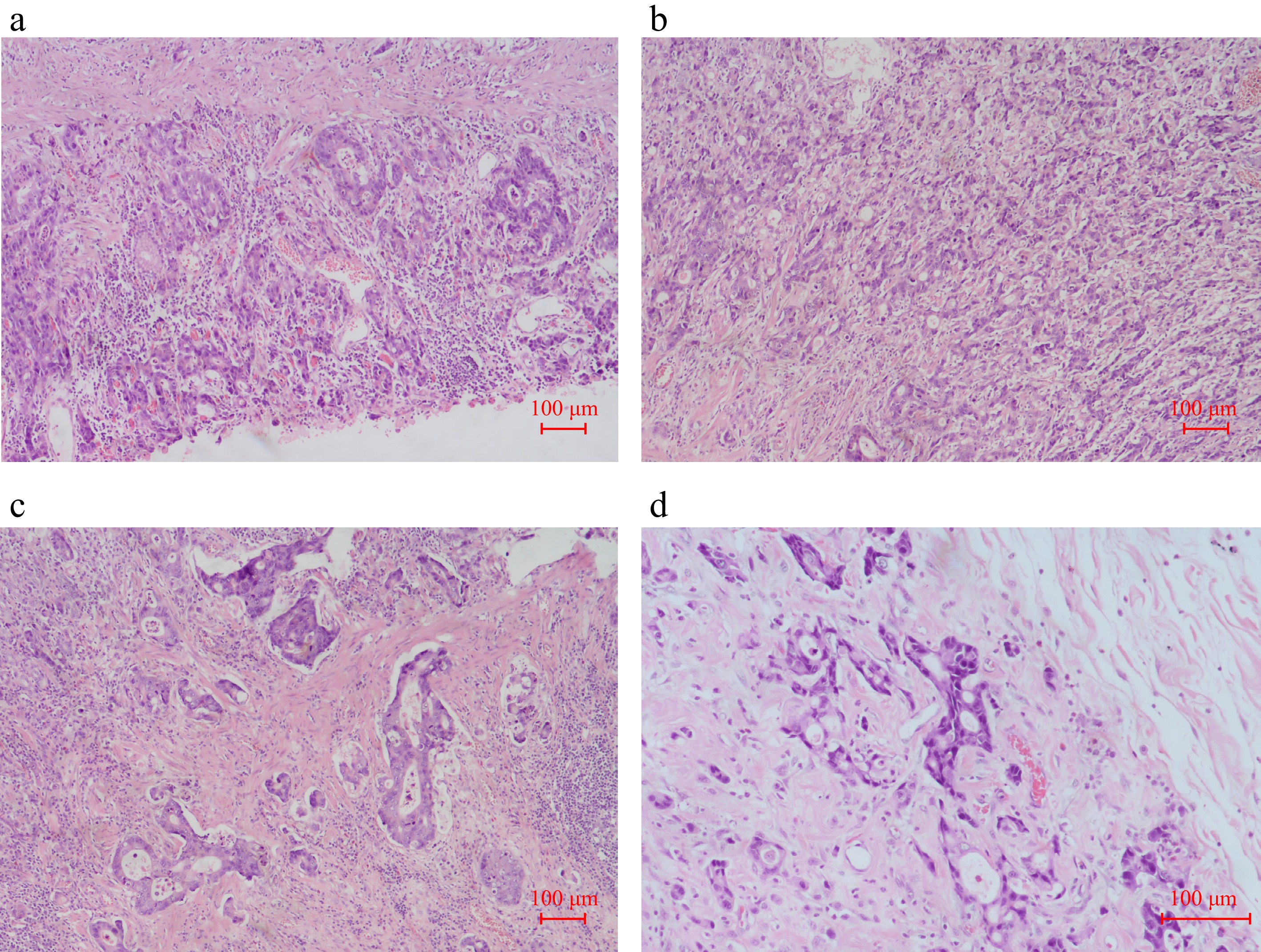
Figure 1.
Pathological images of the gastric mass from the radical distal subtotal gastrectomy with Roux-en-Y gastrojejunostomy on March 20, 2018. (a)–(c) H & E staining (× 10). (d) H & E staining (× 20).
-
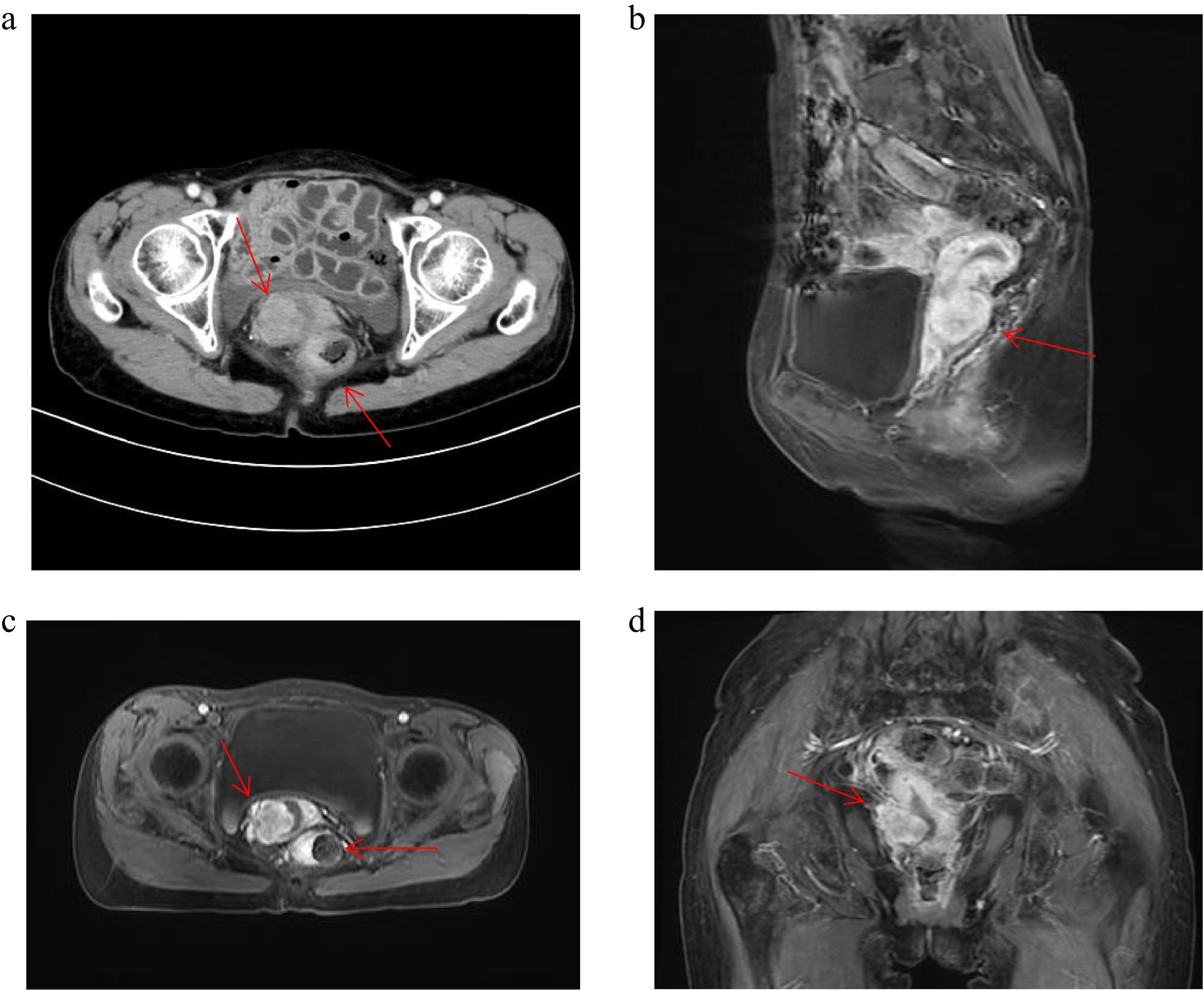
Figure 2.
Patient CT and MRI image. (a) Patient 2024.9.28 CT image. (b)–(d) Patient 2024.10.24 MRI image.
-
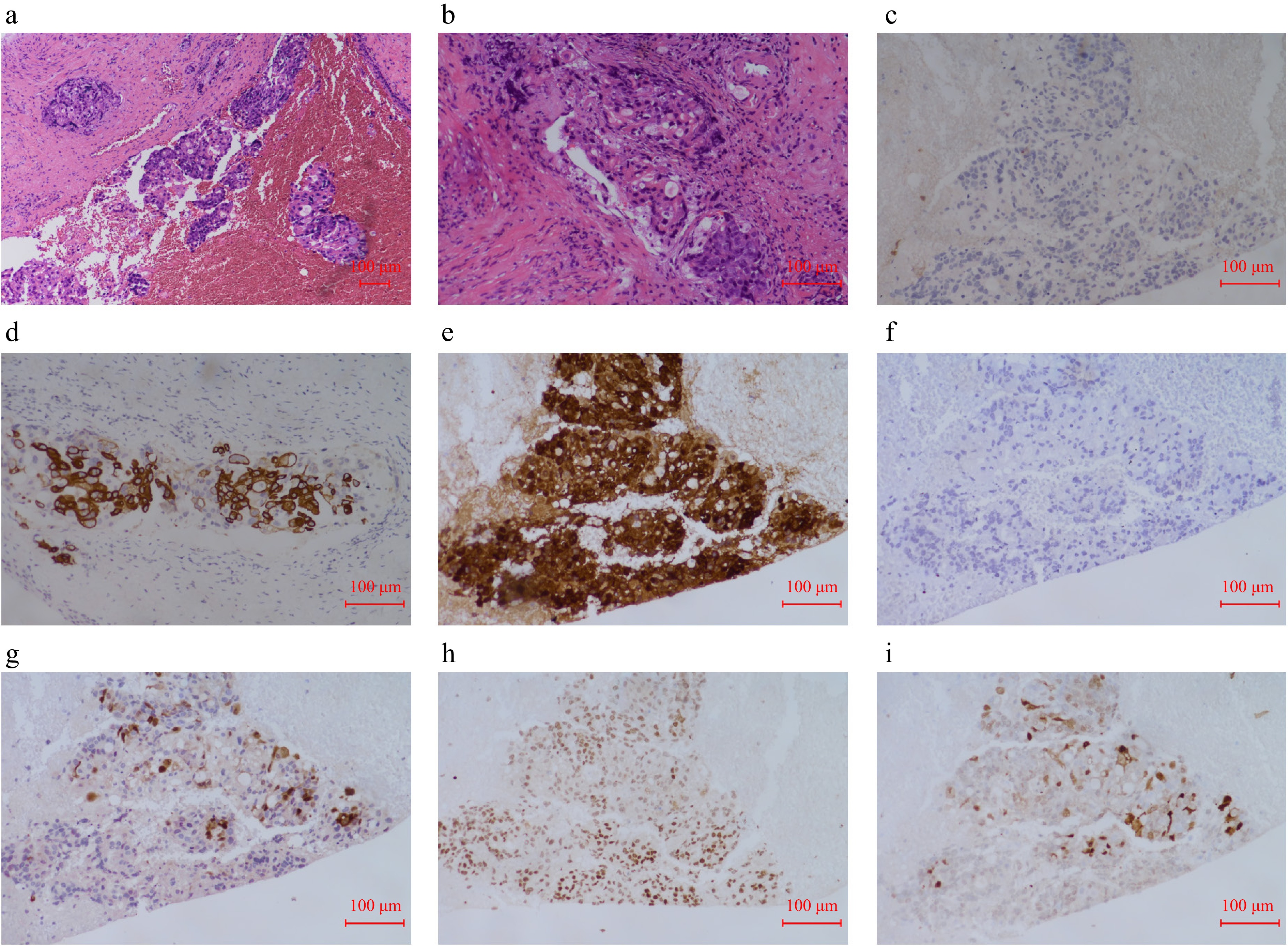
Figure 3.
Cervical pathological images. (a) H & E staining of sections from cervical mass (× 10). (b) H & E staining of sections from cervical mass (× 20). (c) Immunohistochemical CK7 pathological images of the sections from cervical mass. (d) Immunohistochemical CK20 pathological images of the sections from cervical mass. (e) Immunohistochemical Villin pathological images of the sections from cervical mass. (f) Immunohistochemical P40 pathological images of the sections from cervical mass. (g) Immunohistochemical P16 pathological images of the sections from cervical mass. (h) Immunohistochemical PAX8 pathological images of the sections from cervical mass. (i) Immunohistochemical SATB2 pathological images of the sections from cervical mass.
-
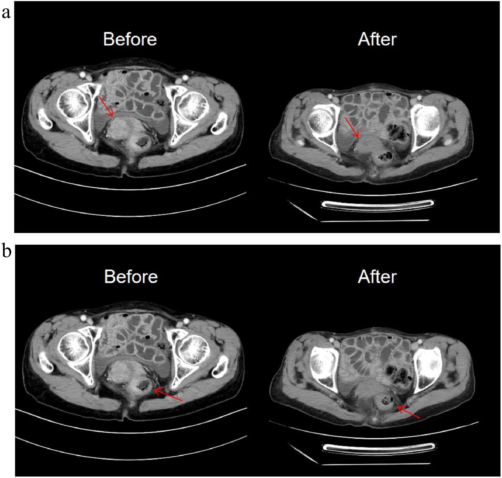
Figure 4.
The changes of CT images after seven treatment cycles. (a) The cervical mass is smaller than before. (b) The rectal mass is smaller than before.
Figures
(4)
Tables
(0)