-
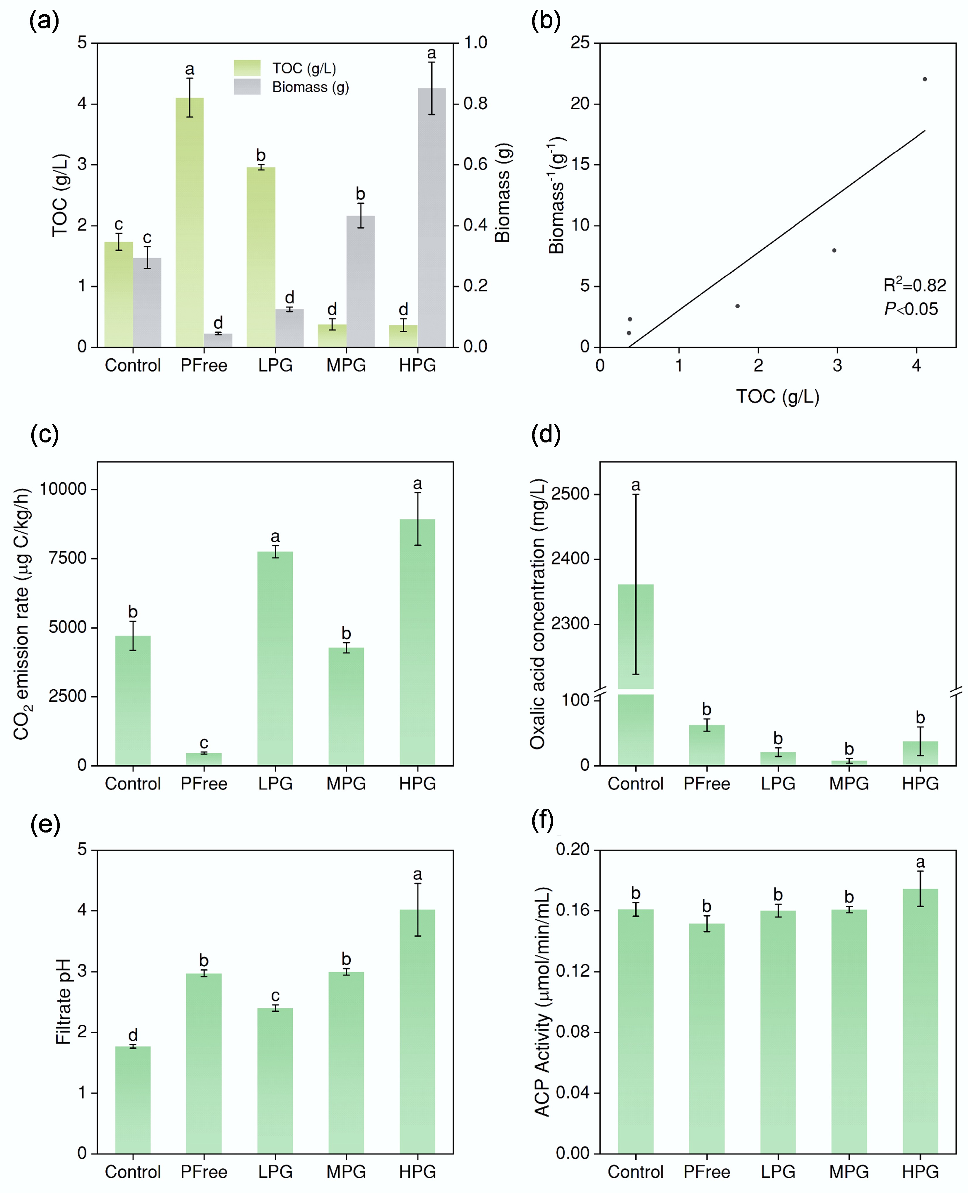
Figure 1.
Incubation effect of A. niger. (a) Biomass, and TOC content of filtrate after six days of incubation under different treatments. (b) The correlation between TOC values and the reciprocal of biomass. (c) CO2 emission rates of the system. (d) Concentrations of oxalic acid secreted by A. niger. (e) pH values of the filtrate after incubation for six days. (f) acid phosphatase activity of filtrate after incubation for six days. (The lowercase letter labels in the figure indicate significant differences among different treatments. If two treatments contained the same lowercase letter, there was no significant difference between them; otherwise, there was a significant difference).
-
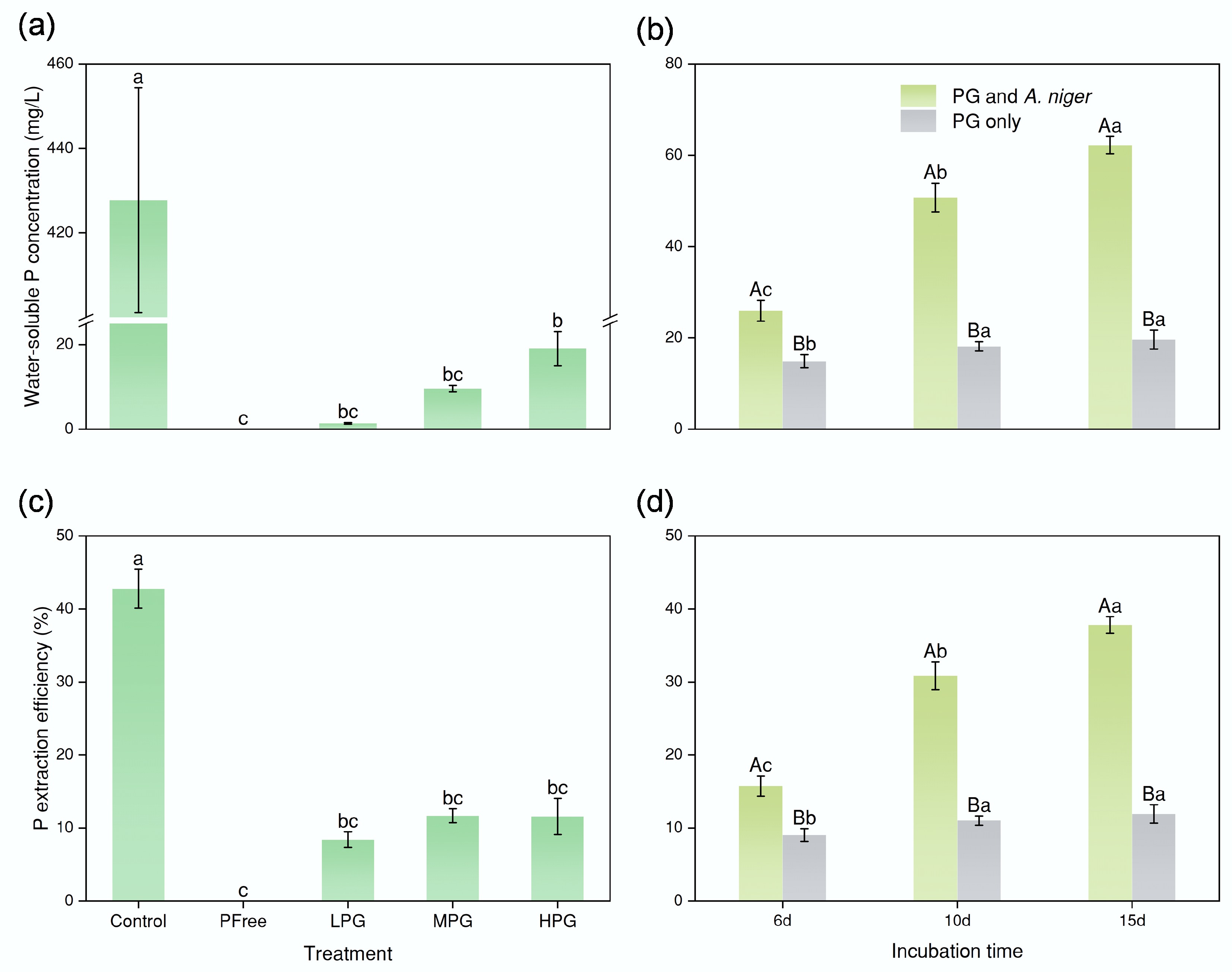
Figure 2.
Effect of bioextraction of P. (a) and (c) concentrations of water-soluble P and P bioextraction efficiency by A. niger after six days of incubation. (b) and (d) concentrations of water-soluble P and P extraction efficiency after six, 10, and 15 d of incubation, respectively (the P involved in the figure pertained solely to P presented in the solution, excluding P in fungal pellets). In (a) and (c), the lowercase letter labels indicate significant differences among different treatments. If two treatments had the same lowercase letter, there was no significant difference; otherwise, there was a considerable difference. In (b) and (d), the capital letter labels indicate whether there was a substantial difference between the treatments PG only or PG and A. niger at the same incubation time. The lowercase letters indicate significant differences among incubation times within each treatment (PG only or PG and A. niger). The method for determining substantial differences was consistent with the above.
-
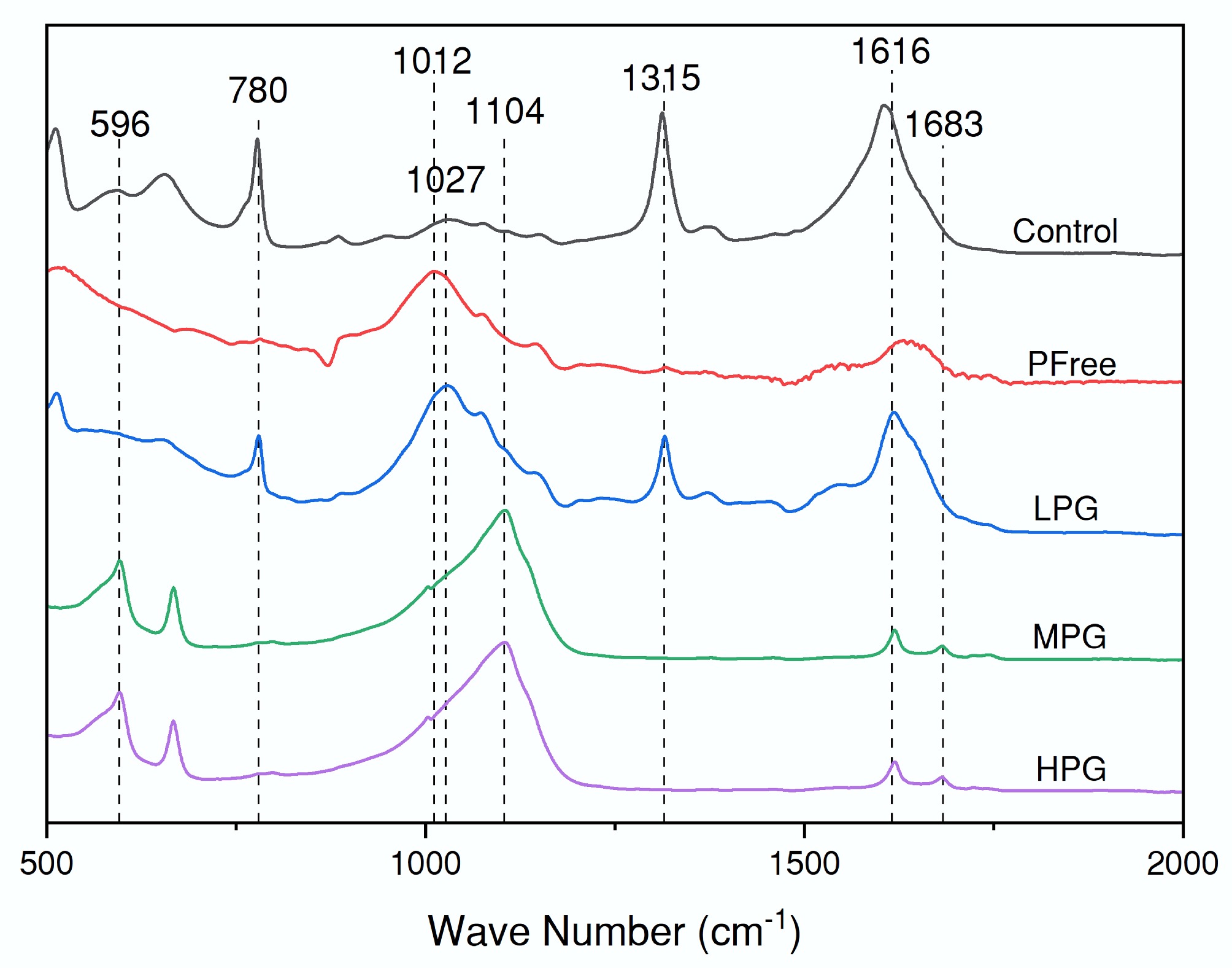
Figure 3.
ATR-IR spectra of the filtered solid phase after incubation for six days.
-
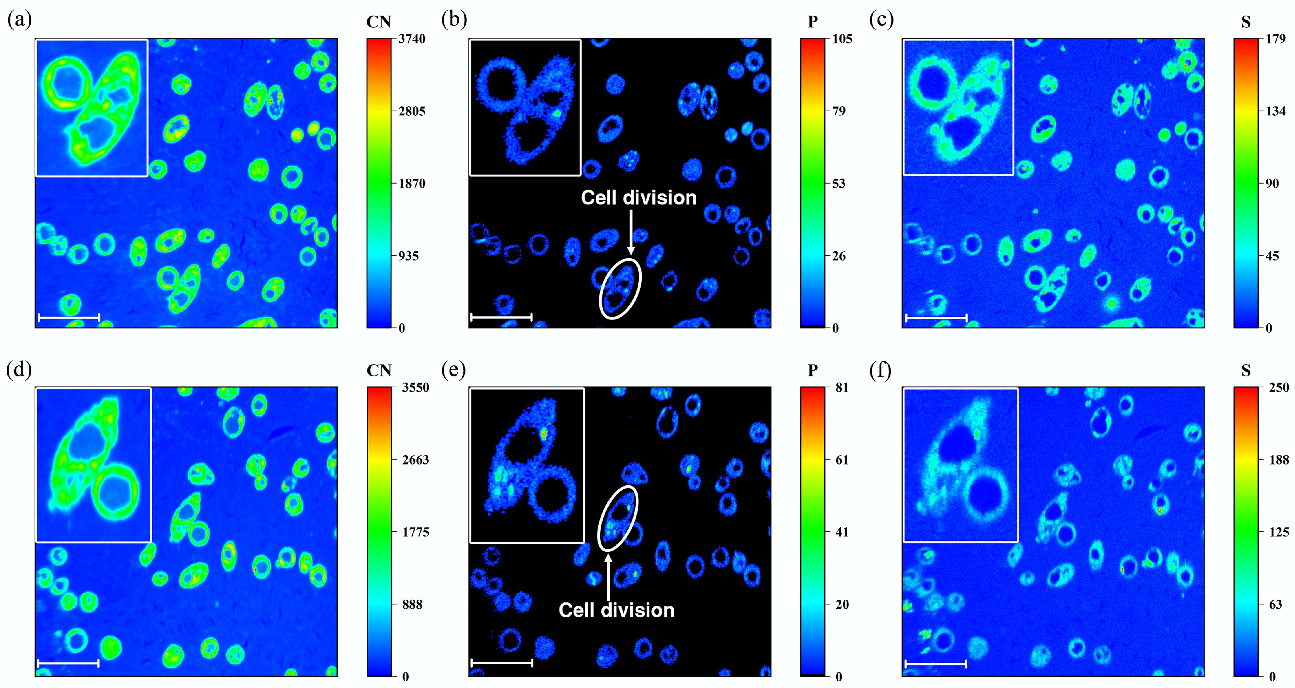
Figure 4.
NanoSIMS images of High PG treatment after 6 days of incubation. (a) and (d) CN element distribution. (b) and (e) P element distribution. (c) and (f) S element distribution. (a)−(c) were from the same region, while figures (d)−(f) were from another region. The scale bar of all the figures was 10 μm.
-
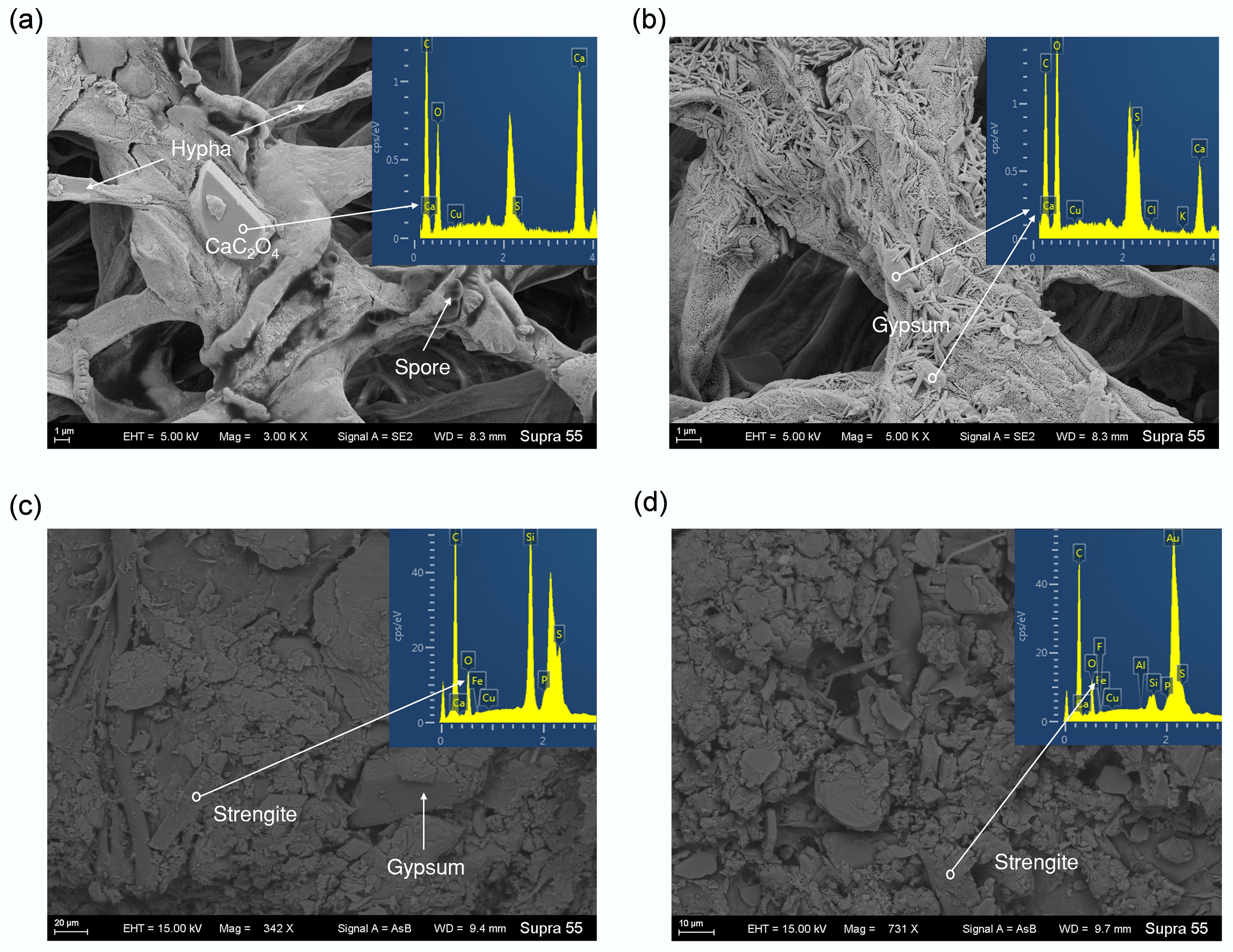
Figure 5.
SEM and EDS images of minerals formed during incubation. (a) Calcium oxalate from Low PG treatment. (b) Gypsum particles adsorbed by A. niger hyphae from High PG treatment. (c) and (d) Strengite from Moderate PG treatment.
-
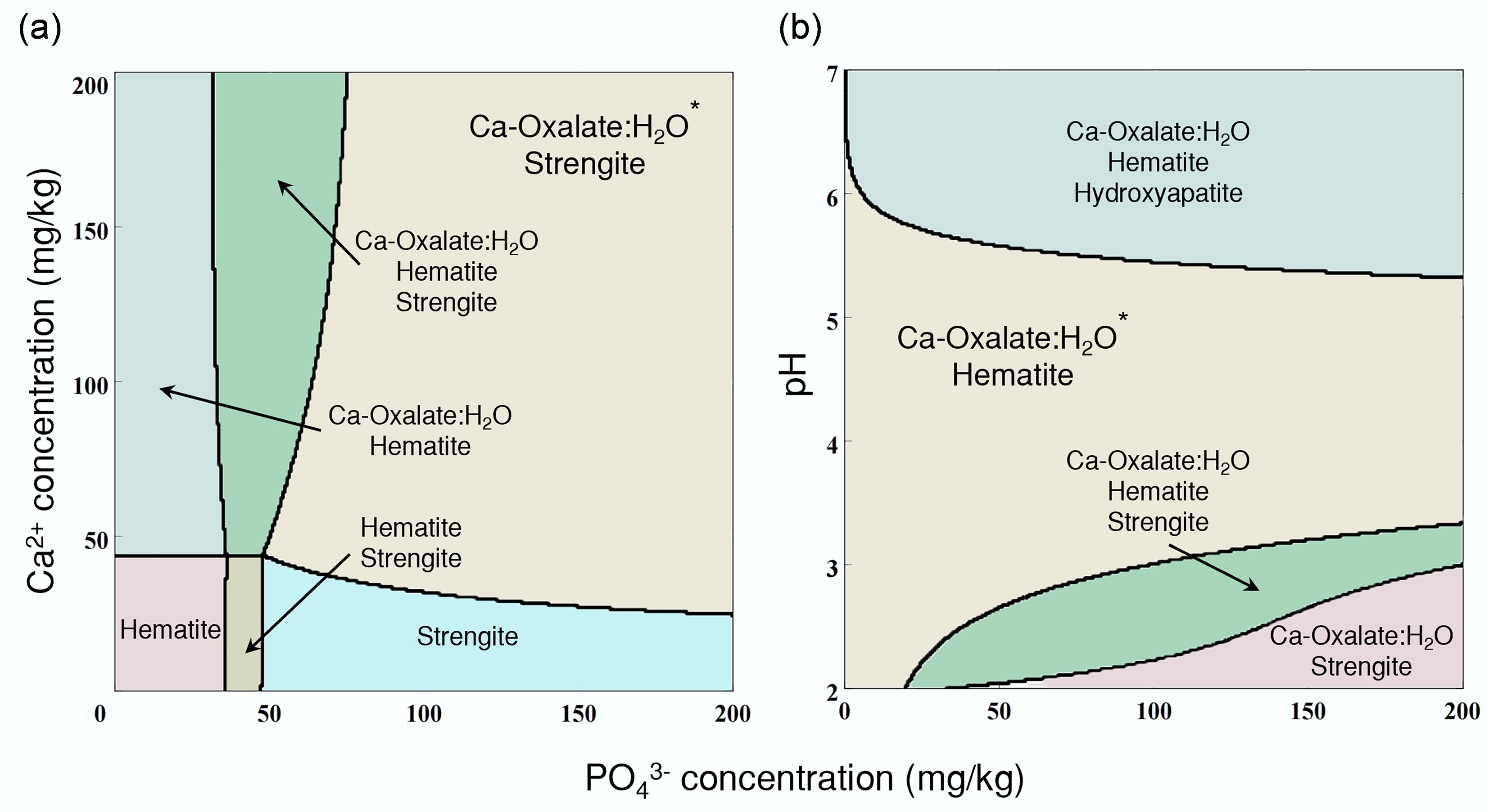
Figure 6.
Simulation of the A. niger-phosphogypsum interaction system. (a) Geochemical simulated mineralization under different concentrations of PO43− and Ca2+ by the GWB Act2 module. (b) Geochemical simulated mineralization under different concentrations of PO43−, and different pH by the GWB Act2 module.
Figures
(6)
Tables
(0)