-
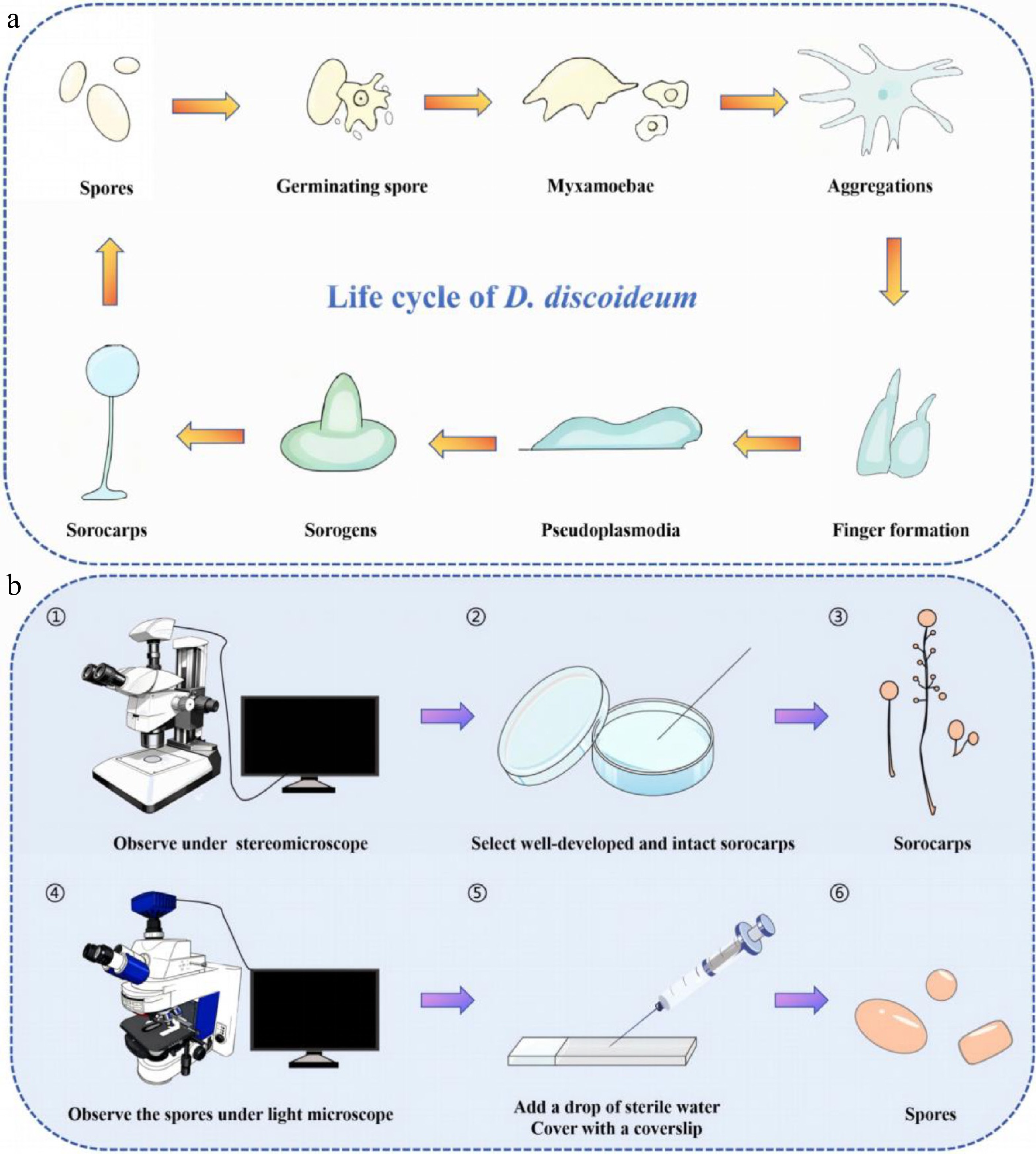
Figure 1.
(a) The amoebae of D. discoideum persist across the organism's complete life cycle. (b) The steps involved for observing spores.
-
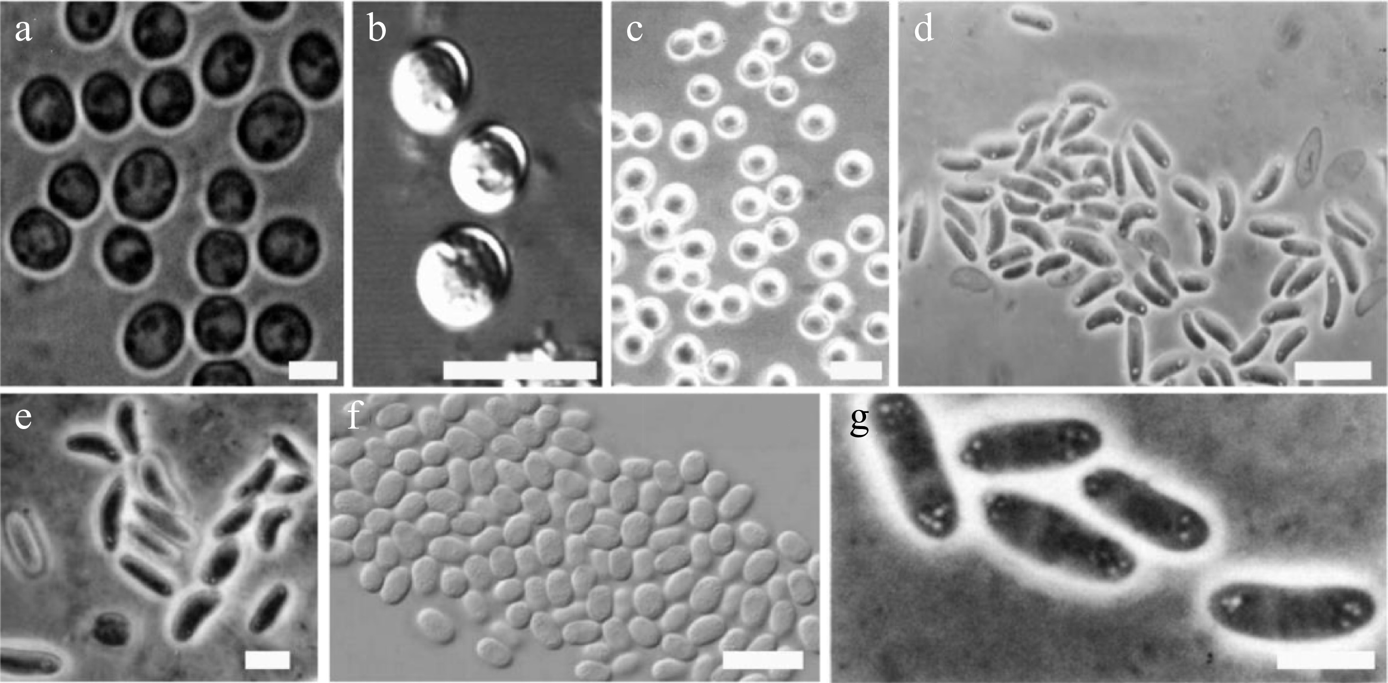
Figure 2.
Examples of different dictyostelid spore shapes (drawn from the corresponding references). (a) Dictyostelium globisporum spores. Bar = 5 μm[18]. (b) D. minimum spores. Bar = 10 µm[19]. (c) Heterostelium equisetoides. Bar = 10 μm[20]. (d) Cavenderia boomerangispora, long and frequently curved PG+. Bar = 10 μm[23]. (e) H. naviculare, elongated navicular spores with consolidated polar granules. Bar = 5 μm[27]. (f) H. flexuosum, relatively small spores, note the widely distributed, numerous unconsolidated granules. Bar = 10 μm[23]. (g) D. polycarpum, group of spores with polar spore granules PG. Bar = 5 μm[30].
-
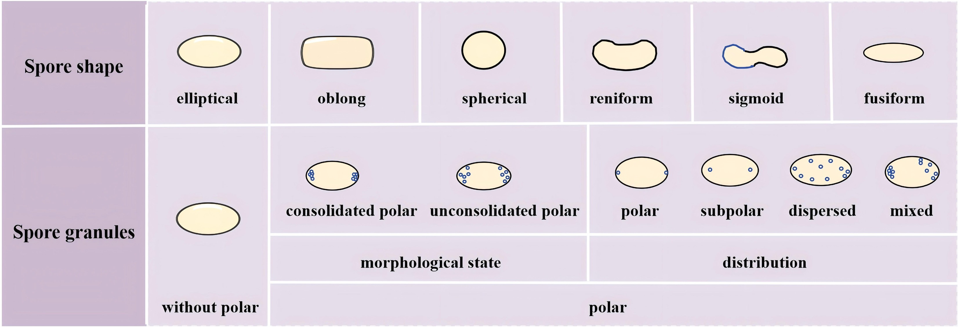
Figure 3.
Microscopic features (shape and granules) of spores used in the identification of dictyostelid species.
-
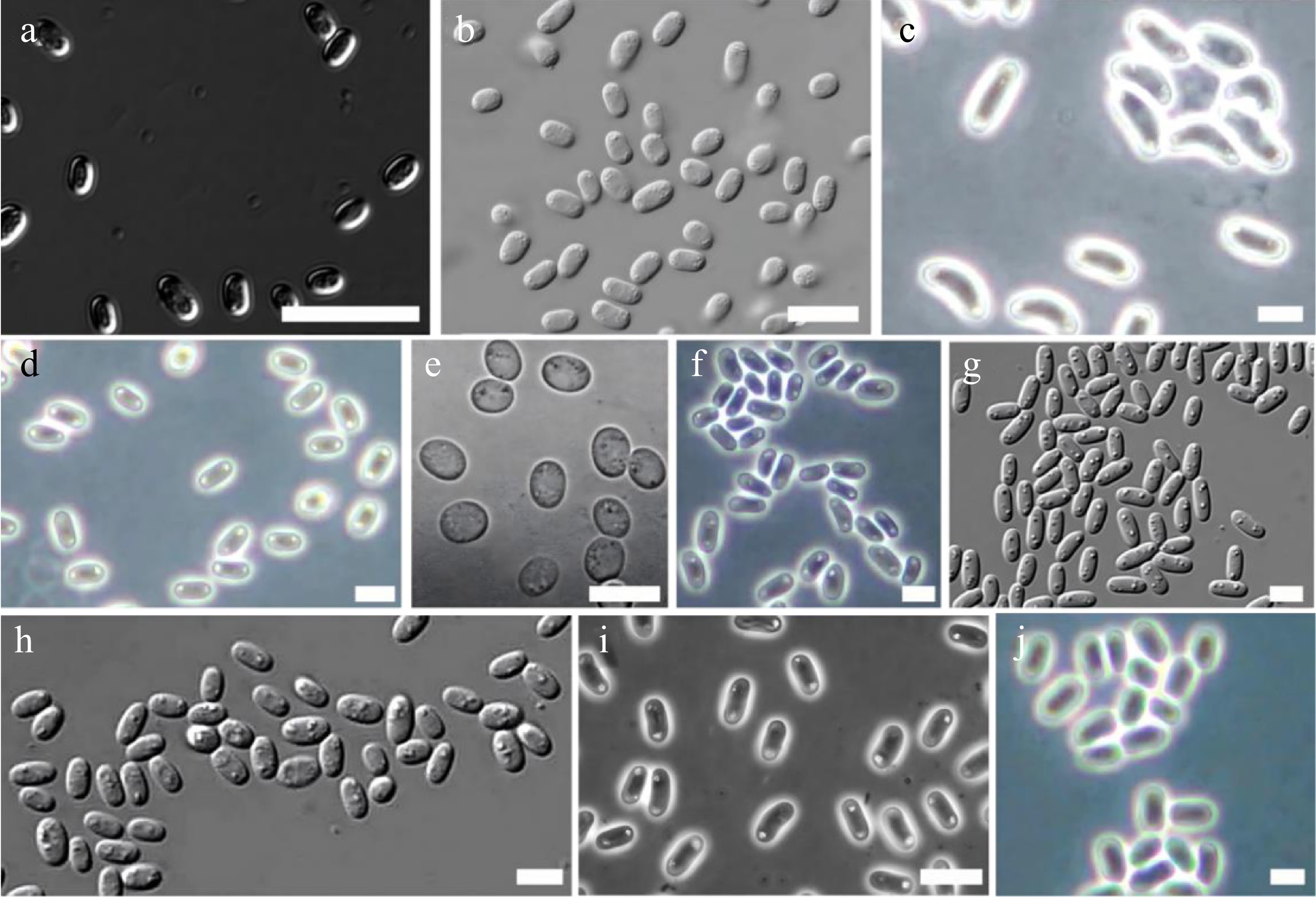
Figure 4.
Examples of different dictyostelid spore granules (drawn from the corresponding references). (a) Dictyostelium robusticaule. Bar = 20 μm[34]. (b) Heterostelium stolonicoideum, oblong spores note the conspicuous unconsolidated polar granules. Bar = 10 μm[23]. (c) Raperostelium cymosum, large elliptical, mostly reniform spores with conspicuous consolidated polar to subpolar granules. Bar = 6 μm[37]. (d) Cavenderia fulva, elliptical spores with prominent refractive consolidated granules at their poles. Bar = 5 μm[37]. (e) R. ibericum. Bar = 10 μm[38]. (f) C. minima, small elliptical irregular spores with polar to subpolar consolidated granules, generally the cluster of granules appears larger at one of the poles. Bar = 6 μm[37]. (g) D. capillare, elliptical spores with conspicuous, consolidated polar granules. Bar = 5 μm[32]. (h) C. bhumiboliana, rather large, elliptical spores with consolidated polar granules. Bar = 10 μm[39]. (i) Hagiwaraea irregularibrachiatum, elliptical short spores with small unconsolidated polar to subpolar granules. Bar = 6 μm[37].
-
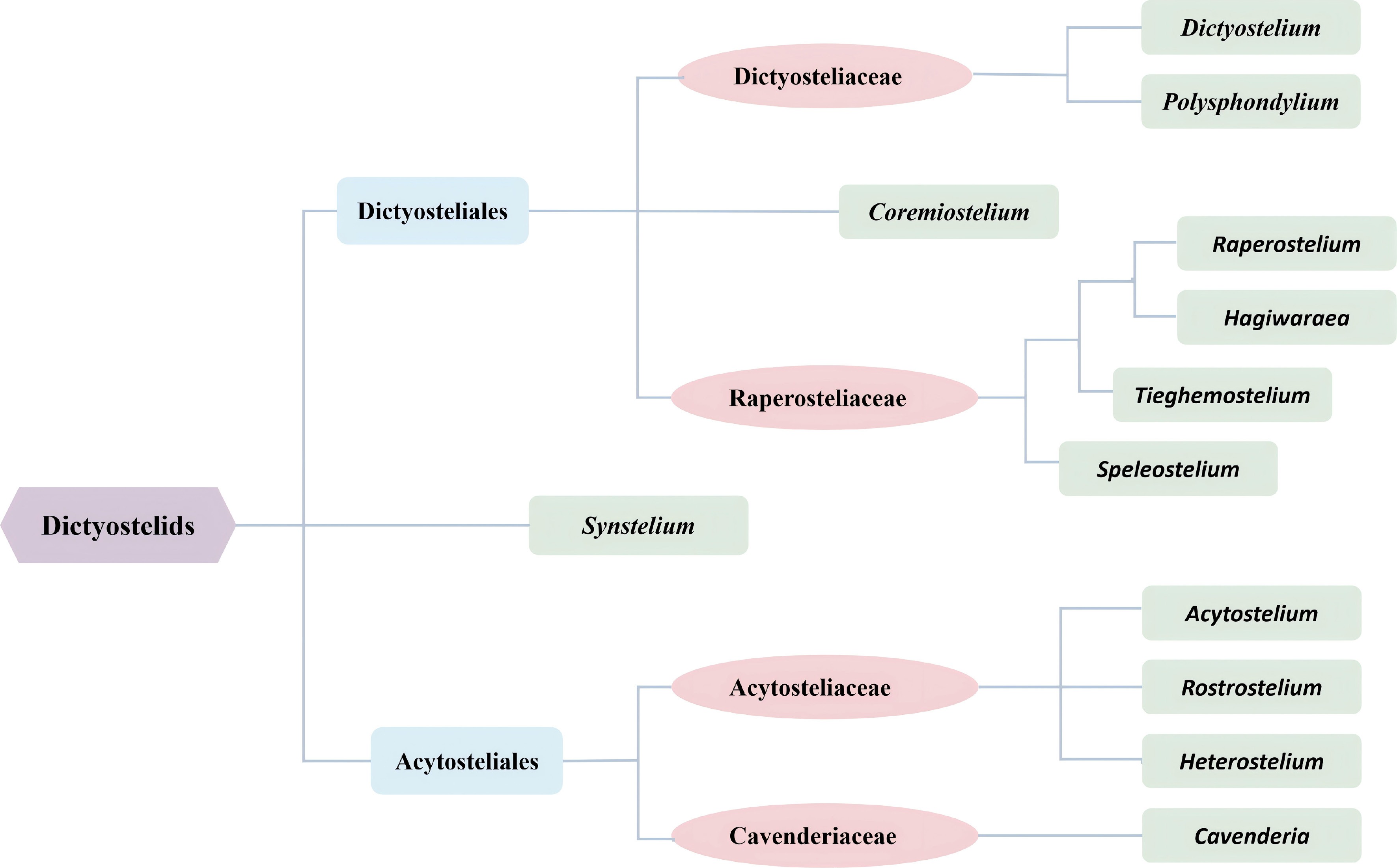
Figure 5.
The current classification used for dictyostelids[36] (drawn from Sheikh et al. 2018).
-
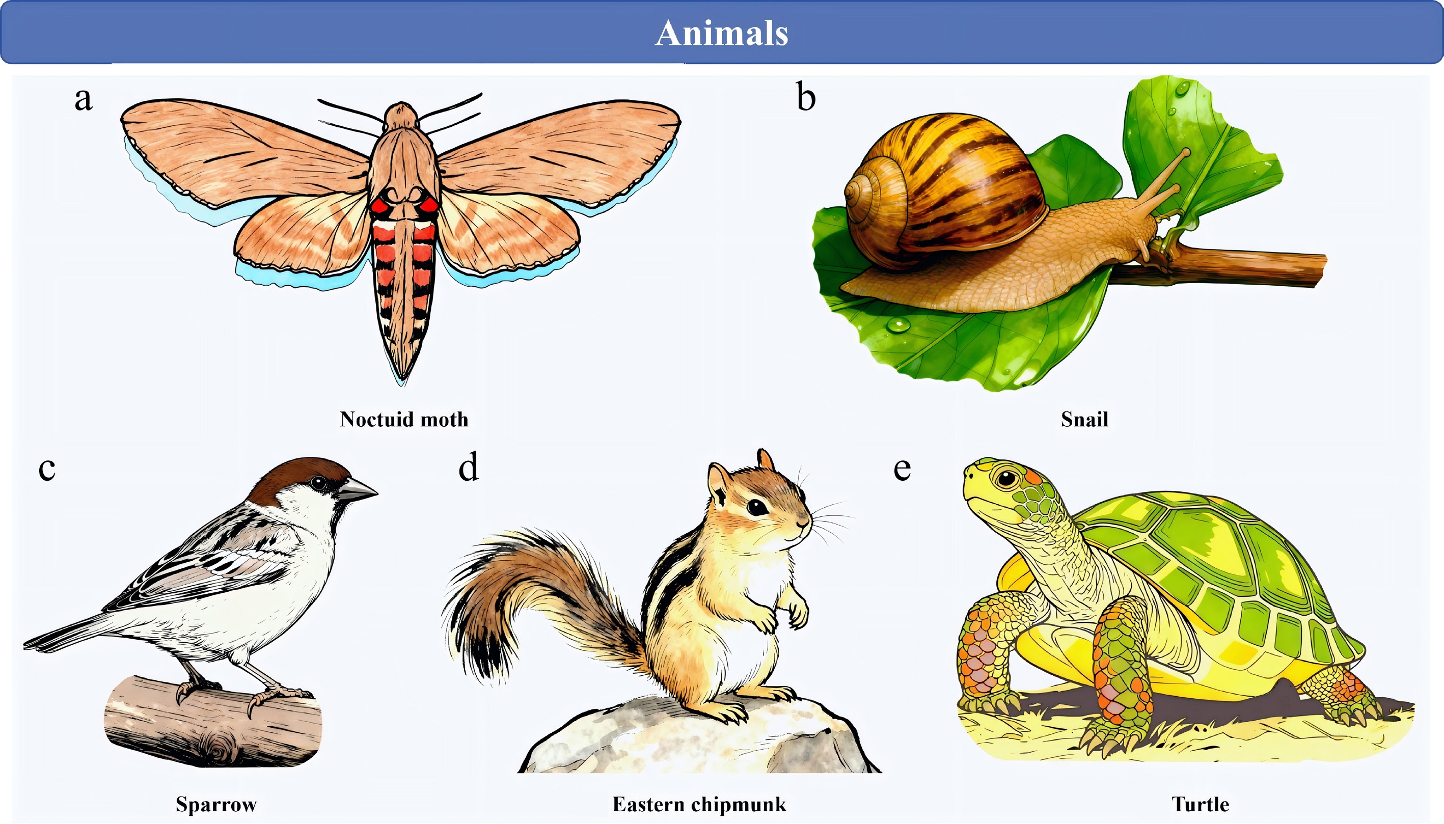
Figure 6.
Dictyostelids rely on animal vectors for spore dispersal in many instances. (a) Noctuid moth. (b) Snail. (c) Sparrow. (d) Eastern chipmunk. (e) Turtle.
-
Species Morphological state (consolidated vs unconsolidated) Distribution
(polar, subpolar,
dispersed)Visibility (conspicuous vs inconspicuous) Special structures
(e.g., halos, refractive features)Others Ref. Dictyostelium ammophilum ! ++ (Occasionally +) Romeralo et al.[40] D. capitatum + – Hagiwaia[41] D. dichotomum Mostly + ++ to + + Vadell & Cavender[14] D. gargantuum – On the surface Vadell et al.[42] D. germanicum Mostly – + On the surface Cavender et al.[43] Polysphondylium violaceum + + Vadell and Cavender[15] P. aureum + + + Hodgson & Wheller[44] P. fuscans – + Perrigo[45] P. patagonicum + Mostly + + Vinaceous Vadell et al.[42] Raperostelium ibericum Mostly +, some – – + One or more relatively
large granulesRomeralo et al.[38] R. australe Polar to subpolar/
dispersed+ Cavender et al.[46] R. cymosum + ++ to + + Cavender et al.[37] R. maeandriforme + ! + Some with a heterogeneous content Cavender et al.[32] Acytostelium anastomosans Central + Cavender et al.[27] *A. subglobosum Distinctively different
from other species due to inconsistently scarce,
minute granulation and clearly recognizable zonationCavender & Vadell[16] Heterostelium anisocaule – + Cavender et al.[46] H. luridum – Mostly throughout
the cytoplasmKauffman et al.[47] H. migratissimum Median + ++ and + + Cavender et al.[8] H. parvimigratum Mostly + Not consistently ++ or +, – Cavender et al.[8] H. radiatum ++ – Perrigo et al.[48] H. rotatum Mostly – – (the largest at
the poles)Landolt et al.[23] H. stolonicoideum Mostly – ++ Landolt et al.[23] H. tikalense – ++ Vadell & Cavender[15] Cavenderia ungulata + ++ + Often surrounded by
a clear narrow haloLarge Cavender et al.[49] C. pseudoaureostipes + Mostly ++, sometimes +
or with – smaller granulesSurrounded by clear halos Many rounded Vadell et al. [39] C. antarctica + ++ to + + Sometimes unipolar, smallest individuals lack granules Cavender et al.[46] C. nanopodia + + Irregular in shape and size Vadell & Cavender[14] C. fasciculata ++ or + + Traub et al.[30] C. fasciculoidea + ++ + Surrounded by a clear halo Visible as angular units Vadell et al.[42] C. fulva 1–2 large + ++ to +,
sometimes –Cavender et al.[37] C. macrocarpa + ++ to + Vadell & Cavender[14] *C. minima Heterogeneous content (often one much larger cluster of granules at one pole with halos, plus tiny dispersed granules) Cavender et al.[37] *C. subdiscoidea + ++ Dense, round, with clear halos Duringdormancy—spores enlarge when in contact with humid substrate, making the spore body heterogeneous and granules larger Vadell et al.[39] C. helicoidea – ++ Cavender et al.[49] Special cases are indicated by an asterisk '*', consolidated '+', unconsolidated '−'; polar '++', subpolar '+', dispersed '−'; conspicuous '+', inconspicuous '−'; concurrence '!'. Table 1.
Species of dictyostelids containing polar particles along with their sources and original literature descriptions.
Figures
(6)
Tables
(1)