-
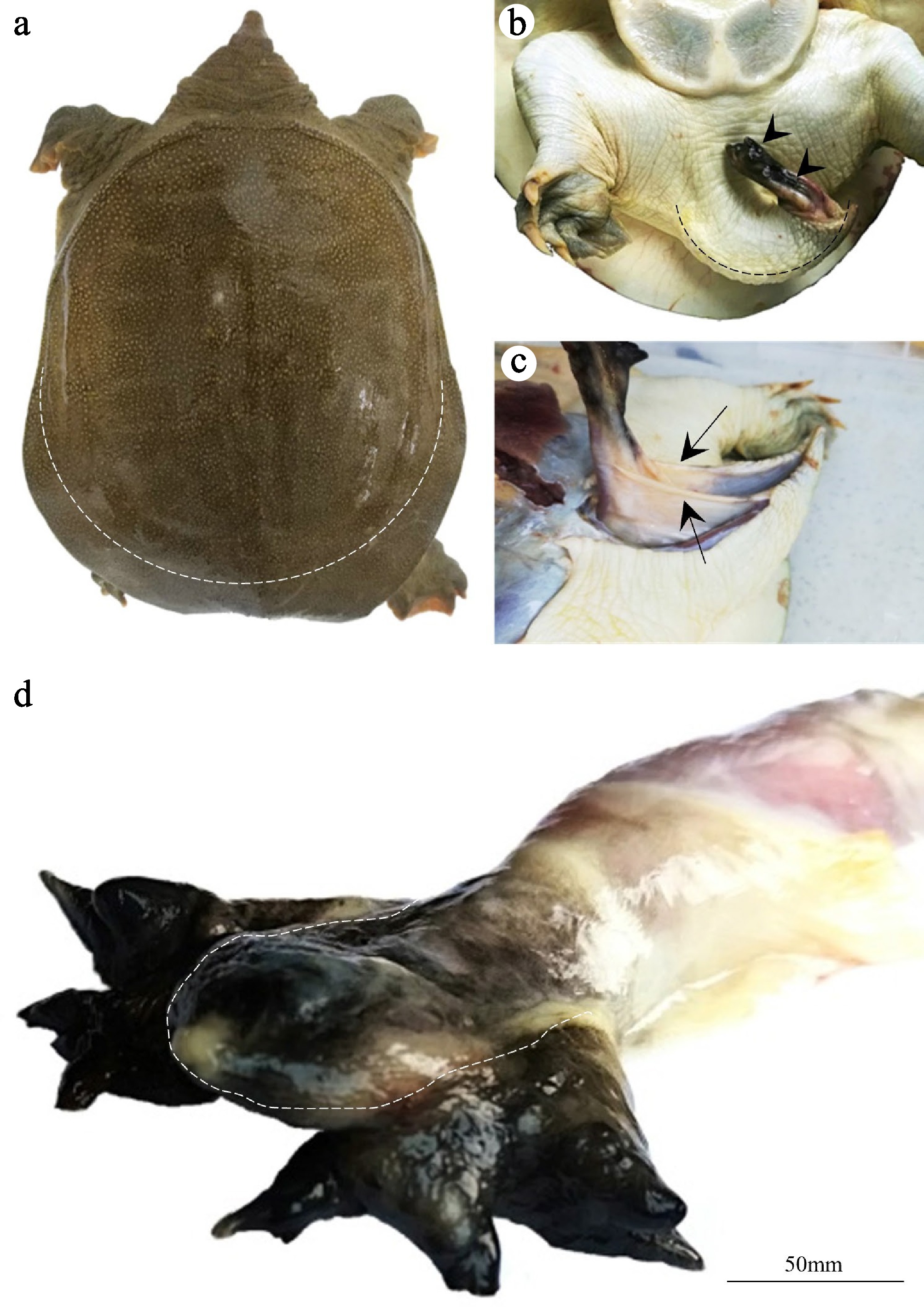
Figure 1.
Observations on the nonerect state of the penis of P. sinensis. (a) Dorsal view of P. sinensis. The penis and the tail were covered by calipish tissue (outside the dotted line). (b) Ventral view of P. sinensis. The hook-shaped penis (dotted line) protruded from the cloaca during sexual arousal and protruded from the cloaca (arrowheads). (c) Observation of the penile mesentery (arrows). (d) The penile glans could be divided into two parts: The hard central cartilage (inside the dotted line) and the soft branches (outside the dotted line).
-
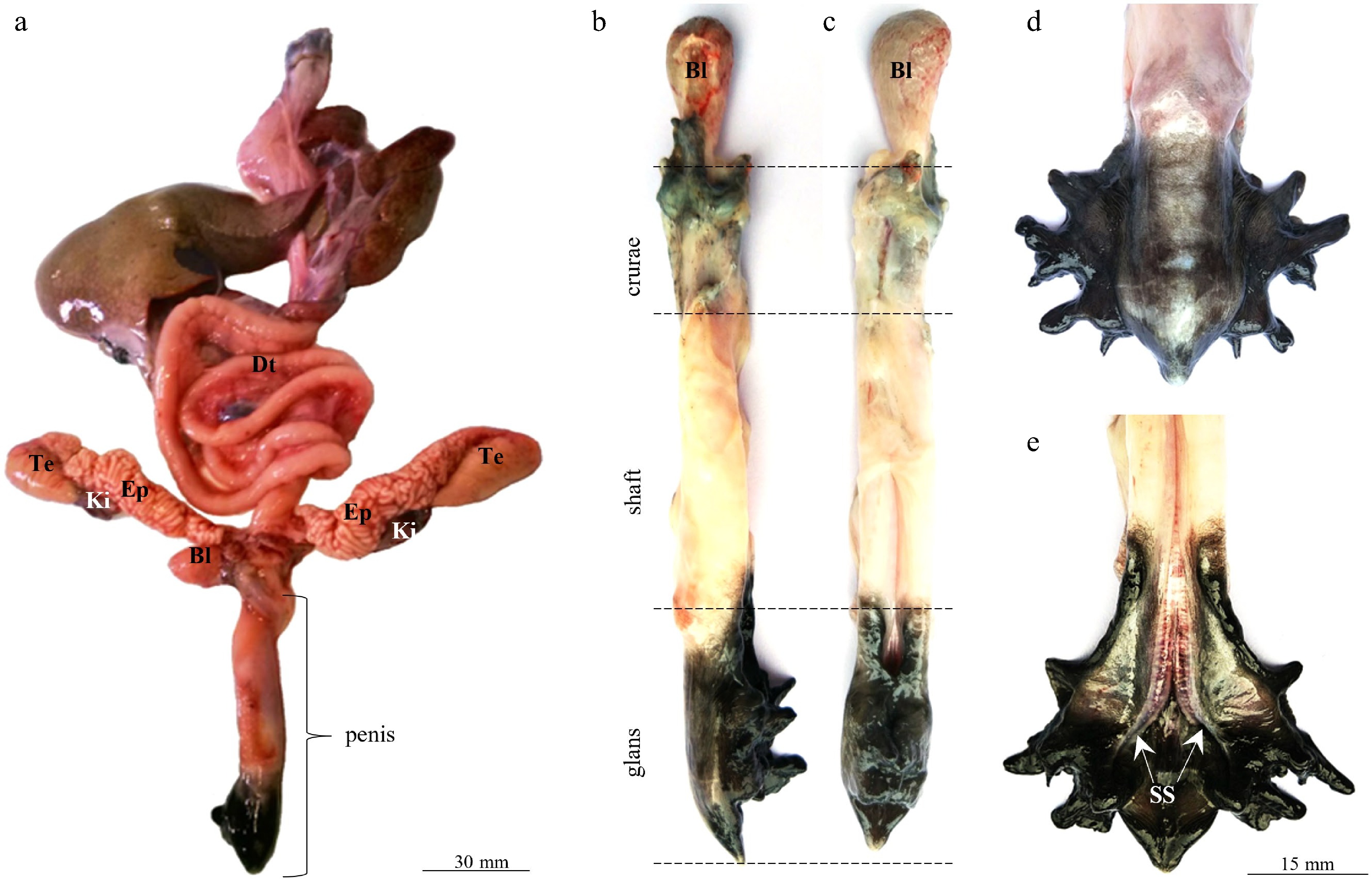
Figure 2.
Anatomical observation of the penis of P. sinensis. (a) The penis and its surrounding organs. (b, c) The penis can be divided into three parts: The crurae, the shaft, and the glans. (d, e) Appearance of the penile glans. Te, testis; EP, epididymis; BL, bladder; DT, digestive tract; Ki, kidney; SS, sulcus spermaticus.
-
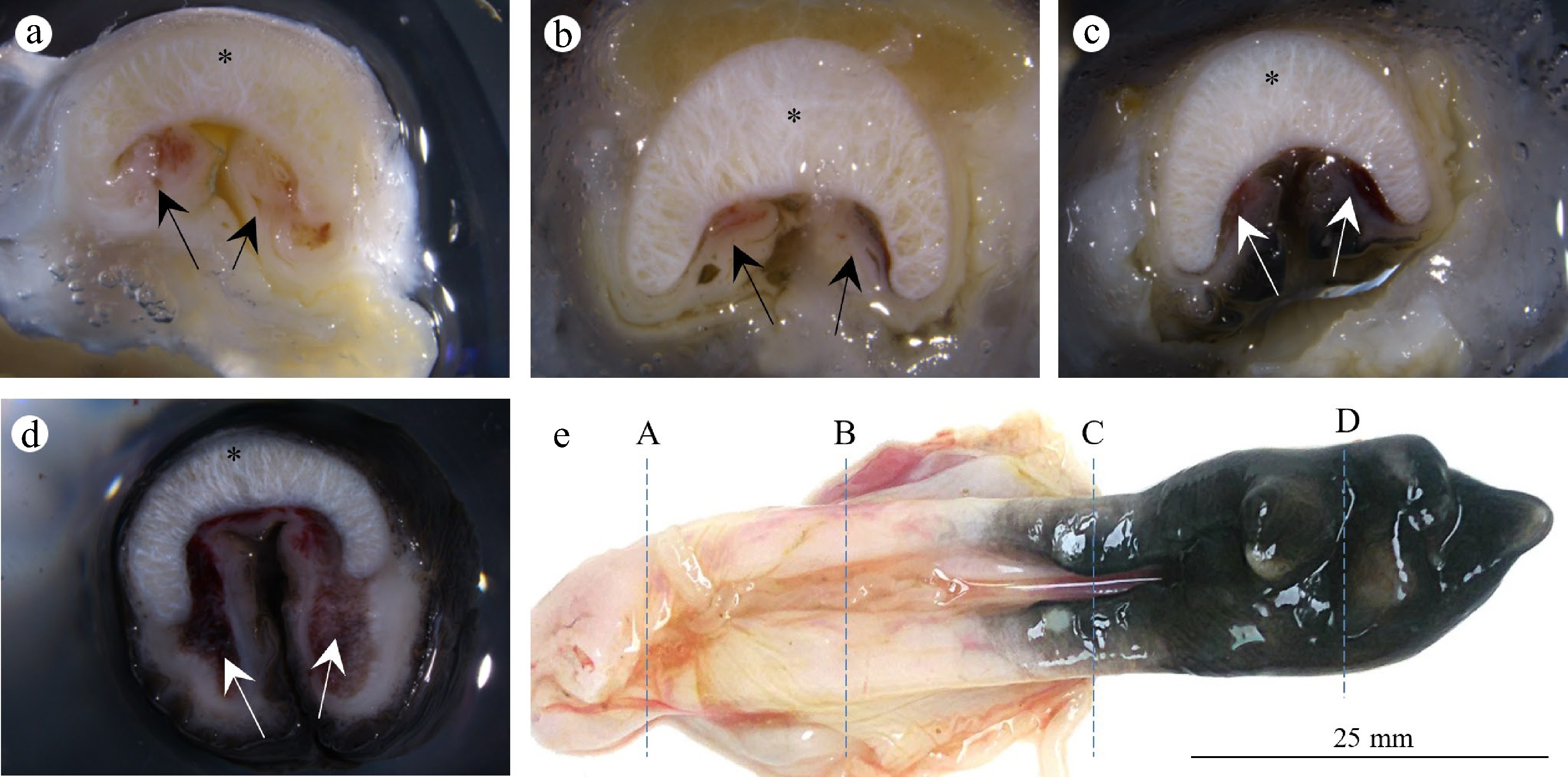
Figure 3.
Collagen cartilage and corpus cavernosum of the penis of P. sinensis. (a–d) Cross-sections of the penis. The penis is mainly occupied by meniscus-shaped cartilage (asterisk) and the corpus cavernosum (arrows). (e) Corresponding sampling locations of (A–D).
-
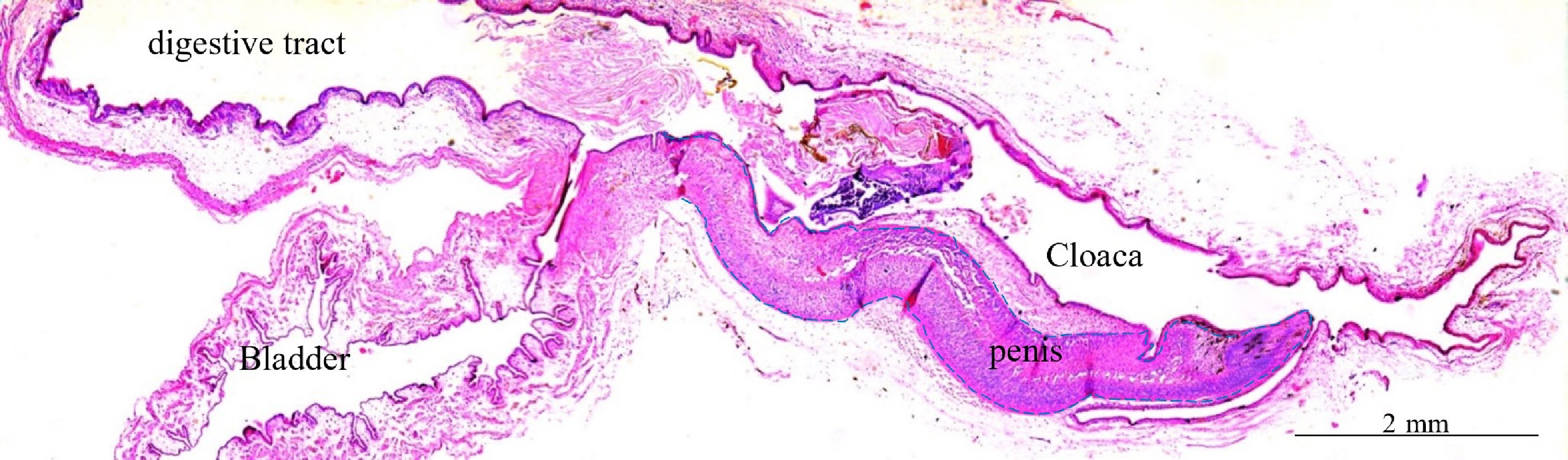
Figure 4.
Observation of the structures around the cloaca of immature P. sinensis revealed by hematoxin and eosin staining.
-
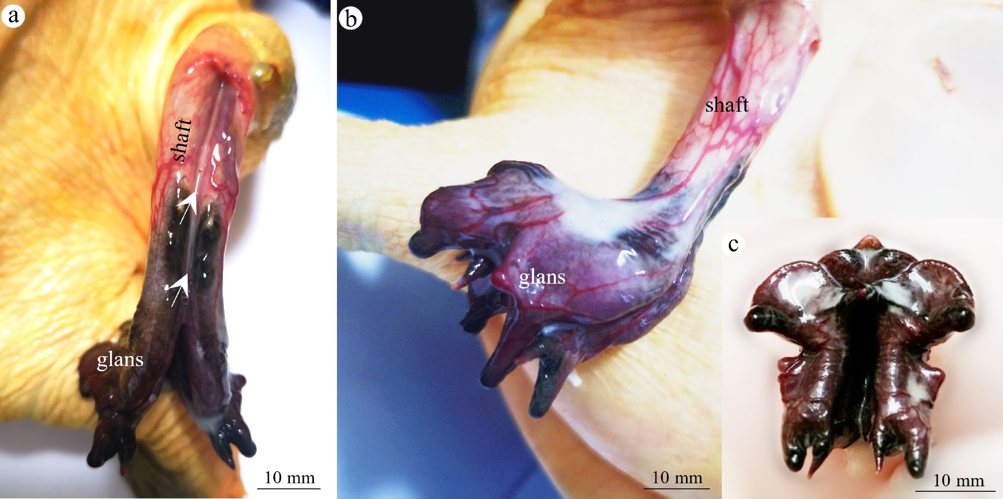
Figure 5.
Observation of the erectile state in the penis of P. sinensis. (a) The penis protruded from the cloaca, with the corpora cavernosa expanding to form a closed channel (arrows). A red vascular network could be seen on the penile surface. (b) The penile glans expands to form a golf club shape. (c) The multibranched structures of the penile glans are blown up.
-
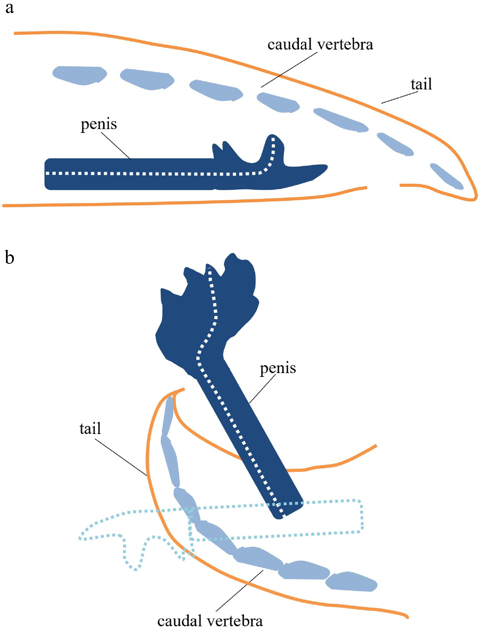
Figure 6.
Key factors in the erect state of P. sinensis. (a) In the natural state, the penis of P. sinensis is hidden in the cloaca, and the tail is in a relaxed state. (b) During sexual arousal, because the tail curved into a hooked shape and the glans expanded extensively, the penis extended out of the cloaca. The dotted outline represents the original position of the penis in the cloaca.
Figures
(6)
Tables
(0)