


-
The Chinese soft-shelled turtle (Pelodiscus sinensis) is a freshwater species with important nutritional and medicinal value in Chinese aquaculture[1]. P. sinensis usually mates and lays eggs from May to August in the middle and lower reaches of the Yangtze River. After mating, sperm can be stored in the reproductive tract of female turtles for six months[2,3]. However, there is a lack of systematic studies on the mating behavior of P. sinensis at present. In particular, there is almost no research on the penis and penile erection in P. sinensis. The structure of the penis is highly differentiated, diverse, and evolving rapidly among different species[4]. In terms of the structure of the penis, there are similarities among turtles, lizards, and crocodiles: The penises of these animals are all fleshy cylinders and contain a hydraulic skeleton that fills with blood before mating. This results in the penis expanding and enhances its bending resistance and strength[5].
The penis has a homologous structure in amniote animals. Penile development in turtles is very similar to that in mice at the tissue, cellular, and molecular levels[6]. Although all amniote animals take part in internal fertilization, not all amniotes have penises. The penises of turtles and mammals are not homologous organs[7,8], so there are some differences in their morphology. If we observe cross-sections of the penises of turtles, birds, mammals, and snakes[9], the penis of each of these animals is not the same. Mammals transport semen through closed pipes, whereas turtles, birds, and snakes transport semen through seminal grooves. In this study, the anatomical structure and erectile state of the penis of P. sinensis were studied by anatomical and histological analyses.
-
Six mature and healthy male P. sinensis (raised under natural conditions, weighing 2–2.5 kg), six 3-month-old male turtles (20–30 g in weight), and six mature and healthy females (raised under natural conditions, weighing 1.5–2 kg) were purchased from Ningbo Wal-Mart Stores in Wanda Mall.
Experimental method
-
Six mature P. sinensis were left in cold water (4–8 °C) for 1 h to keep them quiet while observing the morphological characteristics of the tail and the changes in the sexual reflex when the tail is squeezed. The turtles were rendered comatose using an intraperitoneal administration of sodium pentobarbital (20 mg/kg) and slaughtered. The male reproductive system was carefully separated, and the histological characteristics of the penis, testis, epididymis, and other tissues were observed and photographed. Meanwhile, to study the physiological function of the penis of P. sinensis during mating, the penis in its sexually excited state was also observed and photographed. Meanwhile, the structure of the female cloaca was also observed to analyze the penis's structure and function from a mating perspective.
In order to study the relationship among the penis, digestive system, and urinary system of P. sinensis, six juvenile turtles were slaughtered and used for histological research. Without damaging the cloaca, the carapace was removed to expose the internal organs. The liver, spleen, heart, and most of the intestinal tissues (with the rectum retained) were removed. The tissues surrounding the connection between the rectum and the bladder were left. After removing the excess muscle tissue, the bladder, rectum, penis, and cloaca were separated as a whole. The tissues were fixed in a 4% polyformaldehyde solution for 24 h. After fixation, the tissue was gradually dehydrated using an ethanol gradient (from 70% to 100%) and then treated with xylene for transparency. Next, it was immersed in molten paraffin at 70 °C for 12 h to allow the paraffin to penetrate the tissue. Subsequently, the tissue was embedded and prepared for use as paraffin section (10 μm). After the sections were dried at 42 °C, they were deparaffinized in xylene then hydrated with a gradient alcohol solution. Next, the sections were stained with hematoxylin for 60 s to make the cell nuclei blue, then stained with eosin for 3 s to make the cytoplasm pink. Subsequently, the sections were dehydrated with a gradient alcohol solution and treated with xylene for transparency. Finally, they were sealed with a resin sealant for microscopic examination.
-
In a natural state, the penis and tail of P. sinensis were covered by calipish tissue, the marginal sides of the leathery and pliable carapace (Fig. 1a). The tail of male P. sinensis was thick and strong, and the cloaca opened at the tail. During the sexual response, the tail curved into a hooked shape and the penis extended from the cloaca (Fig. 1b). The black penile glans was entirely free in the cloaca, but both sides of the penile shaft were connected to the cloaca by mesentery tissue and muscle traction. The penile mesenteric tissues and penile traction muscles coordinated the bending of the tail and the actions during the protrusion and retraction of the penis. (Fig. 1c). The black penile glans could be divided into two parts: The hard central cartilage and the soft branches (Fig. 1d).
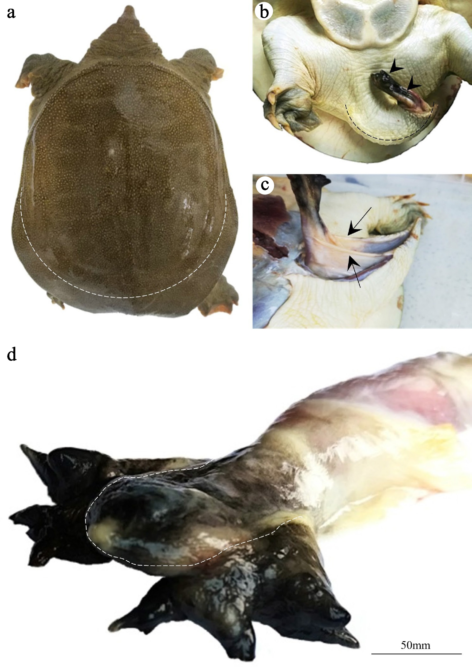
Figure 1.
Observations on the nonerect state of the penis of P. sinensis. (a) Dorsal view of P. sinensis. The penis and the tail were covered by calipish tissue (outside the dotted line). (b) Ventral view of P. sinensis. The hook-shaped penis (dotted line) protruded from the cloaca during sexual arousal and protruded from the cloaca (arrowheads). (c) Observation of the penile mesentery (arrows). (d) The penile glans could be divided into two parts: The hard central cartilage (inside the dotted line) and the soft branches (outside the dotted line).
Anatomical observation of the penis of P. sinensis
-
After dissection, the organs and tissues connected to the penis were separated. The results indicated that that the penile crurae was directly connected to the end of the digestive tract. At the junction between the digestive tract and the penis, the epididymis was connected to the penis through the vas deferens, and the bladder was located on the ventral side of the penis (Fig. 2a). When the redundant tissues were removed, leaving only the bladder and penis, the penis was shaped like a solid rod and could be divided into three parts: The crurae, the shaft, and the glans (Fig. 2b, c). The penile crurae was gray-black, and the penile glans was black; the white middle part was the penile shaft. There was an open tubular structure on the dorsal side of the penile, and the spindle-shaped penile glans looked like a closed flower (Fig. 2c). However, when subjected to external traction, it formed multi-branched structures with small valves, creating a significant synaptic structure (Fig. 2d). The pointed end of the penile glans was solid and hard, and the multibranched structures were soft and elastic. The channels for sperm discharge extended to the side branches of the penile glans in the form of an open seminal groove (Fig. 2e).
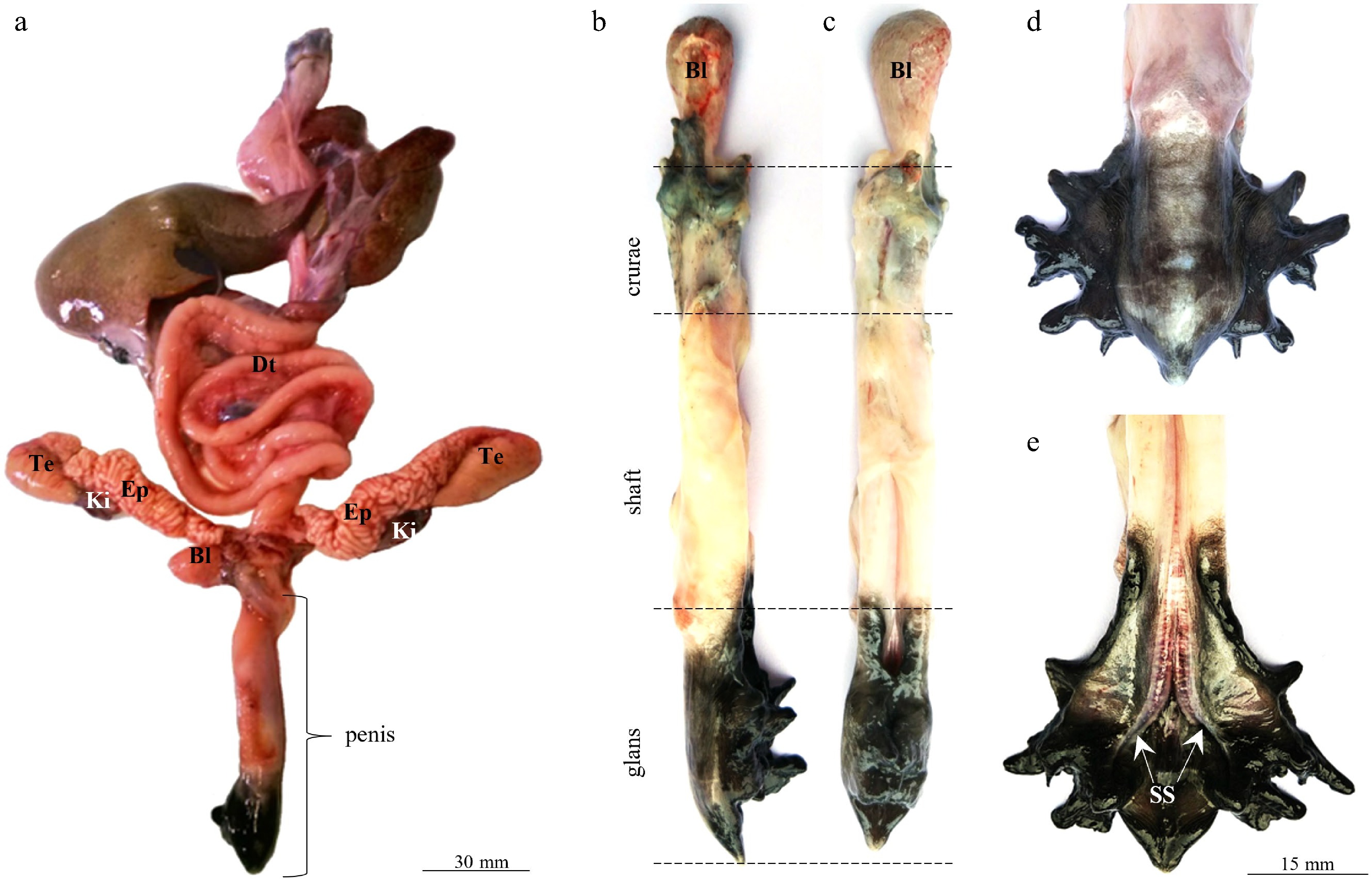
Figure 2.
Anatomical observation of the penis of P. sinensis. (a) The penis and its surrounding organs. (b, c) The penis can be divided into three parts: The crurae, the shaft, and the glans. (d, e) Appearance of the penile glans. Te, testis; EP, epididymis; BL, bladder; DT, digestive tract; Ki, kidney; SS, sulcus spermaticus.
In the cross-sections of the penis at different positions, the most obvious feature was the meniscus-shaped cartilage that occupied a large portion of the penis (Fig. 3). The penile cartilage was located on the ventral side of the penis and has a white fibrous reticular structure. On both sides of the concave surface of the penile cartilage, there was a hollow reticular tissue (Fig. 3a–c). This was the corpus cavernosum, which converged at the penile glans to form a huge blood sinus (Fig. 3d). The penile traction muscle and penile mesentery were located on both sides of the penile shaft (Fig. 3e).
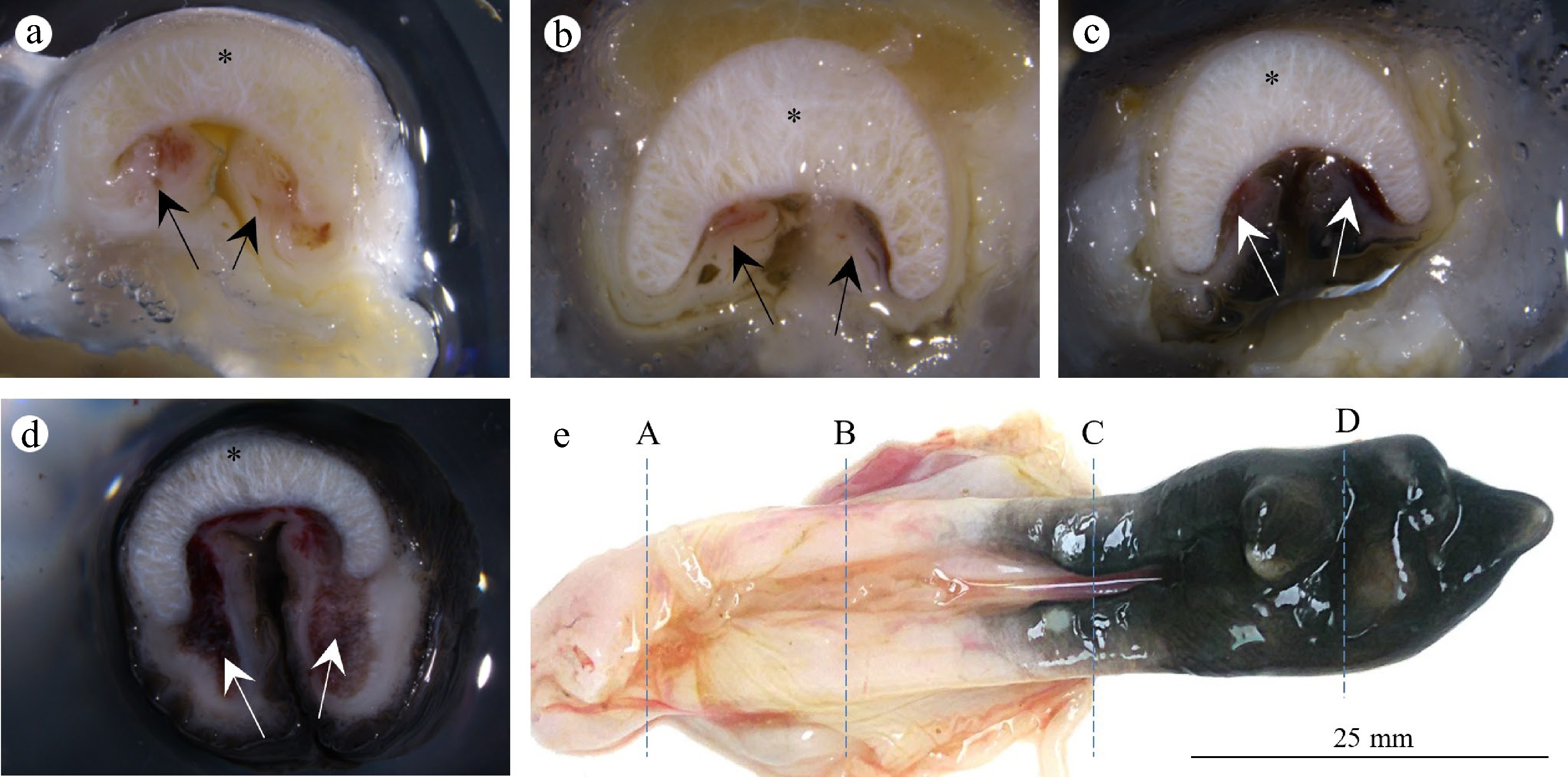
Figure 3.
Collagen cartilage and corpus cavernosum of the penis of P. sinensis. (a–d) Cross-sections of the penis. The penis is mainly occupied by meniscus-shaped cartilage (asterisk) and the corpus cavernosum (arrows). (e) Corresponding sampling locations of (A–D).
Interconnections between the penis, digestive system, and urinary system of P. sinensis
-
The penis, digestive system, and urinary system of P. sinensis were all connected to the cloaca (Fig. 4). The digestive tract and the bladder converged at the base of the penile crurae, and the contents of the digestive tract can enter the cloaca through the dorsal side of the penis for excretion. The bladder of P. sinensis has a sac-like structure containing many folds and opening into the digestive tract. There was a thick layer of muscle tissue at the outlet of the bladder. Although the penis contracted within the cloaca, histological results indicated that the overall structure of the penis was a W-shaped bend, with its distal end separating within the cloaca.
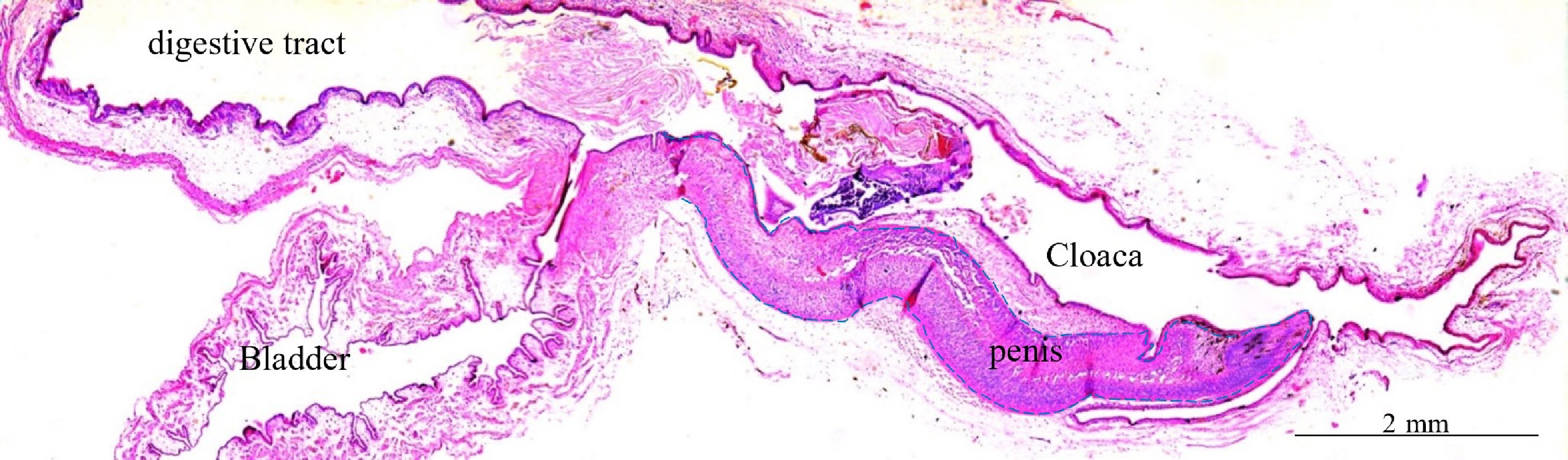
Figure 4.
Observation of the structures around the cloaca of immature P. sinensis revealed by hematoxin and eosin staining.
Observations on the erect state of the penis of P. sinensis
-
In a sexually aroused state, the penis of P. sinensis protrudes from the cloaca, whereas the corpus cavernosum of the penis expanded to form a closed channel for sperm excretion (Fig. 5a). As the glans penis enlarges greatly, a red vascular network could be seen on the penile surface of the penis. At this time, the diameter of the glans penis expanded more than three times, and the penis took on the shape of a golf club (Fig. 5b). The multibranched structures of the glans penis were blown up to form an exaggerated forked structure, and the color changed from black to dark purple (Fig. 5c).
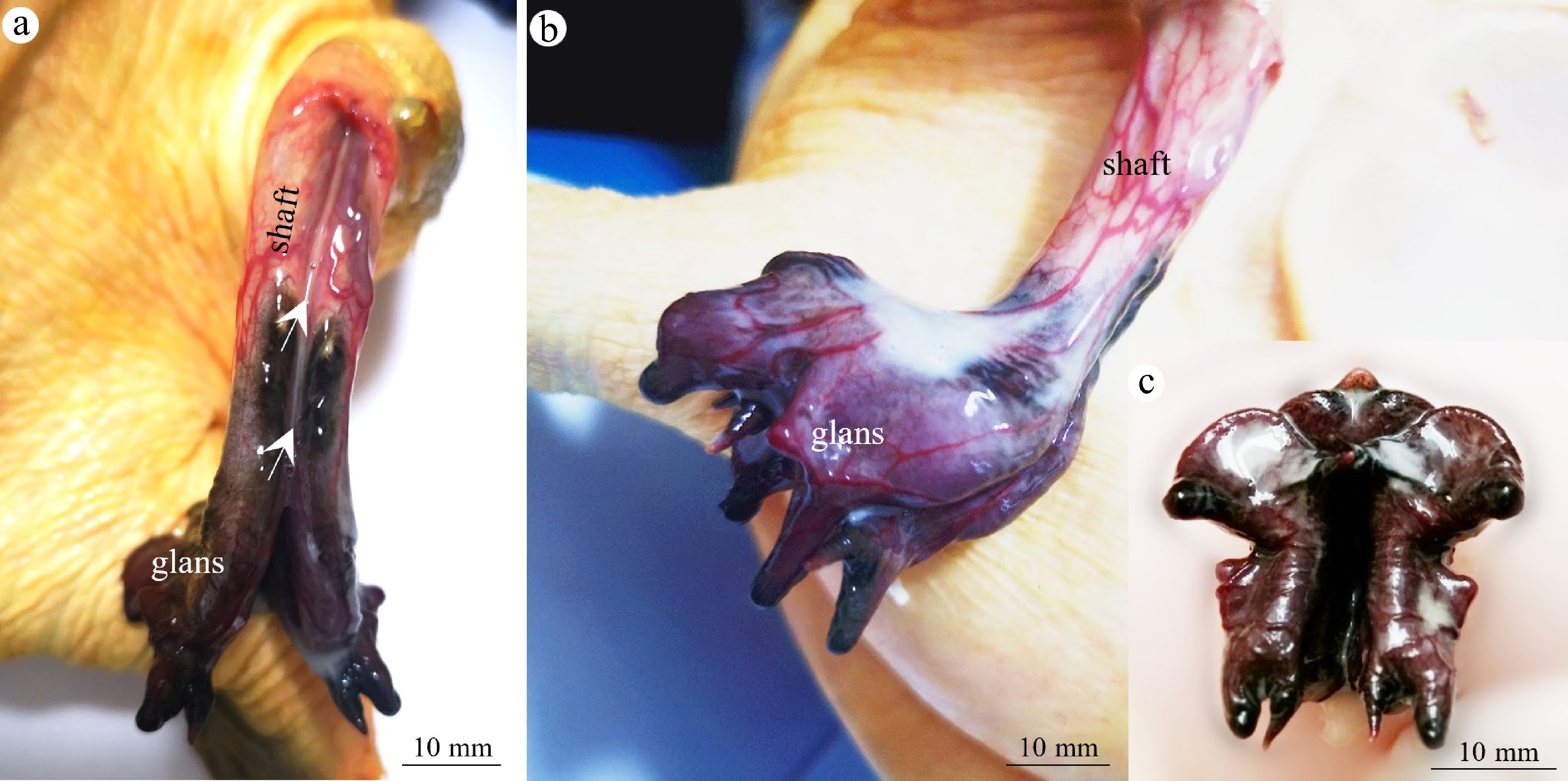
Figure 5.
Observation of the erectile state in the penis of P. sinensis. (a) The penis protruded from the cloaca, with the corpora cavernosa expanding to form a closed channel (arrows). A red vascular network could be seen on the penile surface. (b) The penile glans expands to form a golf club shape. (c) The multibranched structures of the penile glans are blown up.
Analysis of key factors for penile erection in P. sinensis
-
Under normal conditions, the penis of P. sinensis was usually hidden in the cloaca, while the tail was in a relaxed state (Fig. 6a). However, the tail could be bent into a hook shape during sexual excitation. This shortened the distance from the penis to the cloaca opening facilitated the protrusion of the erect penis. The tail curved into a hook shape during erection, which caused the penis to rotate further after erection. At this point, the penile crurae could not move freely, and the penis could not be retracted into the cloaca. In addition, as the corpus cavernosum tissue expanded, a huge glans structure formed, which was so large that the cloaca could not accommodate it. In addition to the other factors (the tail curving into a hook shape and the expanded glans penis), the penile mesentery and the penile traction muscle could also work together to control the spatial position of the penis. Therefore, under the combined effect of these factors, the penis of P. sinensis achieved the function of erection (Fig. 6b).
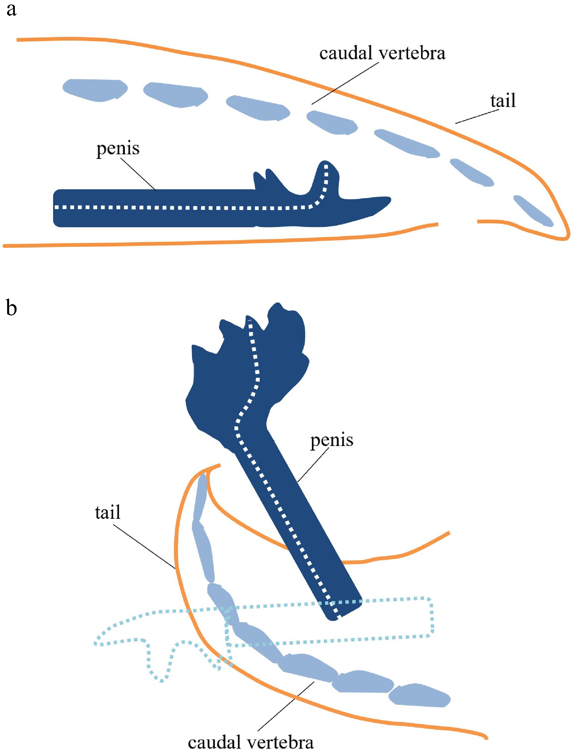
Figure 6.
Key factors in the erect state of P. sinensis. (a) In the natural state, the penis of P. sinensis is hidden in the cloaca, and the tail is in a relaxed state. (b) During sexual arousal, because the tail curved into a hooked shape and the glans expanded extensively, the penis extended out of the cloaca. The dotted outline represents the original position of the penis in the cloaca.
-
This study compares the anatomical structure of the penis in both the nonerect and erect states in P. sinensis and analyzed the main factors influencing penile erection. During sexual excitement, the tail of P. sinensis bends into a hooked shape, forcing the penile tissue to extend out of the cloaca. Similar to many other reptiles, the male's tail may play a crucial role in controlling the direction of the penis during copulation[10]. The male will use its tail to control the penis in order to find the cloacal opening of the female.
The penis evolved and diversified among amniote lineages. For turtles and crocodiles, the end of the penis shows a wide open shape[11,12], which evolved into a glans with branches[7,8,13]. However, unlike the common turtles, the branched glans of P. sinensis is more obvious and special, which may be more conducive to controlling the release of sperm. This branched glans structure is almost absent in mammals and birds, but there is a special structure. the male urogenital mating protuberance in mouse glans[14], which may be an evolutionary coincidence.
Like sea turtles, the penis, which folds and bends in the cloaca, will stick out under the joint action of the mesentery and the penile traction muscle[13]. On the horizontal sides of the penile shaft, mesentery tissue and traction muscles are attached. The penile traction muscle may exist to eject the penis from the cloaca. With cloacal crowding and contraction of the penile traction muscle, the penis is pulled out toward the cloacal outlet, eventually fully exposing the penile shaft.
The morphologic results of the penile cross-section showed that the penis is mainly occupied by cartilage and the corpus cavernosum. Because of the presence of the penile cartilage, the penis maintains good hardness. However, before penis is erect, the cavernous body needs to be sufficiently engorged, similar to most amniote animals. The anatomical structure of the penis in marsupials shows a wide range of morphological variations[15]. Mammals[16], turtles[17], and crocodiles[18] all have their own penis, but only a few existing birds, such as ratites and ducks, have a penis[19,20]. Compared with these animals, the penis of P. sinensis is most similar to that of the tortoise; both consist of a rod-shaped structure with seminal grooves. In mammals, turtles, and crocodiles, the penis enlarges through congestion of the blood vessels[21]. Along with the penis extending from the cloaca, the diameter of the penile glans of P. sinensis can expand more than three times in size. Eventually, the erect penis is exposed to the cloaca and cannot be easily retracted.
In addition, the urinary, digestive, and reproductive systems have a common outlet in P. sinensis. The rectum and bladder of P. sinensis meet at the penile crurae, and the contents of the digestive tract can enter the cloaca through the dorsal side of the penis during excretion. Therefore, the urinary, digestive, and reproductive systems of P. sinensis share the dorsal side of the penis. Although the channel for sperm delivery is fully established in the erect state, the contamination of semen by the urinary and digestive systems needs further study.
This work was supported by the Zhejiang Province New Aquatic Variety Breeding Project (2021C02069-8-3), and the Major Technology Research and Development Project in Ningbo (2021Z009), and the Innovation Yongjiang 2035 Key R&D Programme in Ningbo (2025Z092).
-
The ethical standards for the animal protocol were thoroughly passed and approved by the Experimental Animal Ethics Committee of Zhejiang Wanli University. The approval number for the ethical review is #2020070101. The research followed the 'replacement, reduction, and refinement' principles to minimize harm to animals. This article provides details on the housing conditions, care, and pain management for the animals, ensuring that the impact on the animals was minimized during the experiment.
-
The authors confirm their contributions to the paper as follows: conceived the study: Wang W; conducted the analysis of the results and wrote the manuscript: He X; experimental study: He X, Ren T; participated in the discussion of the results: Ren T, Qian G, Li C; revised and edited the manuscript: Wang W, Li C. All authors reviewed the results and approved the final version of the manuscript.
-
All data generated or analyzed during this study are included in this published article.
-
The authors declare that they have no conflict of interest.
- Copyright: © 2026 by the author(s). Published by Maximum Academic Press on behalf of Nanjing Agricultural University. This article is an open access article distributed under Creative Commons Attribution License (CC BY 4.0), visit https://creativecommons.org/licenses/by/4.0/.
-
About this article
Cite this article
He X, Ren T, Qian G, Li C, Wang W. 2026. Anatomical structure and erectile state analysis of the penis in the Chinese soft-shelled turtle, Pelodiscus sinensis. Animal Advances 3: e014 doi: 10.48130/animadv-0026-0002 

Anatomical structure and erectile state analysis of the penis in the Chinese soft-shelled turtle, Pelodiscus sinensis
- Received: 01 November 2025
- Revised: 21 January 2026
- Accepted: 21 January 2026
- Published online: 29 April 2026
Abstract: The Chinese softshell turtle (Pelodiscus sinensis) is an important aquaculture species. However, there have been few reports on the structure of its penis and reproductive behavior. In study, the structure and function of penis were investigated using anatomical and histological methods. The results showed that the penis of P. sinensis can be divided into three parts: The crurae, the shaft, and the glans. The center of the penis is meniscus-shaped cartilage that provides the hardness of the penis during mating. After the penis becomes erect and filled with blood, the diameter of the glans expands, forming an exaggerated multibranched structure. The penile crurae is close to the bladder, and the dorsal part of the penis is an extension of the digestive tract. Three main factors are required to achieve an erection in P. sinensis: the tail bends to form a hook - like structure, muscle traction, and abnormal enlargement of the glans. These research findings lay the foundation for further studies on the evolution of the male genitalia of the P. sinensis and the development of artificial insemination techniques in the future.
-
Key words:
- Chinese soft-shelled turtle /
- Pelodiscus sinensis /
- Penile anatomy /
- Erectile state