-
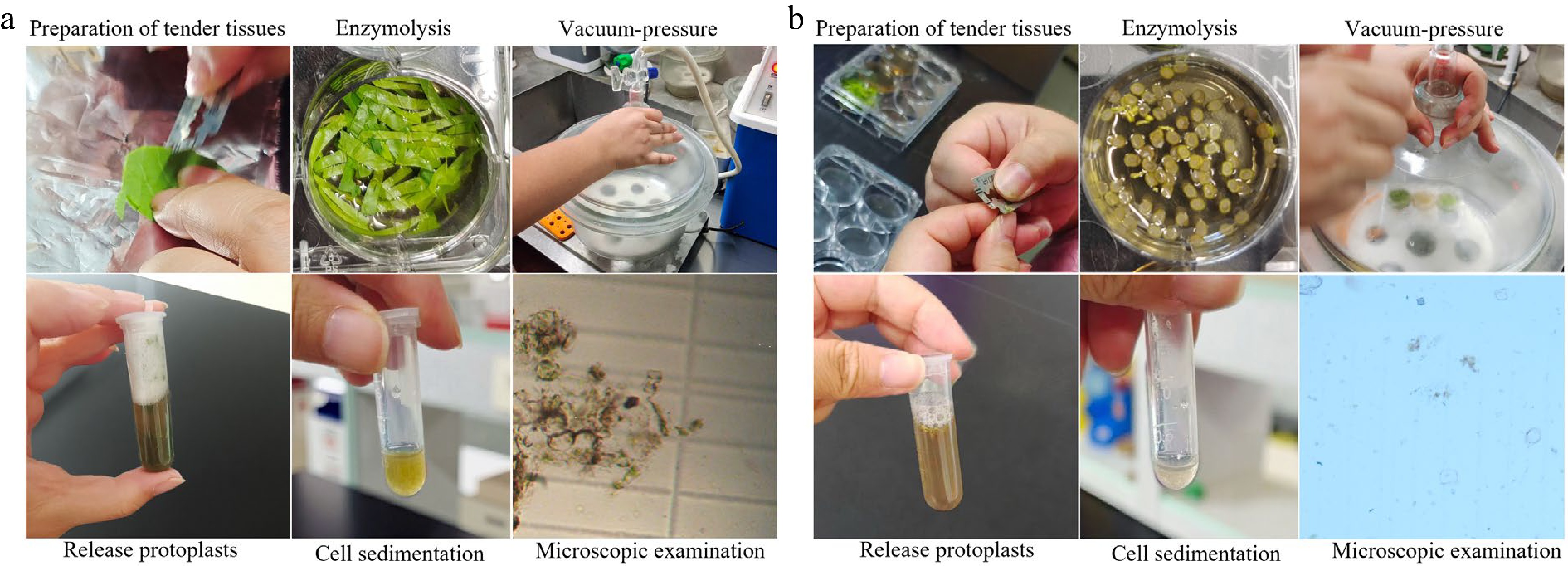
Figure 1.
H. hamabo protoplast isolation from leaf flesh and petiole. (a) The separation process of protoplasts of H. hamabo leaf flesh. (b) The separation process of protoplasts of the H. hamabo petiole. The procedures for isolating protoplasts from the two tissues are as follows; preparation of tender tissues, enzymolysis, vacuum-pressure, release protoplasts, cell sedimentation, and microscopic examination.
-
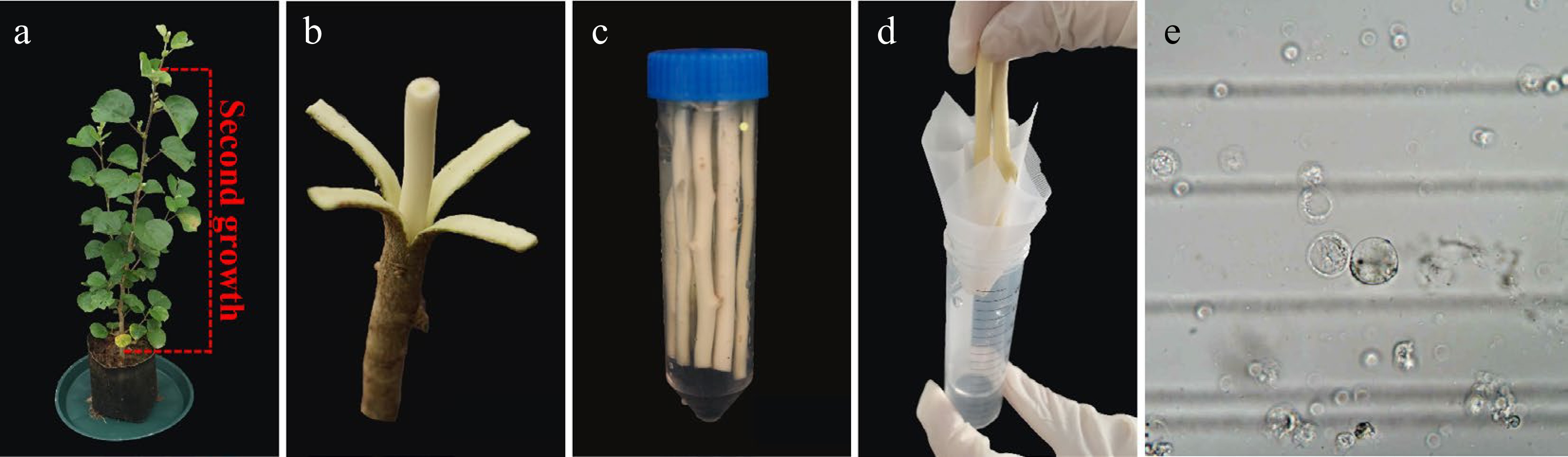
Figure 2.
H. hamabo protoplast isolation from wood-forming. (a) A healthy H. hamabo plant suitable for protoplast isolation. The dotted line indicates the optimal true stem (above the fourth stem segment) used as a xylem protoplast source. (b) Stem segments were cut into 10 cm segments to generate protoplasts from xylem cells. (c) The peeled stem segments were loosely submerged into the cell wall digestion solution in a 50 ml conical tube. (d) To obtain the purified protoplasts, we filtered the released protoplasts through a 75 µm filter membrane. (e) Stem protoplasts.
-
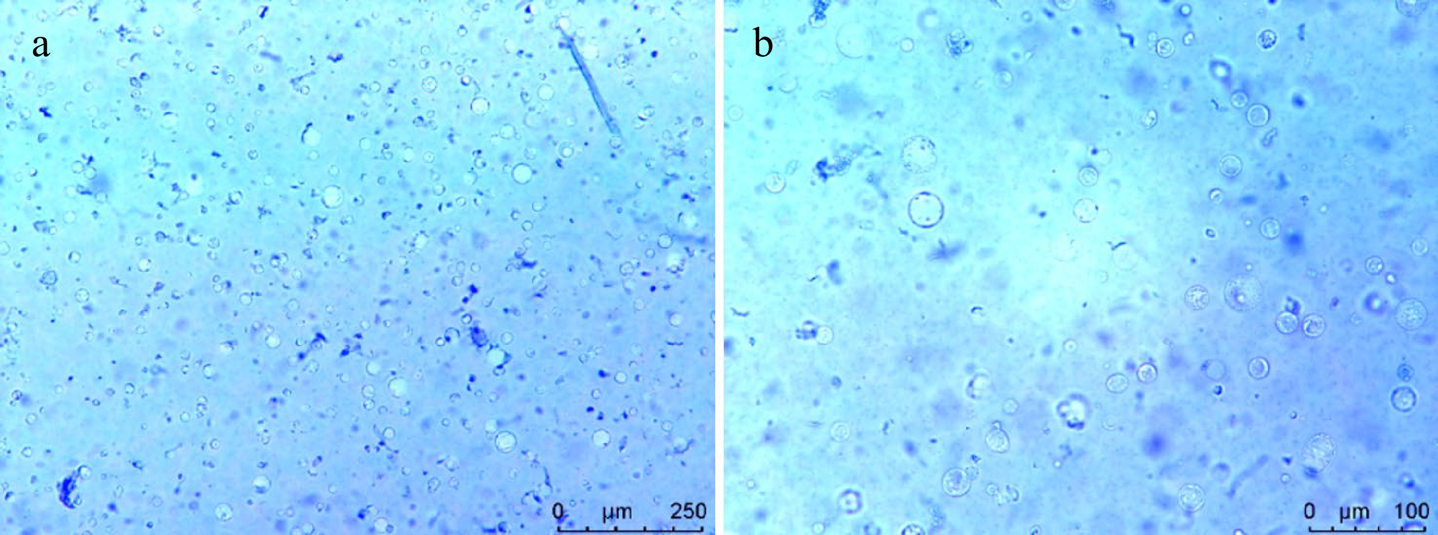
Figure 3.
Viability detection of the protoplasts isolated from H. hamabo by Trypan blue staining. Cell staining under (a) 10×, and (b) 20× microscope.
-
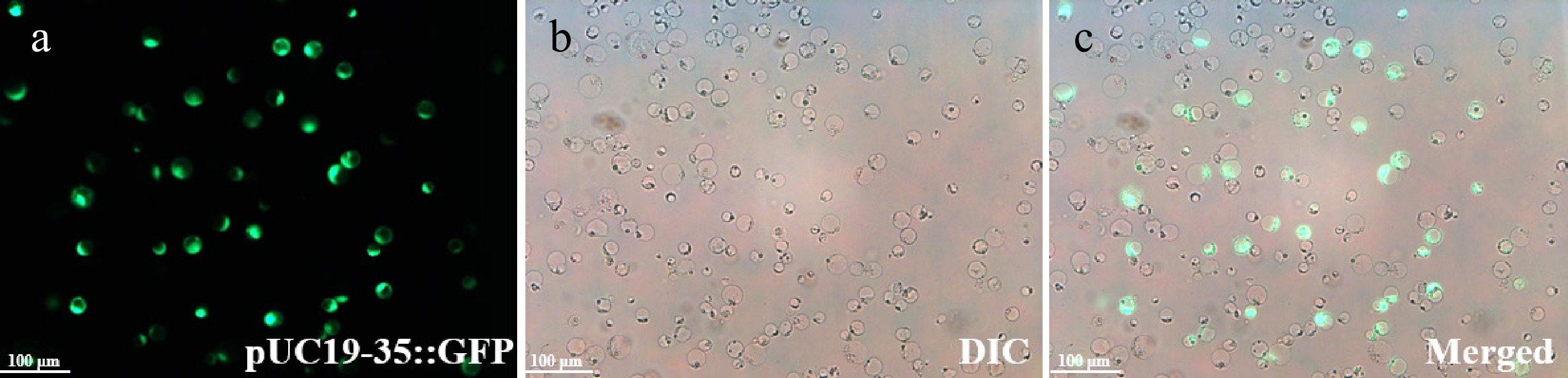
Figure 4.
Protoplasts expressing the pUC19-35::GFP are shown under fluorescence microscopy. The transfection efficiency was 26.03% ± 1.07%. H. hamabo protoplasts in the (a) fluorescence field, (b) bright field with DIC, and (c) a and b superimposed field. Bars = 100 μm.
-
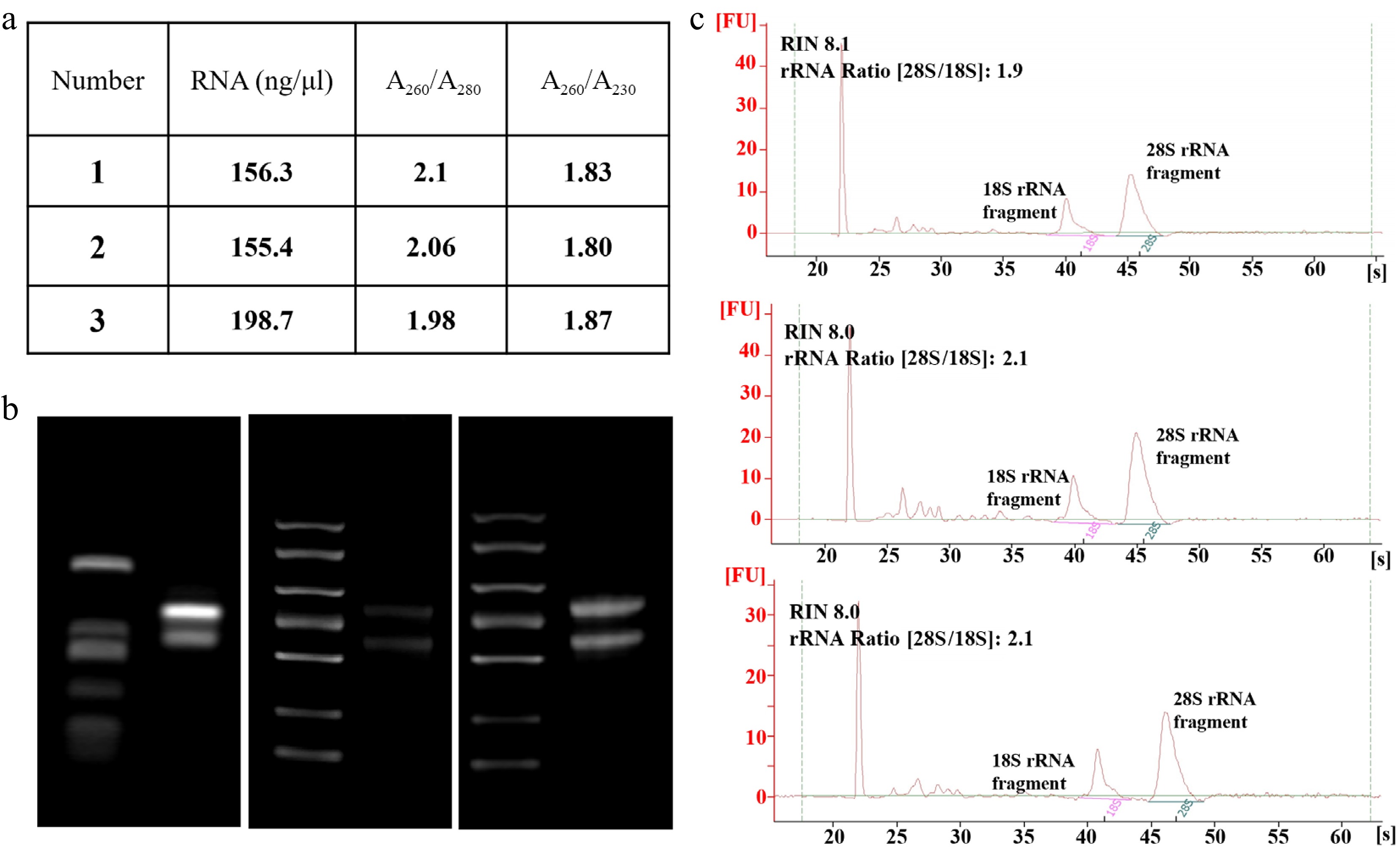
Figure 5.
RNA quality of SDX protoplasts. (a) Total RNA was isolated from stem protoplasts and the quality was examined using the Nanodrop spectrophotometer. (b) 1.2% agarose gel electrophoresis, and (c) Agilent 2100 Bioanalyzer.
-
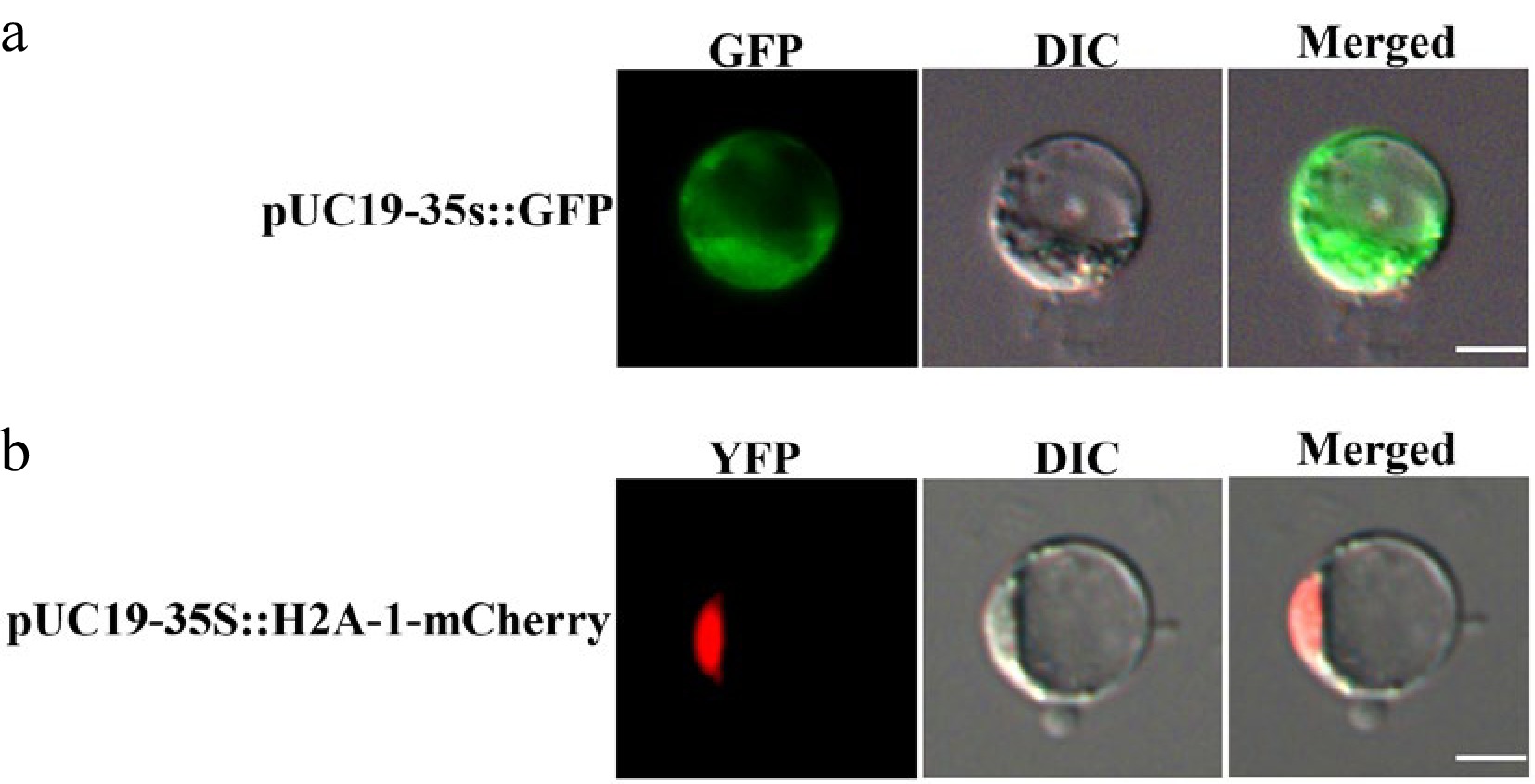
Figure 6.
Subcellular localization of empty vectors in H. hamabo protoplasts. (a) Fluorescence signal of pUC19-35S::GFP; (b) fluorescence signal of pUC19-35S::H2A-1-mCherry. GFP stands for green fluorescent protein and RFP stands for red fluorescent protein. Bars = 10 μm.
Figures
(6)
Tables
(0)