-
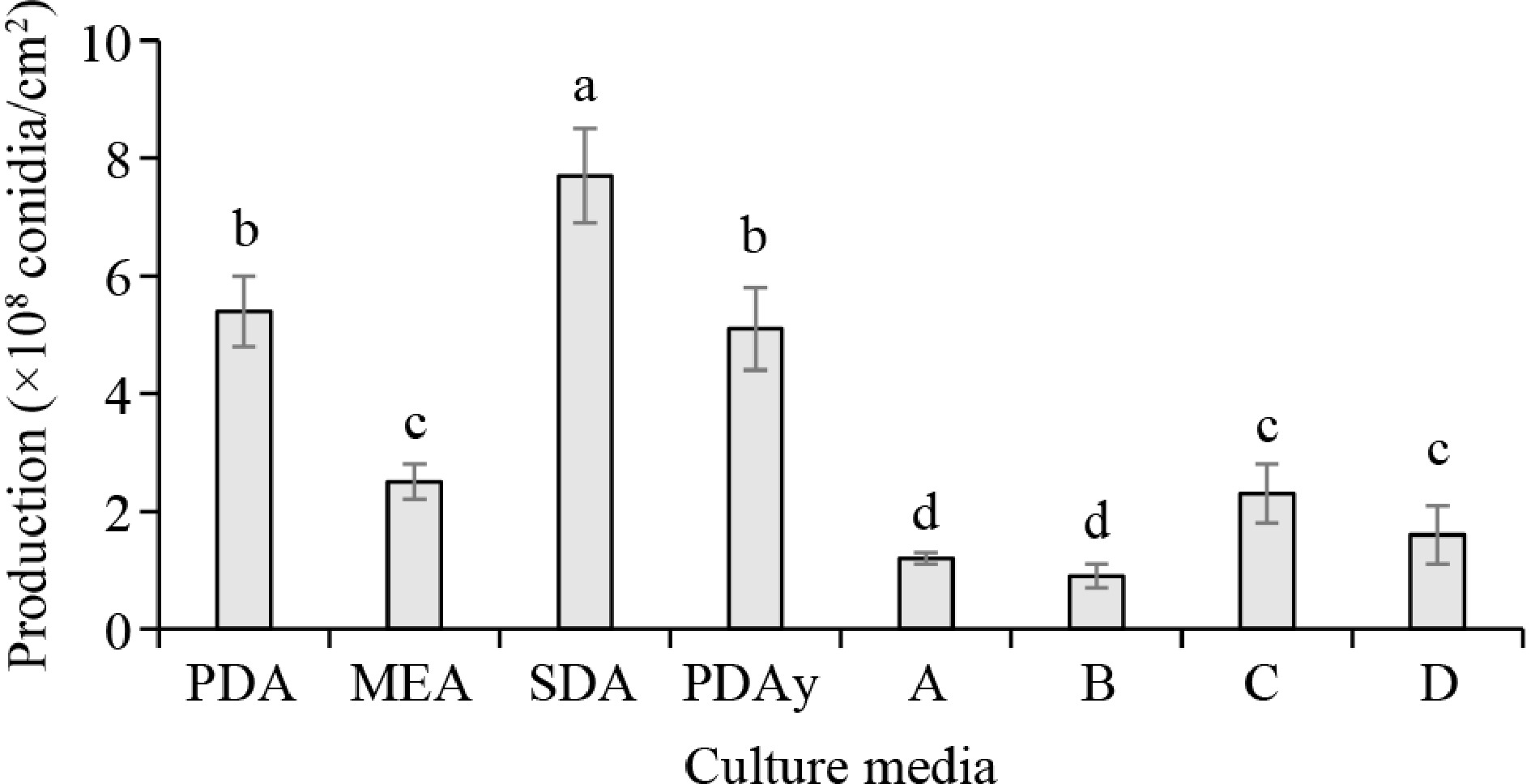
Figure 1.
M. anisopliae conidia production in different culture media. Columns with the same letter do not show significant differences (p < 0.05).
-
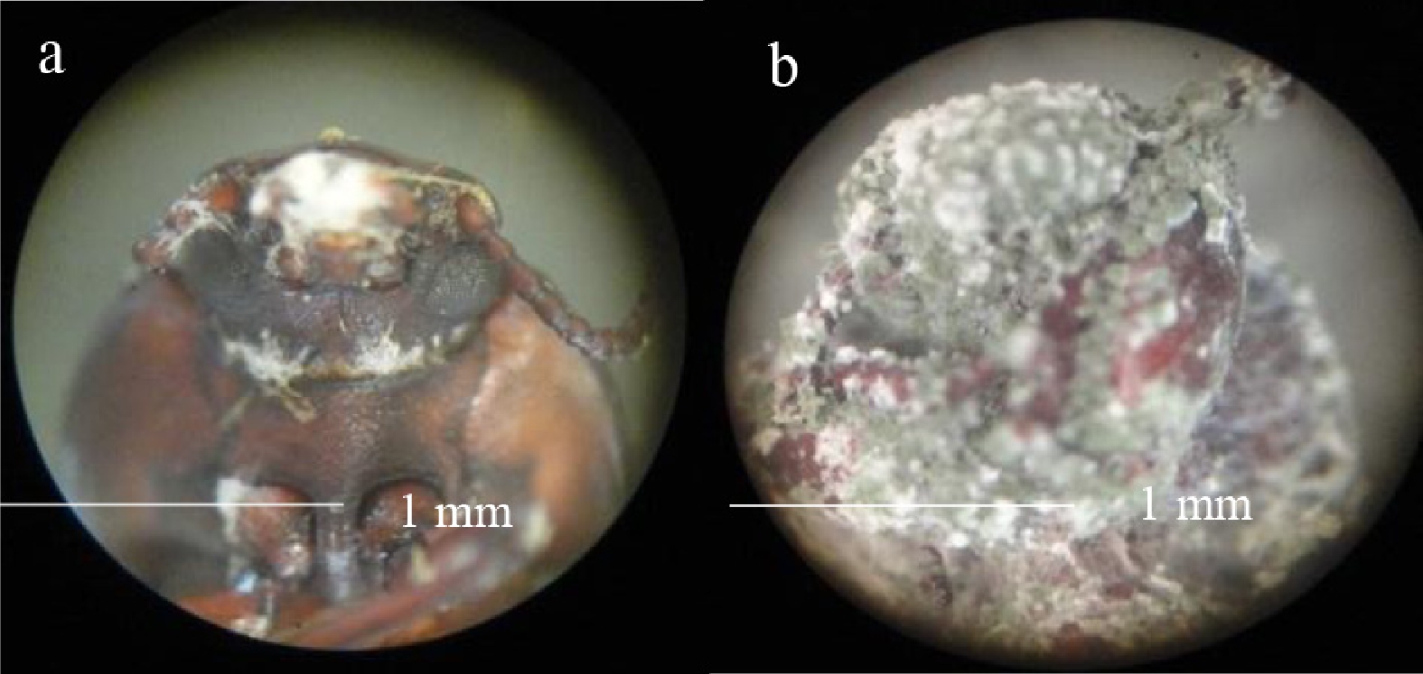
Figure 2.
(a) Mycelial growth of M. anisopliae from inside the insect T. molitor and (b) total invasion over its body.
-
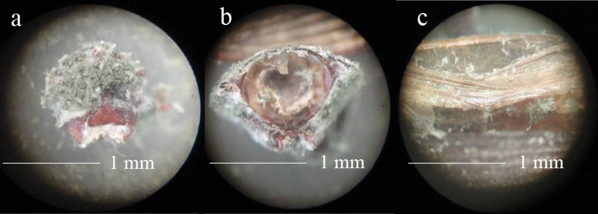
Figure 3.
Mycosis caused by M. anisopliae in T. molitor. (a) Head, (b) cross-section, and (c) intersegmental folds of the cuticle of the insect.
-
Culture medium Composition (g/L) Commercial PDA Potato extract, 4; dextrose, 20 MEA Maltose, 12.75; dextrin, 2.75; glycerol, 2.35; peptone, 0.78 SDA Dextrose, 40; peptone, 10 Laboratory-prepared formulations PDAy PDA with added yeast extract, 10 A Dextrose, 40; yeast extract, 10; meat peptone, 10 B Dextrose, 40; meat peptone, 10 C Malt extract, 30; meat peptone, 5 D Dextrose, 20; potato infusion, 200 Media A, B, C, and D were supplemented with 15 g/L of agar. Table 1.
Culture media used for M. anisopliae conidia production.
-
Treatment t0 (days) k (days−1) LT50 (days) PDA 0.21 ± 0.02e 4.23 ± 0.14d 7.44 ± 0.27a MEA 0.35 ± 0.07d 5.43 ± 0.23b 7.69 ± 0.57a SDA 0.55 ± 0.04b 4.21 ± 0.03d 5.54 ± 0.42c PDAy 0.70 ± 0.09a 6.36 ± 0.23a 7.36 ± 0.20a A 0.54 ± 0.04b 3.96 ± 0.26d 5.27 ± 0.38c B 0.60 ± 0.01b 6.12 ± 0.33a 7.30 ± 0.36a C 0.48 ± 0.04c 4.80 ± 0.09c 6.53 ± 0.25b D 0.34 ± 0.07d 5.32 ± 0.32b 7.63 ± 0.26a The values of the columns with the same letter do not show significant differences (p < 0.05). Table 2.
Parameters of the bioassay with T. molitor infected with conidia of M. anisopliae obtained from different culture media.
Figures
(3)
Tables
(2)