











-
The use of entomopathogenic fungi in the control of agricultural pests caused by insects is considered an alternative that could also help restore functional biodiversity in damaged agricultural ecosystems[1]. It has been suggested that its advantages include zero harmful side effects on other organisms, including humans; pests' resistance to these biological controls is very rare; and the cost/benefit ratio is very favorable, since insecticide treatment is reduced and, in the best cases, eliminated, avoiding contamination that can cause poisoning[2]. Entomopathogenic fungi are the most important group in biological control, because most insects are susceptible to attack by these fungi[1]. These fungi are found in the division Eumycota, and are subdivided into Zygomycota, Ascomycota, Basidiomycota, and Deuteromycota[3]. The attack by entomopathogenic fungi against insects consists of two phases: Infective and reproductive. In the infective phase, the conidia adhere to the insect's cuticle through nonsclerotized regions, as well as wounds, trachea, and mouthparts, subsequently germinating and penetrating them[4]. The adhesion of conidia to the insect depends largely on the secretion of lipase, chitinase, and protease enzymes; therefore, these enzymes are related to the virulence of the fungus, which implies successful infection and penetration[5,6]. In the post-adhesion stage, the fungus produces specialized structures such as the germ tube and penetrating hyphae, allowing it to colonize and develop within the insect, ultimately causing its death[7]. Many studies have been reported on the ability of fungi to infect insects, such as Metarhizium flavoviridae and Beauveria bassiana against the red mite (Dermanyssus gallinae) from poultry farms[8]; Metarhizium pingshaense in combination with deltamethrin against Anhopeles coluzzii, the mosquito that causes malaria[9]; and Metarhizium species on the locusts Locusta migratoria and Schistocerca gregaria[10]. Specifically, Metarhizium anisopliae is a very important entomopathogenic fungus used worldwide. It is characterized as mesophilic, with an optimal temperature for germination and growth of 25–30 °C, although it can grow at 10–35 °C. Optimal spore germination, mycelial growth, and sporogenesis occur at 100% relative humidity at 25–30 °C, growth stops at –6 °C, and the thermal death point is near 50 °C[11]. The insect Tenebrio molitor is the most widely used to evaluate the infectivity of some entomopathogenic fungi used in biological control because of its easy reproduction and survival under laboratory conditions[12−15]. This insect is a pest of stored durable agricultural products, mainly cereals and products rich in starch, which are characterized by having low moisture[16]. Like all beetles, it is a holometabolous insect, meaning it undergoes complete metamorphosis, following a cycle of four different stages during its life: Egg, larva, pupa, and adult[17]. The increased production of highly infectious conidia is a key concern for researchers working with entomopathogenic fungi. Among the established strategies is the use of different culture media to promote fungal growth and conidia production. Culture media based on natural substrates such as sugarcane (Saccharum spp.) bagasse, rice (Oryza sativa), wheat (Triticum aestivum) bran, millet (Panicum miliaceum), sorghum (Sorghum bicolor), and barley (Hordeum vulgare) have been tested[18,19]. Synthetic culture media such as potato dextrose agar (PDA), yeast extract peptone dextrose agar (PDAy)[20−22], Sabouraud dextrose agar (SDA) supplemented with yeast extract[19], and malt extract agar (MEA) have also been tested[23]. In this sense, in the search for accessible culture media that allow the high production of highly infective conidia, in this work, different commercial culture media and other laboratory-prepared formulations were used to evaluate the production and infectivity of M. anisopliae conidia in T. molitor.
-
The CP-Oax strain of M. anisopliae var. lepidiotum (GenBank accession number FJ876298) belonging to the fungal collection of the Postgraduate College, Texcoco, Mexico, was used. The strain, previously stored in glycerol (20%) at −20 °C, was activated on bacteriological agar with grasshopper cuticles at 28 °C. It was then refrigerated at 4 °C until use[24]. Eight culture media were used for conidia production, three of which were commercial (PDA, MEA, and SDA) and five were laboratory-prepared formulations. The composition of all media is reported in Table 1. In all cases, 120-mL serological bottles containing 30 mL of the culture medium were used and inoculated with 1 × 10⁸ conidia/cm2. The bottles were incubated at 28 °C for seven days. Conidia were harvested using 10 mL of a 0.01% Tween 80 solution.
Table 1. Culture media used for M. anisopliae conidia production.
Culture medium Composition (g/L) Commercial PDA Potato extract, 4; dextrose, 20 MEA Maltose, 12.75; dextrin, 2.75; glycerol, 2.35; peptone, 0.78 SDA Dextrose, 40; peptone, 10 Laboratory-prepared formulations PDAy PDA with added yeast extract, 10 A Dextrose, 40; yeast extract, 10; meat peptone, 10 B Dextrose, 40; meat peptone, 10 C Malt extract, 30; meat peptone, 5 D Dextrose, 20; potato infusion, 200 Media A, B, C, and D were supplemented with 15 g/L of agar. Virulence bioassay
-
For virulence tests, T. molitor insects were used, which were acquired from a local pet food store (Tlaxcala, Tlax., Mexico) and subsequently reproduced in the laboratory. Bioassays (four replicates) were performed using the conidia obtained from each culture medium. Adult T. molitor insects were immersed for 3 s in 10 mL of a conidial suspension (1 × 10⁸ conidia/mL). A 0.01% Tween 80 solution was used as a control. Each bioassay was performed in Petri dishes with 12 insects per dish, using oat flakes as food. They were incubated at 28 °C for seven days, with observations every 12 h and the dead insects were transferred to humid chambers to monitor the growth and external sporulation of the fungus on the corpse to confirm death by infection[25]. The time to reach 50% mortality (LT50) was calculated by plotting cumulative mortality against time, using the following equation:
$ Y=(100-S)e ^{{-k(t{\text -}to)} } +S $ Y = 100; 0 ≤ t ≤ t0, where, Y is the survival percentage at time t; k is the specific mortality rate (days−1); t0 is the delay time for the first death to occur (days), and S is the estimated asymptotic survival level (%). This model corresponds to the solution of a first-order differential equation with the indicated delay time, the selected initial condition, and the asymptotic value Y → S for t → ∞[12].
Statistical analysis
-
The means ± standard deviation (SD) are reported. Analysis of variance (ANOVA) and Tukey's test (p < 0.05) were performed using StatView 5.0 software.
-
Figure 1 shows the results for conidia production on the different culture media. In general, the commercial culture media and the PDAy medium yielded the highest values. It is worth noting that the SDA medium was the best, followed by the PDA and PDAy. The number of conidia obtained on the SDA medium was approximately 8.5 times greater than on Medium B, which produced the fewest. It should be mentioned that all the results obtained in this study were higher than those reported by Tlecuitl-Beristain et al.[13], who obtained a maximum conidia value of 4.25 × 10⁷ conidia/cm2 for M. anisopliae using an oat–peptone medium with oxygen-rich pulses (26% O2).
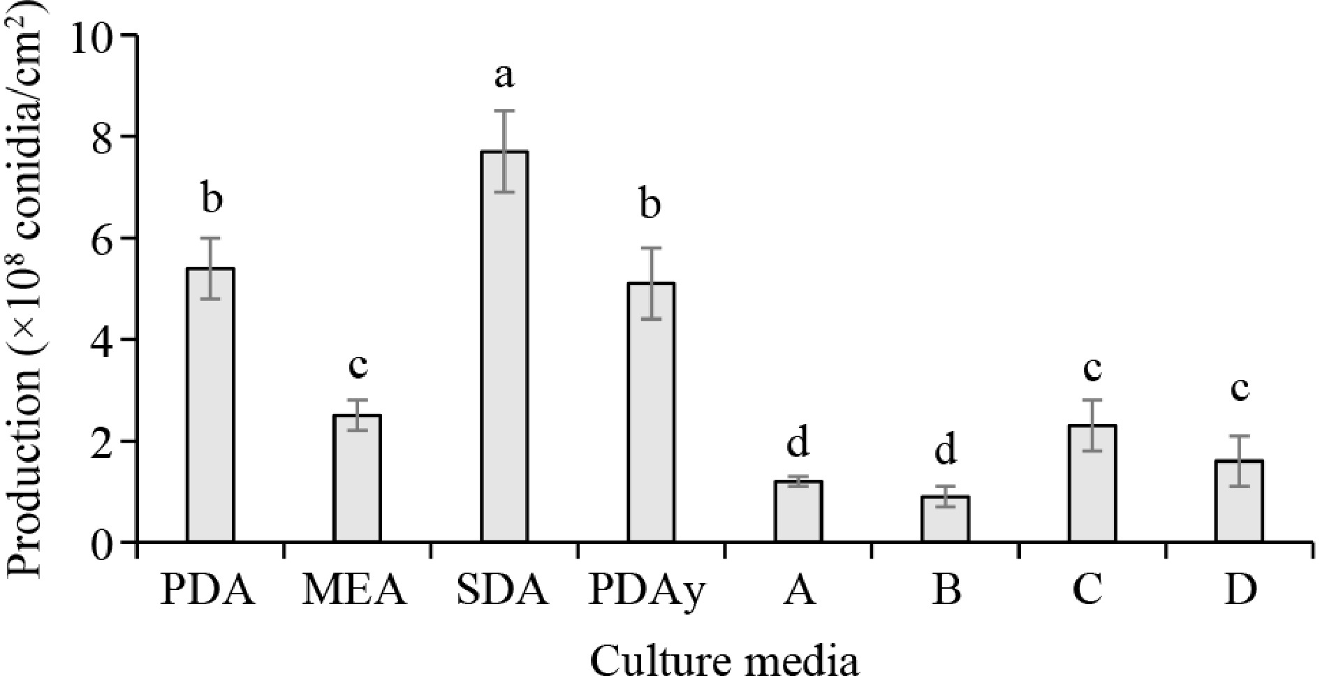
Figure 1.
M. anisopliae conidia production in different culture media. Columns with the same letter do not show significant differences (p < 0.05).
Kamp and Bidochka[20] grew the M. anisopliae strains 2575, 54A-1b, MAA1-2iii, and HAA2-2b for conidia production (×107 conidia/cm2), obtaining 15.33, 5.47, 3.41, and 2.31 on PDA; 5.35, 0.02, 0.00, and 0.04 on SDA; and 3.44, 0.05, 1.59, and 0.05 ofn MEA, respectively. A study tested several agro-industrial waste products (wheat bran, rice bran, tamarind extract, and ground rice) for the production of M. anisopliae. The highest amount of dry biomass (1.15 g/100 mL) was obtained in the ground rice medium, whereas the lowest value was observed in the rice bran medium. The tamarind extract medium exhibited the highest number of conidia (27.8 × 10⁴ conidia/mL)[26]. Bhanu-Prakash et al.[27], using natural substrates, obtained the highest conidial production (×1010 conidia/cm2) with rice, followed by barley and finally with sorghum (6.2, 4.9, and 4.5, respectively). Recently, conidia of Metarhizium robertsii were produced in glass columns with an internal diameter of 2 cm and a height of 20 cm, using 10 g of precooked rice as the initial dry substrate (gssi) in each reactor. The substrate was inoculated with a conidial suspension at a concentration of 1 × 10⁷ conidia/mL and incubated in a water bath at 28 °C for 11 days. Conidia production was highest on Day 8 of culture, reaching 1.3 × 10⁹ conidia/gssi, and remained constant (8.91 × 10⁸ conidia/gssi) from Days 9 to 11 of incubation[28]. In another study, conidia of M. anisopliae were produced on fresh parboiled rice and recycled parboiled rice. Conidia production was optimized using a response surface methodology, considering the variables of temperature, time, and the addition of molasses. Production in all treatments was greater than 1 × 10⁹ conidia/g. Both substrates yielded the highest production at 25 °C for 20 days. The substrate from M. anisopliae production itself was reused, reducing conidia production costs[29]. According to the results obtained, and in comparison with other studies, the conditions used in this study are considered to have favored conidia production, as the values obtained were generally higher than many previously reported. The carbon to nitrogen (C/N) ratio has been considered to have influenced the increase in conidia production; for example, with very high C/N values such as 35, values of 1.6 × 10⁹ and 25.3 × 10⁹ conidia/cm2 were obtained for the M. anisopliae strains V245 and V275, respectively[30].
Figure 2 shows the mycosis caused on the insects. The fungus begins to invade through the natural openings of the host insect and its growth starts from the inside out (Fig. 2a); subsequently, the invasion is completed throughout the insect's body (Fig. 2b). Infection has been described as occurring through the contact and adhesion of spores via the cuticle, mouthparts, intersegmental membranes, or spiracles, all of which are areas with high humidity that favor spore germination and allow hyphal penetration[31]. After adhesion, the fungus produces specialized structures such as the germ tube and penetration hyphae[32].
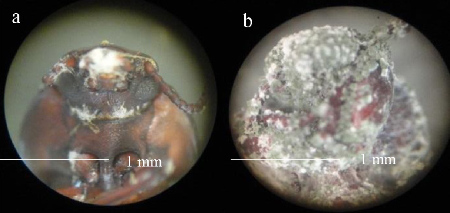
Figure 2.
(a) Mycelial growth of M. anisopliae from inside the insect T. molitor and (b) total invasion over its body.
Figure 3a shows the mycosis-infected head of T. molitor. Figure 3b shows a longitudinal section revealing the mycosis inside the insect. Figure 3c shows the intersegmental folds of the host's cuticle with mycosis. These images show different areas where the fungus has invaded the insect host, causing its death, which has already been reported previously[12,31,32].
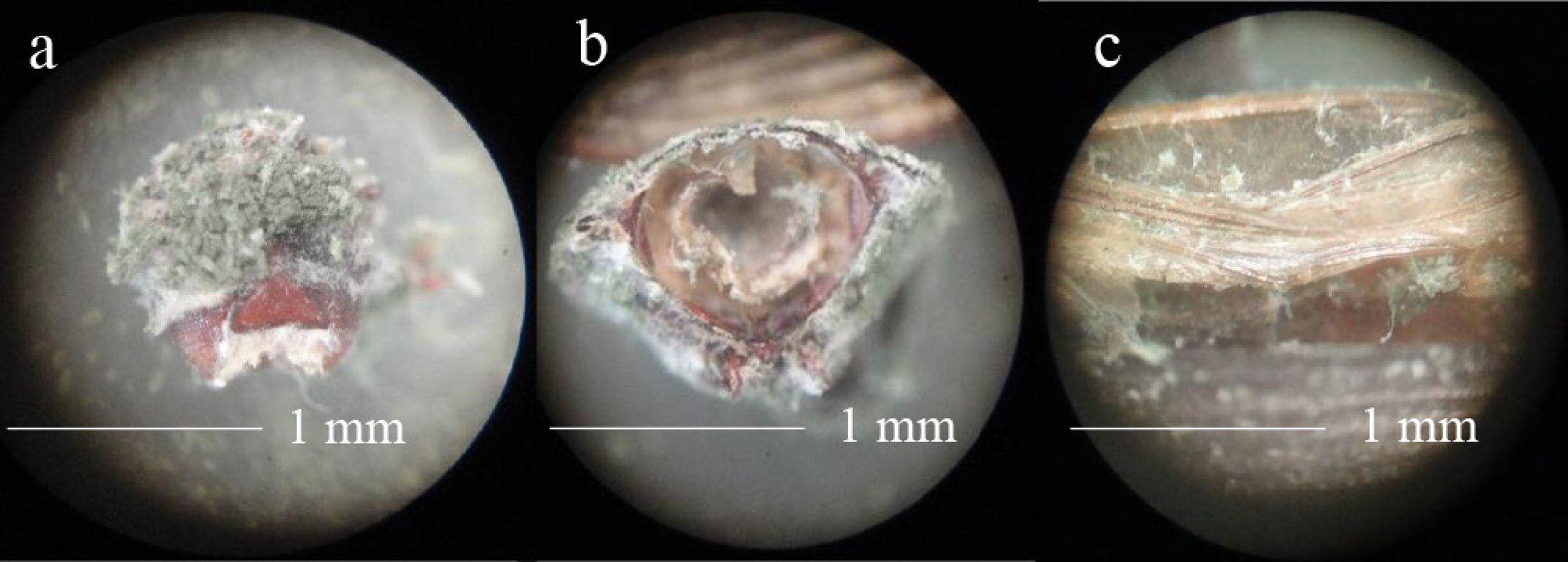
Figure 3.
Mycosis caused by M. anisopliae in T. molitor. (a) Head, (b) cross-section, and (c) intersegmental folds of the cuticle of the insect.
The kinetic parameters of the M. anisopliae virulence bioassay against T. molitor are shown in Table 2. Regarding the t0 parameter, the lowest value was observed in the PDA medium, followed by the MEA and D media, whereas the highest value was observed in the PADy medium. The PDA, SDA, and A media showed the lowest k values, whereas the PDAy and B media showed the highest values. Regarding the LT50 parameter, the SDA and A media showed the lowest values. Moreover, 100% mortality (S = 0) of adult T. molitor was obtained at 11–15.4 days post-infection. According to the observed values of k and LT50, it was determined that the SDA and A media produced the most virulent conidia, which implies that these conidia caused the death of the insects in less time; however, considering the number of conidia produced (Fig. 1), the SDA medium was the most productive (7.7 × 10⁸) and Medium A was one of those that produced the fewest conidia.
Table 2. Parameters of the bioassay with T. molitor infected with conidia of M. anisopliae obtained from different culture media.
Treatment t0 (days) k (days−1) LT50 (days) PDA 0.21 ± 0.02e 4.23 ± 0.14d 7.44 ± 0.27a MEA 0.35 ± 0.07d 5.43 ± 0.23b 7.69 ± 0.57a SDA 0.55 ± 0.04b 4.21 ± 0.03d 5.54 ± 0.42c PDAy 0.70 ± 0.09a 6.36 ± 0.23a 7.36 ± 0.20a A 0.54 ± 0.04b 3.96 ± 0.26d 5.27 ± 0.38c B 0.60 ± 0.01b 6.12 ± 0.33a 7.30 ± 0.36a C 0.48 ± 0.04c 4.80 ± 0.09c 6.53 ± 0.25b D 0.34 ± 0.07d 5.32 ± 0.32b 7.63 ± 0.26a The values of the columns with the same letter do not show significant differences (p < 0.05). Tlecuitl-Beristain et al.[13] used an oat–peptone medium with oxygen-rich pulses to induce conidia production in M. anisopliae, and in the virulence assay with T. molitor, they observed k values of 2.22 and 1.26 days−1, whereas the LT50 values were 3.90 and 4.31 days, for 26% O2 pulses and a 21% O2 atmosphere, respectively. The pathogenicity of Metarhizium rileyi was evaluated in T. molitor larvae and pupae for 12 days. The pupal stage was highly susceptible, showing 100% mortality rates after 12 days with a median lethal concentration (LC50) of 7.8 × 10⁶ conidia/mL, and the larvae showed 92% mortality rates 12 days after treatment with an LC50 of 1.0 × 10⁶ conidia/mL. These results suggest that fungal virulence varies between the larval and pupal stages of T. molitor[33]. The whitefly Bemisia tabaci was infected with B. bassiana and M. anisopliae, achieving LT50 values between 3.36 and 9.26 days[5]. Recently, it was reported that M. pingshaense, in synergy with deltamethrin, exhibited an LT50 of 8 days against A. coluzzii[9]. In another study, the virulence of two Metarhizium spp. strains was evaluated using T. molitor, resulting in 100% mortality 7 days after inoculation[34]. The virulence of four M. anisopliae strains against last-stage larvae of Galleria mellonella and T. molitor was evaluated in vitro at four conidial concentrations: 1 × 10⁶, 1 × 10⁷, 1 × 10⁸ and 1 × 10⁹ conidia/mL. The strains with the best performance were Ma58MI and Ma10MI, both for T. molitor (LT50 = 4.06 and 5.22 days, respectively) and for Galleria mellonella (LT50 = 5.18 and 6.37 days, respectively)[35]. Altahawi et al.[14] isolated nine strains of M. anisopliae and tested them against T. molitor larvae at concentrations of 1 × 10⁸ spores/g, observing a maximum mortality rate of 71% at 10 days of inoculation, and the LT50 was 6.28 days. Another study evaluated the pathogenicity of M. rileyi conidia against Spodoptera litura and Spodoptera frugiperda prepupae at 3, 6, 9, and 12 days post-infection. The fungus was found to cause mortality rates of 61%–90% and 46%–73% in S. litura and S. frugiperda pupae, respectively, 12 days after treatment. The LC50 against S. litura and S. frugiperda was 3.4 × 10¹⁴ conidia/mL and 6.6 × 10⁵ conidia/mL, respectively[36]. It is worth mentioning that T. molitor has been used as a host in conidial infectivity assays of other entomopathogenic fungi, such as one where the pathogenicity of B. bassiana was evaluated against different developmental stages of T. molitor. The fungus exhibited pathogenicity levels of 97.33%, 82.66%, and 65.33% against pupae, larvae, and adults, respectively. The LC50 was 2.7 × 10⁴, 1.8 × 10⁵, and 1.3 × 10⁷ conidia/mL for pupae, larvae, and adults, respectively. According to these results, the authors suggest that this strain of B. bassiana shows potential in controlling the various developmental stages of T. molitor[37]. Recently, the virulence of B. bassiana against Halyomorpha halys, T. molitor, and Popillia japonica was evaluated, showing 100% mortality in the first two insects within 10 days of infection. The LC50 values were very low: 9.5 × 10³ conidia/mL in H. halys, 2.6 × 10³ conidia/mL in T. molitor, and 8.3 × 10⁴ conidia/mL in P. japonica. The LT50 values for H. halys, T. molitor, and P. japonica were 6, 5.3, and 6.9 days, respectively. A previous study showed that B. bassiana conidia were effective against three major insect pests[38].
-
The quantity and virulence of M. anisopliae conidia differed depending on the culture medium used, with commercial agars favoring higher values for both parameters evaluated in this study, noting that the values were higher than those previously reported in many studies. These results suggest that the production of highly virulent M. anisopliae conidia is more efficient with the use of commercial culture media.
We thank the Polytechnic University of Tlaxcala and the Biotechnology Laboratory of the Research Center for Biological Sciences of the Autonomous University of Tlaxcala.
-
The authors confirm their contributions to the paper as follows: study conception and design, analysis and interpretation of results: Tlecuitl-Beristain S, Díaz-Godínez G; data collection: Gutiérrez-Lara C, García-Barrientos R, García-Dávila J; draft manuscript preparation: Díaz-Godínez G. All authors reviewed the results and approved the final version of the manuscript.
-
The data that support the findings of this study are available on request from the corresponding author.
-
The authors declare that they have no conflict of interest.
- Copyright: © 2026 by the author(s). Published by Maximum Academic Press, Fayetteville, GA. This article is an open access article distributed under Creative Commons Attribution License (CC BY 4.0), visit https://creativecommons.org/licenses/by/4.0/.
-
About this article
Cite this article
Gutiérrez-Lara C, García-Dávila J, García-Barrientos R, Tlecuitl-Beristain S, Díaz-Godínez G. 2026. Production and infectivity of Metarhizium anisopliae conidia obtained from different culture media. Studies in Fungi 11: e019 doi: 10.48130/sif-0026-0014 

Production and infectivity of Metarhizium anisopliae conidia obtained from different culture media
- Received: 13 December 2025
- Revised: 01 April 2026
- Accepted: 07 April 2026
- Published online: 02 July 2026
Abstract: Some of the most important agricultural pests are insects, and their control using biological methods is an environmentally friendly alternative that also contributes to the protection of biodiversity and human health. The use of entomopathogenic fungi has shown effectiveness against different insect crop pests. It is worth mentioning that fungi such as Beauveria bassiana and Metarhizium anisopliae are widely studied for their entomopathogenic capacity, with many studies being conducted to increase their production and the virulence of their conidia. The mealworm (Tenebrio molitor) is the most commonly used insect to evaluate the infectivity of some entomopathogenic fungi because of its ease of reproduction in the laboratory. In this study, the conidia production of M. anisopliae cultivated in different culture media was investigated; subsequently, the infectivity of the conidia against T. molitor was evaluated. Eight culture media were used for conidia production, three of which were commercial, namely potato dextrose agar, malt extract agar, and Sabouraud dextrose agar (SDA), and five were laboratory-prepared formulations. In general, commercial culture media showed the best results in terms of conidia production. The SDA medium was the best, as it showed the highest conidia production (7.7 × 10⁸ conidia/cm2) and was one of the media that showed the lowest median lethal time (LT50) value (5.54 days). The quantity and virulence of M. anisopliae conidia differed depending on the culture medium used, with commercial agars favoring higher values for both parameters evaluated in this study.
-
Key words:
- Conidia /
- Biological control /
- Metarhizium anisopliae /
- Tenebrio molitor /
- Virulence