-
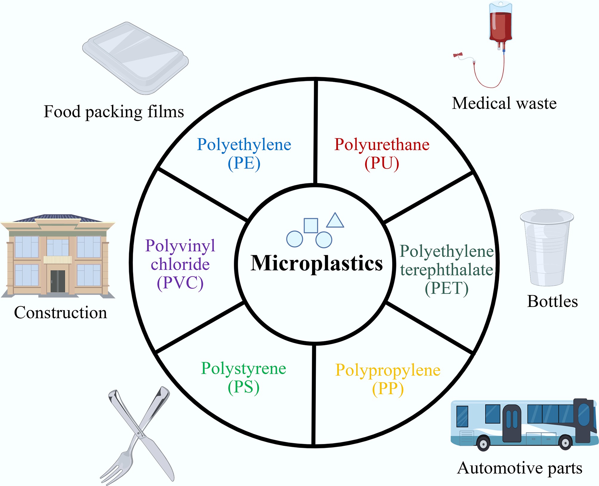
Figure 1.
Sources and types of micro- and nano-plastics.
-
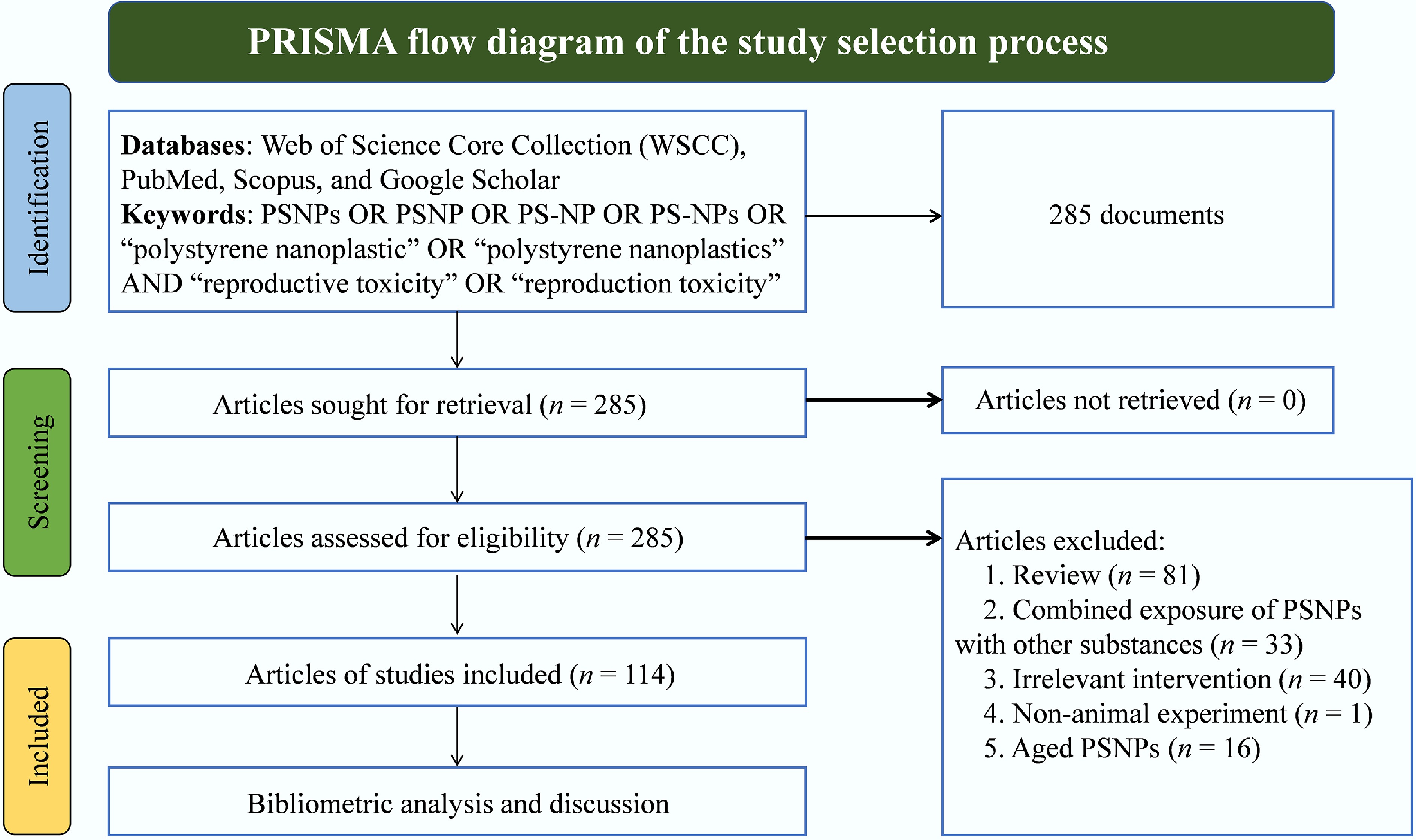
Figure 2.
PRISMA flow diagram of the study selection process.
-
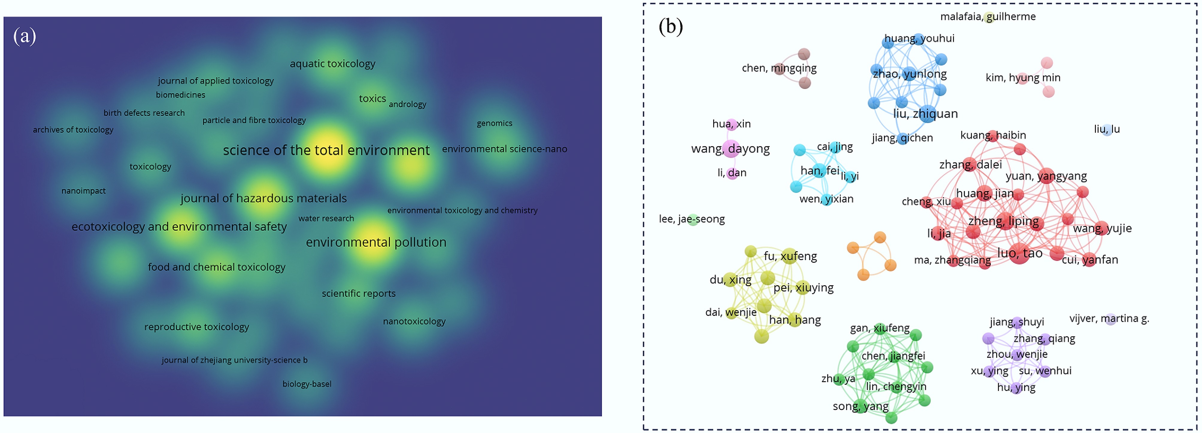
Figure 3.
Network map of journals and authors on the reproductive toxicity of PSNPs. (a) Publication volume of articles on the reproductive toxicity of PSNPs across various journals, with darker colors indicating higher publication counts. (b) Publication output of authors in this field, where larger nodes indicate more publications, and lines represent collaborative relationships.
-
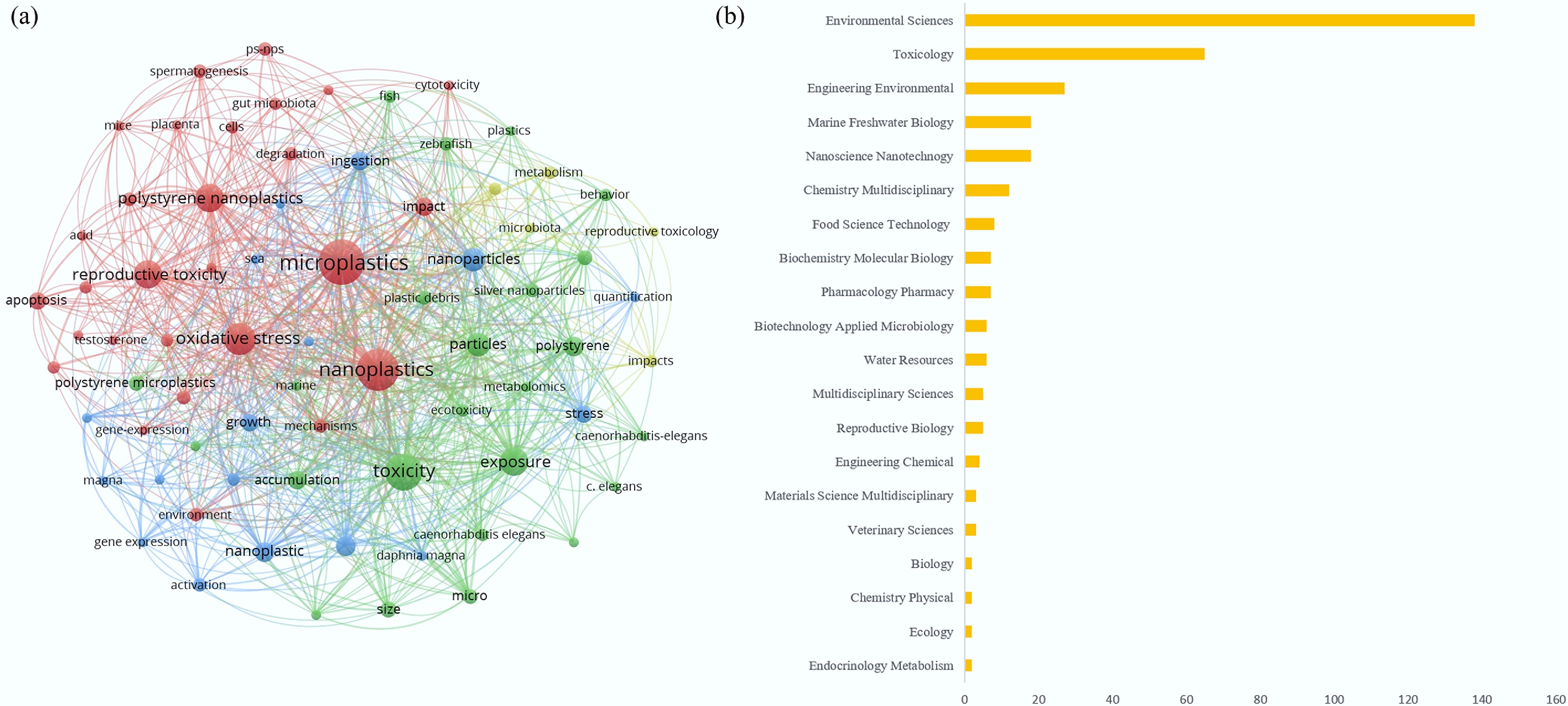
Figure 4.
Network map of keywords and subject areas in the field of reproductive toxicity of PSNPs. (a) Keyword co-occurrence map, where larger nodes indicate a higher frequency of mention, and connecting lines represent the co-occurrence associations between keywords. (b) Interdisciplinary intersection counts for PSNP-related reproductive toxicity research.
-
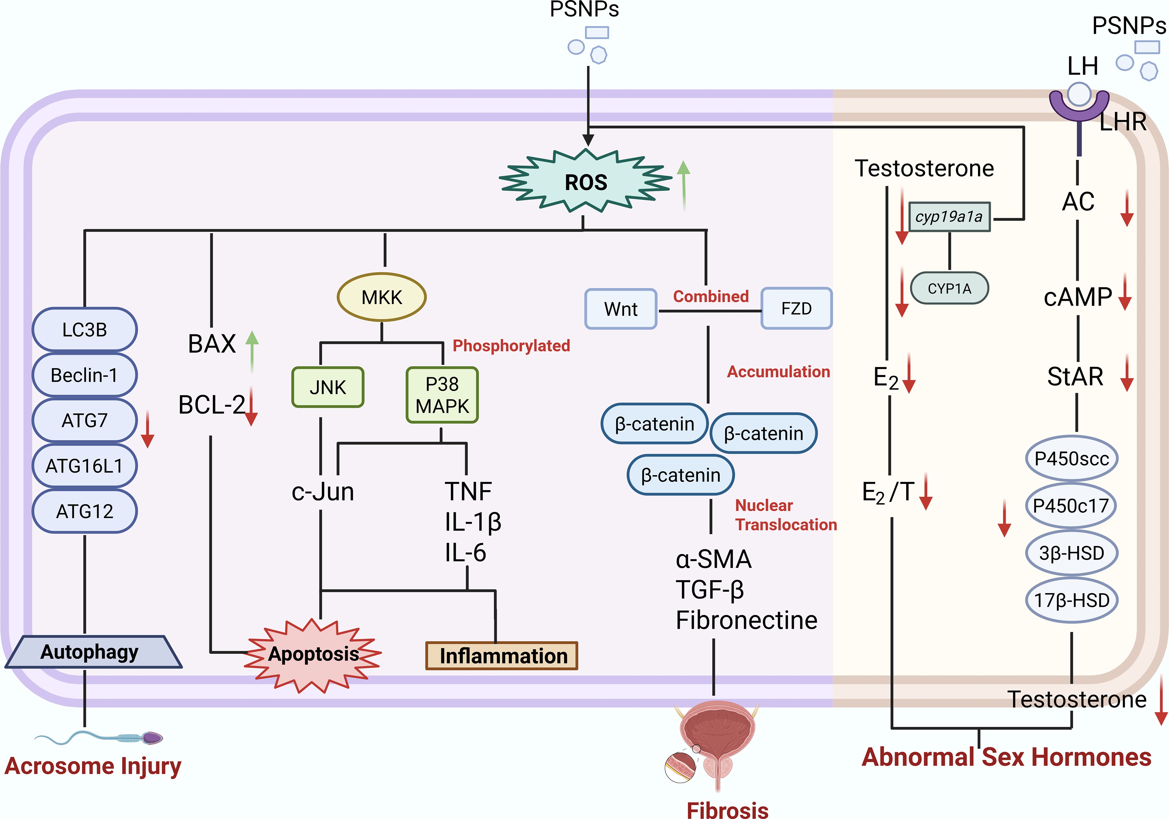
Figure 5.
Mechanisms of PSNPs-induced reproductive toxicity.
-
Sex Species Route of exposure Affected signaling pathway Dose/size of exposure Outcome Ref. Male Mice Orally exposed PSNPs induce oxidative stress and affect the expression of genes associated with apoptosis and inflammation. 25-, 50-, and
100-nmDamage the testicular structure and function, but also decrease sperm count and quality, ultimately reducing male fertility. [38] Rats Gavage Endocrine disruption 1, 3, 6, and
10 mg/(kg·d)
25-, 50-nmReduce serum LH and testosterone (T) levels. [57] Zebrafish Water Meiosis and reproduction, DSB repair mechanisms. 5 mg/L of 45 nm Induced histopathological lesions in the testis. Sperm cells showed anomalous aggregation and condensed chromatin. Although the sperm remained viable, their motility was markedly impaired. [54] Oysters Configured as a suspension and exposed to the environment Perturbation of antioxidant defenses. 50 nm polystyrene beads with amine (50-NH2 beads) or carboxyl (50-COOH beads) functions Caused a decrease in the percentage of motile spermatozoa (66%) and in the velocity (38%). [41] Female Mice Gavage Endocrine disruption: E2 and P were elevated, while FSH and LH levels were diminished. 2.5% w/v, 10 mL
50–90 nmDecreases the live birth rate and neonatal crown-rump length, embryo implantation sites, and uterine wet weight. [30] Rats Direct drinking Wnt/β-catenin signaling pathways 0, 0.015, 0.15, and 1.5 mg/d
0.5 μmOvarian fibrosis in rats, along with apoptosis of granulosa cells, ultimately leads to a decline in ovarian reserve capacity. [58] Zebrafish Water PPAR signaling pathway, dysregulated lipid transport, binding and activity processes 5 mg/L
80, 200, 500 nmPolycystic ovary syndrome-like ovulatory dysfunction. [59] Oysters Oral ingestion An alteration in glucocorticoid response, insulin pathway, and fatty-acid metabolism 14% ± 2% of the
2 μm and 69% ± 6% of the 6 μmSignificant decreases in oocyte number (−38%), diameter (−5%). [47] Fruit fly Mixed with the standard cornmeal fly feed Several metabolic routes were perturbed.
These included steroidogenesis and the turnover of arginine and proline.
At the systemic level, cholesterol homeostasis was affected.
Longevity-related signalling also showed changes.
Regulation of lipolysis in fat cells was disrupted. Ovarian steroid production and progesterone-driven oocyte maturation were similarly influenced.1, 10, 50, and
100 mg/L
100 nmAccumulate in the crop and gut and might even cross the intestinal barrier to reach ovarian tissue; the number of eggs produced and the eclosion rate decrease. [46] Table 1.
Comparison of sex-specific damage in PSNPs among different species
Figures
(5)
Tables
(1)