









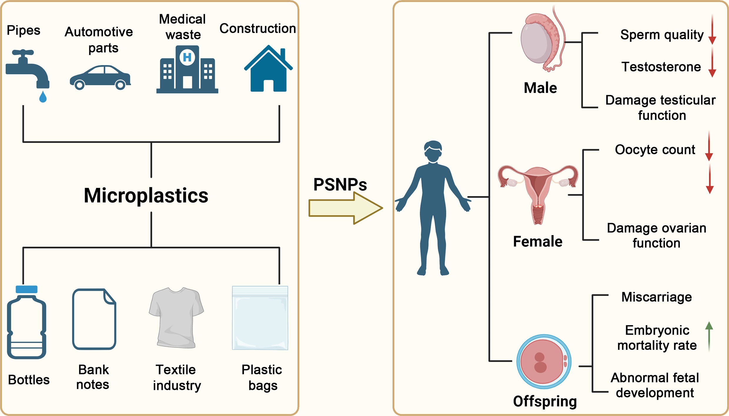


-
Plastic products are widely utilized in daily life owing to their versatility, practicality, low cost, and lightweight properties. Common plastic types include polyethylene (PE), polypropylene (PP), polyurethane (PU), polyethylene terephthalate (PET), polystyrene (PS), and polyvinyl chloride (PVC)[1,2] (Fig. 1). Since 1950, the global output of plastics has grown exponentially[3]. It is estimated that by 2050, global plastic production will reach between 902 and 1,124 Mt[4]. Meanwhile, most plastics are used as disposable consumer goods and have limited recyclability. Only about 9% of global plastic is recycled, while the remaining 79% accumulates in natural ecosystems[5]. Extensive use and improper disposal of plastics have led to plastic pollution of terrestrial and aquatic systems. Even with immediate and concerted action, 710 Mt of plastic waste will enter terrestrial and aquatic systems[6]. Plastic pollution thus constitutes a pressing and substantial environmental challenge.
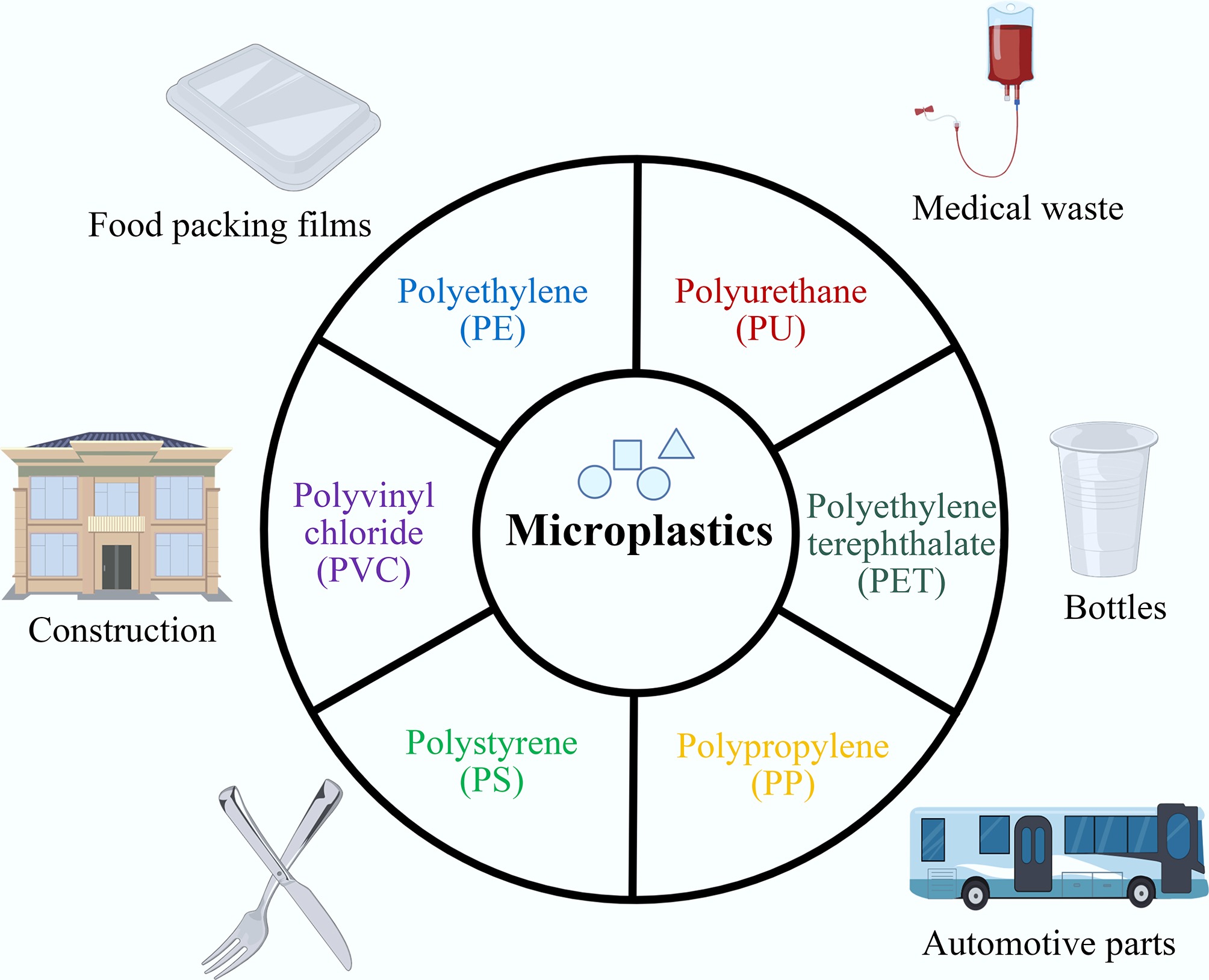
Figure 1.
Sources and types of micro- and nano-plastics.
Upon entering the environment, plastics undergo continuous degradation into MNPs[7]. MNPs, the microscopic plastic fragments first described in 2004[8], are persistent, mobile, and ubiquitous in terrestrial and aquatic environments. Varied sources and weathering patterns give rise to diverse morphological characteristics of microplastics (MPs), encompassing fragments, fibers, pellets, spheres, films, and foams[2]. MPs are now broadly defined as plastic particles ≤ 5 mm in diameter, and research has shown that micron-sized particles can be further degraded into nanoplastics (NPs), which are < 100 nm[9]. MNPs are pervasive in daily life, having been detected in the water we drink, the air we breathe, and the food we eat, including seafood, table salt, honey, sugar, and beverages such as beer and tea[10]. Four primary exposure pathways for MNPs have been identified: the first and most unavoidable is via inhalation; MNPs in the air are transmitted to the lungs through the respiratory tract and then enter the bloodstream[11]. Second is via ingestion; the ingested MNPs enter the stomach through the esophagus and, with the help of microfold cells, cross the intestinal barrier and enter the lymphatic tissues[12]. Third is via dermal contact; MNPs accumulate in the dermis and subcutaneous tissue after contact with the skin and then enter the organism[13]. Fourth is medical intervention, a newly recognized pathway involving exposure to MNPs from medical procedures and devices (e.g., infusion tubes, syringes, masks, and implants), which remains understudied[14].
Recent experimental evidence has demonstrated the adverse effects of MNPs on multiple organ systems in humans. MNPs have been detected in various biological samples, including blood[15], urine[16], sputum[17], feces[18], and breast milk[19]. In 2021, the research by Ragusa et al.[20] first reported the presence of MPs in the human placenta and meconium[19]. Research by Wan et al.[21] indicates that PSNP exposure can induce abortion in mice. These findings demonstrate direct exposure to the fetus, raising concerns for reproductive toxicity and long-term health consequences for the offspring.
While MPs and NPs share similar characteristics and biological effects, NPs exhibit higher bioavailability and mobility due to their smaller size, enabling them to cross biological membranes more readily[22]. PS is the most commonly used material in nanoplastics research, attributed to its widespread use, accessibility, and compatibility with fluorescence labeling and various surface modifications[23,24]. There are more than 50 distinct types of plastics, yet only a few are employed in large-scale production, with polystyrene being one of them[25]. Given that PS is extensively used in food and cosmetic packaging, it is more prone to being incorporated into food as tiny plastic fragments and subsequently ingested by the human body[26], or released into the environment, thereby causing nonnegligible hazards to both human health and ecosystems[27]. Therefore, this review focuses specifically on the reproductive toxicity of PSNPs.
-
Relevant literature was retrieved from the Web of Science Core Collection (WOSCC), PubMed, Scopus, and Google Scholar using a combination of keywords such as (PSNPs OR PSNP OR PS-NP OR PS-NPs OR polystyrene nanoplastic OR polystyrene nanoplastics) AND (reproductive toxicity OR reproduction toxicity). A total of 285 papers were initially identified, and further screening was conducted based on titles and abstracts. The inclusion criteria were peer-reviewed journal publications of original research that focused on the reproductive toxicity of PSNPs in various organisms. Studies with low-quality animal experiments were excluded from the analysis. The literature search was completed on January 30, 2026, and the retrieved results were subjected to a systematic review and evaluation. Ultimately, 114 papers meeting the predefined criteria were included for subsequent relevant analyses (Fig. 2).
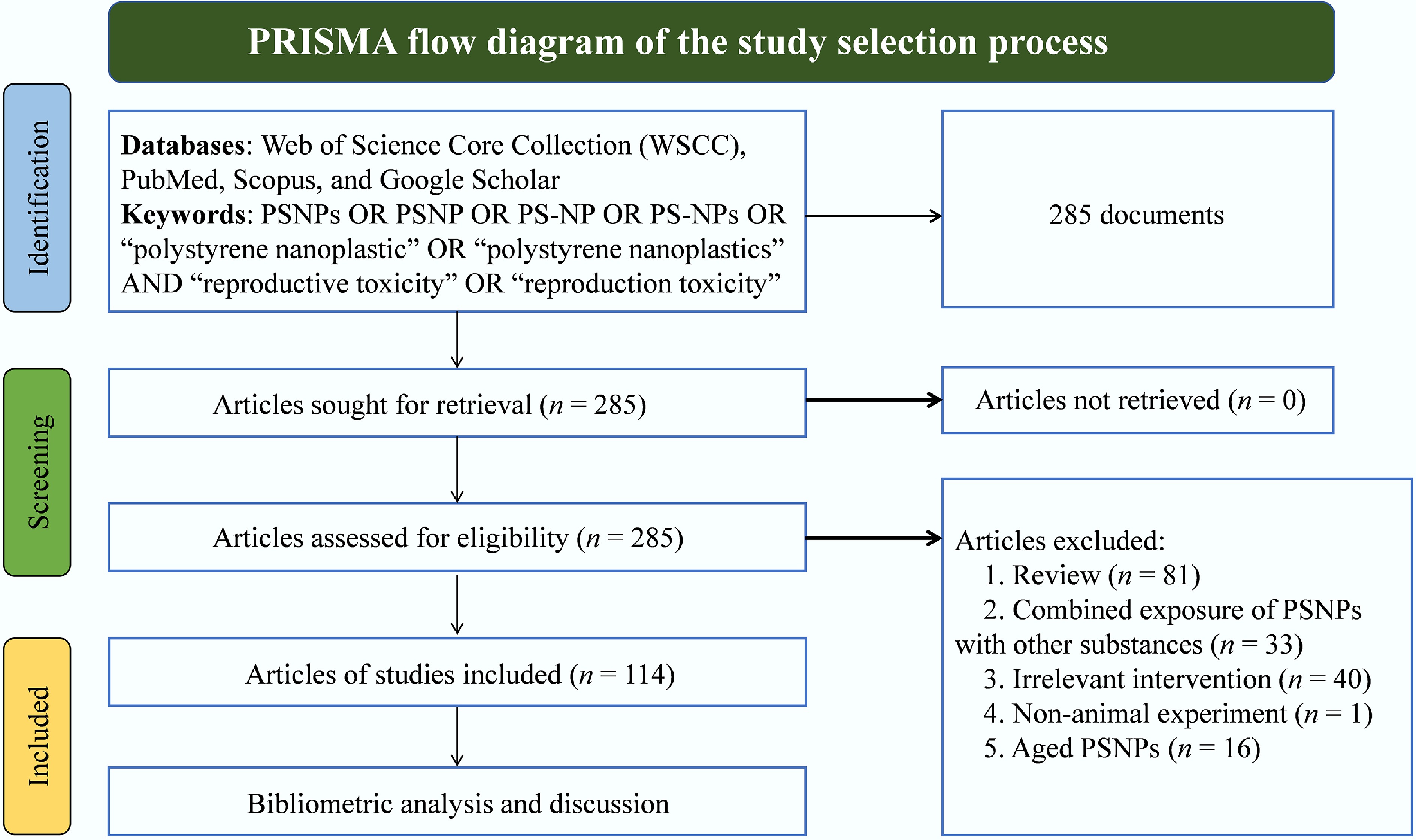
Figure 2.
PRISMA flow diagram of the study selection process.
Analysis method
-
Bibliometric analysis is defined as a tool for statistical and quantitative analysis of research publications that can quantify the literature growth on specific subjects and the impact of individual research results, and it is a suitable choice for assessing trends in research activities[28].
VOSviewer (v1.6.20;
www.vosviewer.com ) was used for cluster analysis to produce a network map. The co-occurrence network visualization of content is based on keywords, and the size of the circle is proportional to the frequency of occurrence in publications; that is, the larger the size, the more frequently the keyword appears in the selected articles. Keywords that co-occur frequently are situated close to each other. Accordingly, this analytical approach enables the visualization of research trends pertaining to the reproductive toxicity of PSNPs through the co-word network generated by this method. -
We investigated the publication output of journals focusing on the reproductive toxicity of PSNPs. The publication status of studies on the reproductive toxicity of PSNPs is presented in Fig. 3a. Science of the Total Environment featured the largest number of relevant reports (17 articles), followed by Environmental Pollution (13 articles), while the Journal of Hazardous Materials and Chemosphere each had nine publications. Notably, the majority of these papers were published in environment-related journals, providing valuable insights for researchers when selecting appropriate journals for their future submissions on this topic. The journal analysis ranks journals by publication count, providing a quantitative overview of research activity in this field. However, publication count alone does not guarantee topic suitability; therefore, when selecting a target journal, authors should also consider the journal’s scope, recent publications on related topics, and other practical factors such as review speed and open access policies.
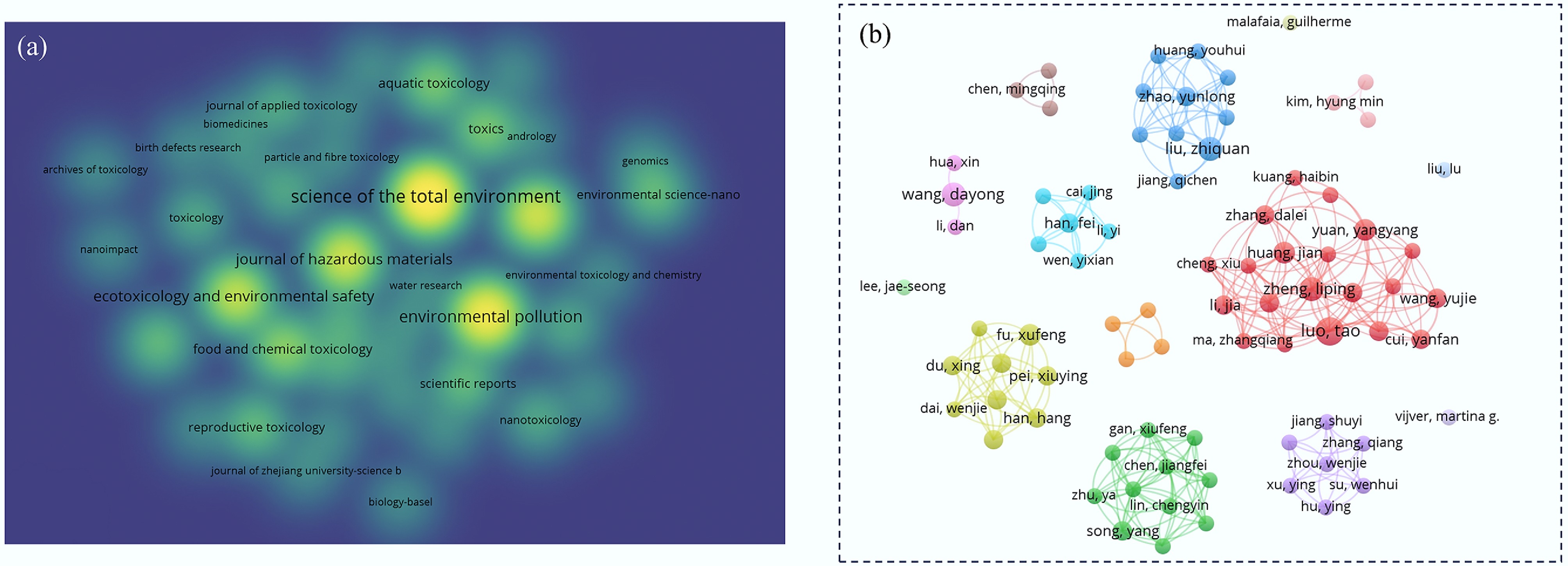
Figure 3.
Network map of journals and authors on the reproductive toxicity of PSNPs. (a) Publication volume of articles on the reproductive toxicity of PSNPs across various journals, with darker colors indicating higher publication counts. (b) Publication output of authors in this field, where larger nodes indicate more publications, and lines represent collaborative relationships.
Active authors
-
As shown in Fig. 3b, a total of 798 contributors have been involved in research on the reproductive toxicity of PSNPs. In terms of the number of papers and citation frequency, Chinese authors rank first, followed by researchers from South Korea. Tao Luo, Liping Zheng, and Zhiquan Liu are the top three researchers in this field. This is evidenced by their publication volume and citation count, which underscore their contributions to this field. The size of the nodes represents the number of papers, and the connections between nodes indicate the collaboration among researchers.
Keyword analysis
-
The co-occurrence network in Fig. 4a prominently identifies four primary research themes, which are distinctly clustered into separate groups. Cluster 1 (red) encompasses the keywords: microplastics, nanoplastics, reproductive toxicity, oxidative stress, apoptosis, autophagy, mechanism, spermatogenesis, and gut microbiota, with a core focus on the mechanisms of reproductive toxicity and its target effector organs. Cluster 2 (green) includes zebrafish (Danio rerio), marine environment, Daphnia magna, polystyrene nanoplastics, exposure, and accumulation, indicating that zebrafish and Caenorhabditis elegans (C. elegans) are the most commonly used model organisms, and that PSNPs have been the subject of extensive investigation in the field of microplastic-induced reproductive toxicity. In addition, neurotoxicity (cluster 3, yellow) and gene expression (cluster 4, blue) are frequently cited in the relevant literature, highlighting these as the current research hotspots in this domain.
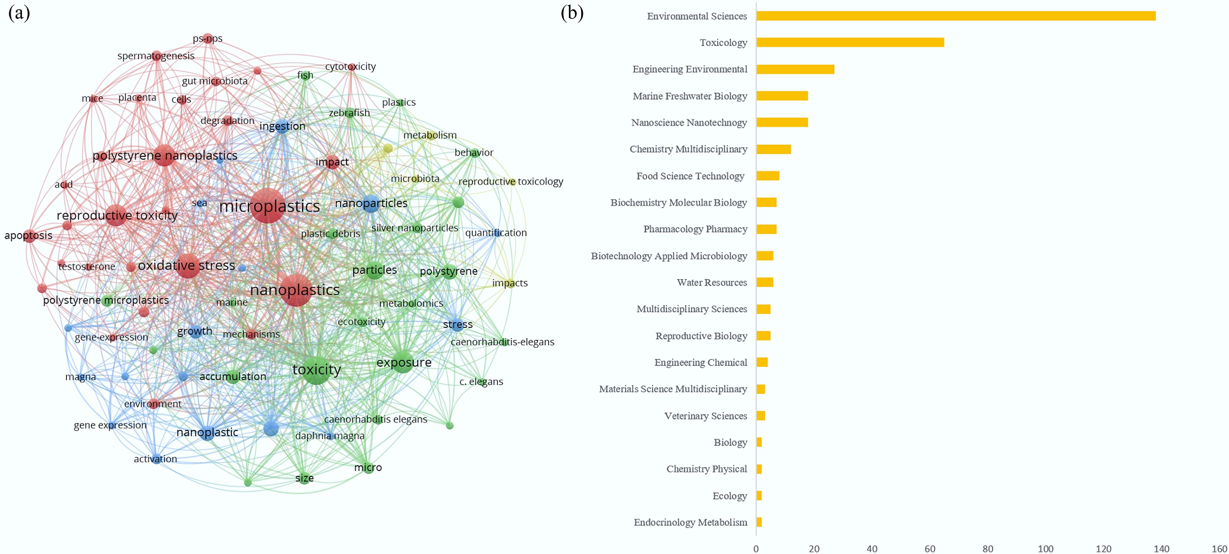
Figure 4.
Network map of keywords and subject areas in the field of reproductive toxicity of PSNPs. (a) Keyword co-occurrence map, where larger nodes indicate a higher frequency of mention, and connecting lines represent the co-occurrence associations between keywords. (b) Interdisciplinary intersection counts for PSNP-related reproductive toxicity research.
Field of study analysis
-
Analytical results reveal that the reproductive toxicity of PSNPs intersects with 41 distinct disciplines, confirming that this research topic is inherently interdisciplinary in nature. Among these disciplines (Fig. 4b), Environmental Sciences is the most closely related field with 139 publications, followed by Toxicology with 65 publications and Environmental Engineering with 27 publications. This disciplinary distribution reflects the diversity of the field and demonstrates the need for cross-disciplinary collaboration among environmental, toxicological, and engineering fields to conduct more comprehensive research and risk prevention on reproductive toxicity caused by PSNPs.
Experimental evidence for the reproductive toxicity of PSNPs
-
Declining fertility rates represent a critical public health concern[29], with impaired fecundity being a key driver of low birth rates. Accumulating evidence has indicated that environmental contaminants adversely affect reproductive health and pregnancy outcomes[30]. Recent studies have detected MNPs in neonatal meconium[18], human semen, and breast milk[31]. The male reproductive system is particularly susceptible to pollutants[32], and semen is more sensitive than blood to the accumulation of toxicants[33]. Research by Montano et al.[32] demonstrated the presence of MNPs in human semen and proposed that MNPs likely enter semen through the epididymis and seminal vesicles, which are prone to inflammation. Additionally, MNPs can cross the placental barrier and accumulate within placental tissue[20]. Most of the MNPs present in the placenta are composed of PE and PS. The presence of MNPs in the human placenta is associated with adverse pregnancy outcomes, such as intrauterine growth restriction (IUGR). Inverse associations between MNP exposure and birth outcomes have been observed in terms of birth weight, length, head circumference, and one-minute Apgar score[34], suggesting the potential impact of MNPs on fetal development.
Miscarriage is defined as an abnormal pregnancy loss before viability, which is related to environmental pollutants. PSNP exposure leads to increased incidence of preterm birth and miscarriage, reduced litter size, and enhanced apoptosis in placental tissues, suggesting reproductive toxicity in mammals[35]. In chorionic villus tissue samples from pregnant women with unexplained recurrent miscarriage (RM), higher levels of PS plastic fragments were detected compared with controls, and the PS fragment content was positively correlated with miscarriage[36]. Meanwhile, PSNPs can cross the placental barrier and are readily internalized into trophoblast cells, inhibiting migration/invasion and migrasome formation in human trophoblast cells and inducing miscarriage[21]. Direct studies on the effects of PSNPs in humans are limited. Instead, toxicological evaluations were performed using cultured human cells and animal models. In different animal models, the male and female reproductive systems sustained distinct types of damage (Table 1).
Table 1. Comparison of sex-specific damage in PSNPs among different species
Sex Species Route of exposure Affected signaling pathway Dose/size of exposure Outcome Ref. Male Mice Orally exposed PSNPs induce oxidative stress and affect the expression of genes associated with apoptosis and inflammation. 25-, 50-, and
100-nmDamage the testicular structure and function, but also decrease sperm count and quality, ultimately reducing male fertility. [38] Rats Gavage Endocrine disruption 1, 3, 6, and
10 mg/(kg·d)
25-, 50-nmReduce serum LH and testosterone (T) levels. [57] Zebrafish Water Meiosis and reproduction, DSB repair mechanisms. 5 mg/L of 45 nm Induced histopathological lesions in the testis. Sperm cells showed anomalous aggregation and condensed chromatin. Although the sperm remained viable, their motility was markedly impaired. [54] Oysters Configured as a suspension and exposed to the environment Perturbation of antioxidant defenses. 50 nm polystyrene beads with amine (50-NH2 beads) or carboxyl (50-COOH beads) functions Caused a decrease in the percentage of motile spermatozoa (66%) and in the velocity (38%). [41] Female Mice Gavage Endocrine disruption: E2 and P were elevated, while FSH and LH levels were diminished. 2.5% w/v, 10 mL
50–90 nmDecreases the live birth rate and neonatal crown-rump length, embryo implantation sites, and uterine wet weight. [30] Rats Direct drinking Wnt/β-catenin signaling pathways 0, 0.015, 0.15, and 1.5 mg/d
0.5 μmOvarian fibrosis in rats, along with apoptosis of granulosa cells, ultimately leads to a decline in ovarian reserve capacity. [58] Zebrafish Water PPAR signaling pathway, dysregulated lipid transport, binding and activity processes 5 mg/L
80, 200, 500 nmPolycystic ovary syndrome-like ovulatory dysfunction. [59] Oysters Oral ingestion An alteration in glucocorticoid response, insulin pathway, and fatty-acid metabolism 14% ± 2% of the
2 μm and 69% ± 6% of the 6 μmSignificant decreases in oocyte number (−38%), diameter (−5%). [47] Fruit fly Mixed with the standard cornmeal fly feed Several metabolic routes were perturbed.
These included steroidogenesis and the turnover of arginine and proline.
At the systemic level, cholesterol homeostasis was affected.
Longevity-related signalling also showed changes.
Regulation of lipolysis in fat cells was disrupted. Ovarian steroid production and progesterone-driven oocyte maturation were similarly influenced.1, 10, 50, and
100 mg/L
100 nmAccumulate in the crop and gut and might even cross the intestinal barrier to reach ovarian tissue; the number of eggs produced and the eclosion rate decrease. [46] The impact on the male reproductive system
-
The influence of PSNPs on the male reproductive system is mainly manifested in the testes, sperm motility, spermatogenic cells, the BTB, testosterone levels, etc. The BTB is one of the tightest blood-tissue barriers in the mammalian body, regulating the transport of nutrients (e.g., glucose, amino acids), essential molecules (e.g., hormones, electrolytes), and harmful toxicants (e.g., environmental pollutants, drugs, chemicals), while also serving as an immunological barrier[37].
In the mouse (Mus musculus) model, PSNPs could disrupt the BTB and then enter the testicular tissue. Testosterone levels in serum were significantly decreased. PSNPs accumulated in the testes, induced oxidative stress, affected the expression of apoptosis- and inflammation-related genes, and led to damage to testicular microstructure and functions[38]. The environment for spermatogenesis is disrupted, resulting in a decline in sperm count and motility. At the same time, abnormal sperm morphology occurs, such as acrosome loss, cephalic (small head), acephalia (no head), cervical folding, and tailless. Exposure of male mice to PSNPs of varying sizes impaired their reproductive performance and even led to infertility. Notably, this study demonstrated that spermatozoa serve as a toxic target of PSNPs, precisely because PSNPs are sufficiently small to penetrate sperm cells and inhibit capacitation, thereby causing male infertility[39].
Regarding the definition of sperm quality, motility (e.g., the percentage of motile spermatozoa and the swimming speed) and reproductive success rate are predominant[40]. In the oyster (Crassostrea virginica) model, oyster spermatozoa were exposed to PSNPs, and their fertilization ability was hindered, reducing the percentage of motile spermatozoa and velocity[41]. What is noteworthy is that PSNPs that simulated solar aging could lead to the downregulation of reproduction-related genes (SOX-8, SOX-E, Piwi1, TGF-β), delaying male gonadal development[42]. This might be closer to the result caused by the environment.
The impact on the female reproductive system
-
In addition to concerns about toxicity in the male reproductive system, potential toxicity to the female reproductive system has also received widespread attention. Interestingly, studies have shown that female mice are more susceptible to the adverse effects of MPs on reproductive capacity than male mice, indicating that there may be sex-specific differences in pollutant toxicity[31]. The antral follicle stage is a decisive determinant of female reproductive success. At this juncture, the follicle gains robust responsiveness to the pituitary hormones FSH and LH. This endocrine sensitivity initiates a cascade of events: granulosa cell proliferation, steroidogenesis, and antral cavity formation. These processes are essential for the oocyte to complete meiotic maturation and for the follicle to undergo ovulation. Thus, antral follicle integrity is fundamental to fertility[43]. Mechanistically, reduced antral follicle size serves as one of the first identifiable morphological endpoints for ovarian toxic injury. After ovulation, the corpus luteum becomes the primary source of progesterone. This steroid hormone primes the endometrium to receive the implanting embryo and preserves early pregnancy[44]. The number of eggs produced is the most representative indicator for evaluating ovarian function[45].
In the mouse model, long-term exposure to PSNPs disrupted the ovarian function of female mice; embryo implantation numbers and uterine wet weights were significantly reduced. Consistent with this ovarian impairment, multiple adverse changes occurred in a dose-dependent manner: antral follicle size declined, corpus luteum density dropped, atretic follicle density rose, estrous cycles lengthened, and serum progesterone concentrations decreased[30].
In the fruit fly (Drosophila melanogaster) model, PSNPs could accumulate in the crop and gut, and might even cross the intestinal barrier to reach ovarian tissue[46]. In a study by Sussarellu et al.[47], oysters exposed to PSNPs for 2 months exhibited significant reductions in oocyte number (−38%) and diameter (−5%). Consequently, offspring production and offspring development were reduced by 41% and 18%, respectively.
The impact on the development of offspring
-
PSNPs not only damage parental reproductive organs but also exert intergenerational toxic effects on offspring development. In this context, intergenerational toxicity is defined as the adverse effects on offspring resulting from direct parental exposure to PSNPs. Exposure induces a marked decrease in fertilization success, impairs embryo and larval development, causes multiple malformations, and even leads to complete developmental arrest[48]. In the mouse model, PSNP exposure decreased the live birth rate and neonatal crown-rump length[30]. Chronic exposure to PSNPs even alters the sex ratio of Daphnia pulex neonates[49]. In oysters, offspring of parents subjected to exposure exhibited diminished movement. Additional observations included morphological defects, developmental cessation, and stunted growth. These phenotypes collectively indicate that PSNPs are a toxic insult affecting early-life neurobehavioral and somatic development[50]. In C. elegans, PSNP exposure increases embryonic and larval lethality[51]. The larval yield and larval development of offspring derived from exposed parents decreased by 41% and 18%[47].
Zebrafish
-
Zebrafish are common model animals for the reproductive toxicity of PSNPs. As a vertebrate model, zebrafish feature rapid embryonic development, low maintenance cost, and high fecundity[52], which are similar to mammals in terms of physiology, development, metabolism, and signaling pathways. Their response to toxic substances can well predict the response of mammals[53]. In zebrafish, both male and female germ cells are directly affected to different degrees after exposure to PSNPs, and accumulation occurs within the germ cells. In males, PSNPs induce testicular histological alterations with delayed spermatogenesis, abnormal sperm clustering and chromatin compaction, and reduced motility of spermatozoa. Moreover, abnormal oocyte growth and follicular growth were observed in females[54].
PSNPs exert adverse effects on the fish reproductive system both in the directly exposed generations and in their unexposed offspring. Studies exploring maternal exposure in P0 zebrafish and corresponding F1 offspring yielded consistent findings. Exposure to PSNPs drastically suppressed egg production in P0 females. PSNPs were also detected in multiple organs of both P0 and F1 zebrafish. Mechanistically, PSNPs disrupt the expression of hypothalamic-pituitary-gonadal (HPG) axis-related genes. This disruption impairs the reproductive performance of female P0 zebrafish, hinders F1 development, and disturbs endocrine homeostasis in F1 larvae[55]. Our previous research showed that long-term parental PSNPs exposure reduces offspring fertilization and survival rates, increases deformity rates, and induces premature embryo hatching[56]. It is worth noting that when observing the gene expression of the high-concentration PSNPs maternal exposure group, a decrease in foxl2a and an increase in sox9a were found, indicating that PSNPs have the potential to affect the sex differentiation of zebrafish, and further research is needed[55].
Toxicity mechanisms and signaling pathways
-
PSNPs pose a significant threat to reproductive capacity, with toxicity mediated through diverse molecular mechanisms, including oxidative stress, endocrine disruption, inflammation, autophagy, and apoptosis (Fig. 5). Clarifying these mechanisms plays a vital role in assessing the comprehensive hazards induced by PSNP exposure and establishing approaches to reduce their adverse outcomes on reproductive and systemic health.
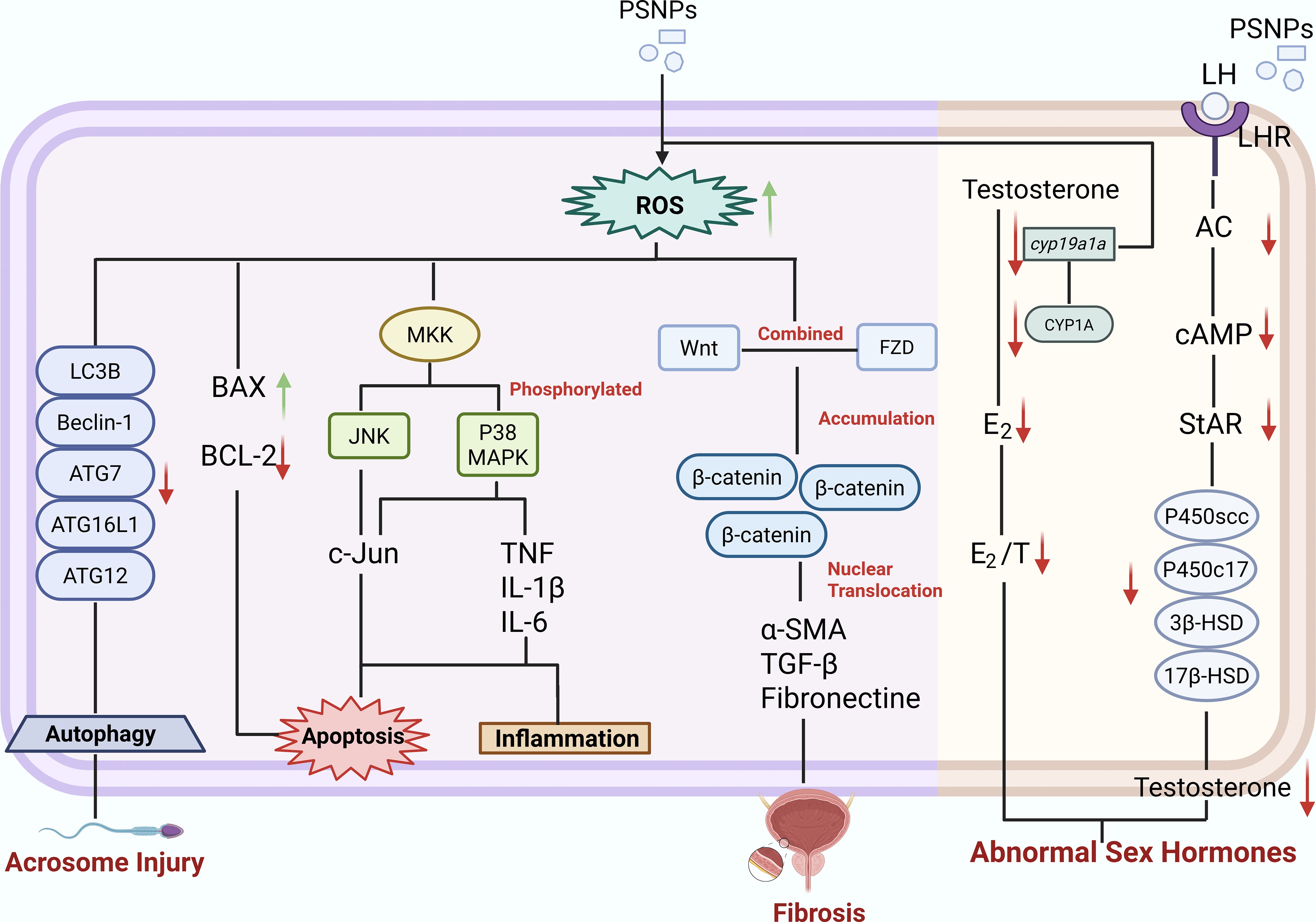
Figure 5.
Mechanisms of PSNPs-induced reproductive toxicity.
Oxidative stress
-
Oxidative stress is the core mechanism underlying the reproductive toxicity induced by PSNPs, which involves multiple cellular processes and signaling pathways. Understanding the mechanism by which oxidative stress induces reproductive toxicity of PSNPs is crucial for strategies to intervene and mitigate the adverse effects of PSNPs on reproductive and developmental health. Cellular function depends on a tightly regulated oxidant-antioxidant equilibrium. This balance, if perturbed, generates oxidative stress that impairs tissue performance. Therefore, maintaining redox stability is a prerequisite for normal organismal physiology[14]. PSNPs have been proven to trigger excessive production of reactive oxygen species (ROS)[60] and impair the redox balance. Studies have shown that PSNPs activate the cellular antioxidant defense system by increasing intracellular ROS levels, causing oxidative stress and activating Jun NH2-terminal kinase (JNK) and P38 Mitogen-Activated Protein Kinase (p38 MAPK) signals. The MAPK signaling pathway constitutes a large superfamily including p38, JNK, and extracellular signal-regulated kinase (ERK). Existing studies have confirmed that MAPK acts as a redox-sensitive signaling mediator. It is tightly linked to sperm maturation. p38 MAPK is involved in the synthesis of inflammatory cytokines at the transcription and translation levels, thus elevating their production in the testis, causing injury to seminiferous tubules and spermatogenic cell apoptosis. Subsequently, sperm motility and concentration decrease, the sperm abnormality rate increases, and spermatogenesis is disrupted[61]. With the increase in exposure concentration, the phosphorylation degree of p38 MAPK also increases accordingly. Phosphorylation of p38 MAPK initiates a signaling cascade. This cascade governs multiple physiological outcomes. These outcomes encompass inflammatory responses, lineage specification, senescence, cytokine secretion, and programmed death[62]. Therefore, in the male reproductive system, it is suggested that PSNPs impede energy supply, interfere with spermatogenesis, lead to a decline in sperm quality, cause deformities, and subsequently result in reproductive toxicity.
The Hippo signaling pathway participates in both physiological and pathological ovarian aging. PSNP exposure triggers oxidative stress and disrupts Hippo pathway activity. These alterations further induce granulosa cell apoptosis and eventually impair mammalian fertility[63]. The Wingless/integrase-1 (Wnt) cascade is a central pathway governing multiple processes during embryonic development[64]. As an evolutionarily conserved signal transduction cascade, Wnt signaling relays signals via cell–cell interactions. It modulates cell fate, polarity, differentiation, and migration, thus orchestrating organogenesis and fetal development[65]. In females, exposure to PSNPs upregulated ovarian protein levels of Wnt, beta-catenin, and its phosphorylated form. This upregulation drove activation of the Wnt/beta-catenin signaling axis. Pathway activation induced both fibrotic lesions and apoptosis of granulosa cells. Oxidative stress served as the trigger for apoptosis, and together these insults compromised ovarian reserve[58].
Endocrine disruption
-
Testosterone, the most important androgen in males, plays an important role in spermatogenesis, and reduced testosterone levels are strongly correlated with testicular and Leydig cell dysfunction[66]. PSNP exposure impairs reproductive function by disrupting hormonal homeostasis via interference with the hypothalamic-pituitary-gonadal (HPG) axis. Rat animal experiments demonstrated that high-dose PSNP treatment markedly reduced serum LH and testosterone (T) levels in male rats[57].
In the female reproductive system, PSNPs can disrupt the synthesis of estrogen and progesterone, impairing ovarian function and pregnancy conditions[67]. The synthesis and metabolism of sex hormones regulate the expression of genes related to the HPG axis. Cyp19a1a is a key rate-limiting enzyme that converts androgen precursors into estrogen. PSNP exposure leads to the inhibition of the Cyp19a1a gene, resulting in a decrease in estrogen levels in zebrafish, which affects zebrafish reproduction, gonadal development, and sex differentiation[55].
Inflammation
-
Inflammation is closely associated with oxidative stress and elevated ROS. Exposure to PSNPs in mouse ovaries led to increased levels of proinflammatory cytokines, such as interleukin-6 (IL-6), indicating that PSNPs induce ovarian inflammation[68]. Furthermore, studies by Hou et al.[69] have demonstrated that oxidative stress triggers NLRP3 inflammasome-mediated activation of caspase-1, which subsequently promotes the formation of effector proinflammatory cytokines, ultimately resulting in ovarian inflammation and culminating in apoptosis of ovarian granulosa cells.
The p38 MAPK kinase pathway shares many similarities with other MAPK kinase cascades, being associated with inflammation, cell growth, cell differentiation, and cell death[70]. Given its role in controlling the transcription and translation of inflammatory mediators, p38 MAPK is a potential target for anti-inflammatory therapies[71]. A study by Xie et al.[62] indicated that PSNP exposure can increase the production of pro-inflammatory factors (IL-1β, TNF-α, and IL-6) in the testicles of mice through the activation of the p38 MAPK signaling pathway, leading to a decline in sperm quality in mice.
Autophagy
-
As an evolutionarily conserved survival mechanism, autophagy represents the major intracellular catabolic process that degrades and recycles misfolded proteins and damaged organelles via autophagosomes[72]. Spermatogenesis, sperm motility, and fertilization all rely on abundant energy provision[73,74]. Mitochondria serve as the primary cellular energy generators. Their dysfunction impairs normal sperm physiological performance. Mitochondria can slow down the deterioration of tissue damage through autophagy during aging and damage, and excessive autophagy may lead to apoptosis[75]. Studies have shown that autophagy is associated with sperm acrosome injury induced by PSNPs. The acrosome is a large secretory vesicle in the head of sperm and plays a key role in gamete interactions. Its dysfunction is recognized as an important cause of fertilization failure and is an essential underlying mechanism of male reproductive damage[76]. Autophagy mediates PSNPs-induced acrosome damage by regulating two key acrosome-forming proteins (GOPC and DPY19L2) and reducing the expression of autophagy-related proteins (e.g., LC3B, Beclin1, ATG7, ATG16L, and ATG12). Notably, autophagy inhibition induced by PSNPs is involved in the depolymerization of vesicles associated with the Golgi apparatus. This represents a suppression of protective autophagy, resulting in an increase in irregular Golgi complexes, which ultimately leads to acrosome defects in spermatozoa[77].
Apoptosis
-
Programmed cell death, also known as apoptosis, is an orderly, frequently energy-consuming process. It relies on the activation of cysteine proteases known as caspases and involves an intricate signaling cascade. This cascade transduces initial triggering signals and ultimately drives programmed cell death[78].
Trophoblasts, the key cells at the maternal–fetal interface, play an essential role in placental implantation and reproduction[79]. In the study of the effect of PSNPs on human trophoblast cells, it was found that PSNP exposure upregulates cleaved caspase-3 or downregulates Bcl-2. By activating the Bcl-2/caspase-2/caspase-3 signaling pathway, it induced human trophoblast cell apoptosis through the mitochondrial pathway[36]. Similarly, mouse testicular Leydig cells could also undergo apoptosis through this pathway, causing damage to the male reproductive system[80]. Granulosa cells play an important role in the development and maturation of follicles[81]. PSNPs could cause apoptosis of granulosa cells through oxidative stress-triggered NLRP3/caspase-1 pathways, thereby reducing ovarian reserve in rats[69]. Yu et al.[22] found that PSNPs induced ROS production in C. elegans and regulated cell apoptosis via the CED-9/CED-4/CED-3 pathway.
Apoptosis induced by PSNPs has also been observed in aquatic organisms. In zebrafish, staining with Acridine orange revealed higher apoptotic rates in the exposed cohorts. This was accompanied by elevated p53, caspase-3, and caspase-9, along with reduced Bcl-2 transcript levels. The oxidative stress elicited by PSNPs triggered the apoptotic machinery[82].
Combined toxicity of PSNPs with coexisting pollutants and toxicity of aged PSNPs
-
The combined reproductive toxicity of PSNPs with other pollutants is another research hotspot. MNPs have been confirmed to serve as carriers for these pollutants, and the combined reproductive toxicity may be more severe than that of a single pollutant. We should also pay attention to the impact of aging PSNPs on reproductive toxicity. The above situations may be closer to the toxic effects that PSNPs exert in daily life.
Co-exposure to arsenic and PSNPs exacerbated oxidative stress, leading to disruption of the blood-testis barrier in mice[83]. Combined subchronic exposure to PSNPs and dibutyl phthalate (DBP) caused greater damage to the male reproductive system than exposure to each substance individually, leading to vascular congestion of the testes, Leydig cell hyperplasia and injury, disturbed testosterone production, widespread congestion of the tunica albuginea, decreased sperm development, and compromised antioxidant defense systems[84]. Combined exposure to PSNPs and diclofenac exacerbated oxidative stress, apoptosis, and lipid peroxidation, significantly upregulated the expression of TNF-α, COX-2, and IL-1β in adult zebrafish, reduced the hatching rate, and increased the malformation rate of larvae[85]. Exposure to PSNPs-UV resulted in reduced reproductive fitness in C. elegans and elevated mortality among progeny embryos and larvae, an effect mediated by the DAF-16 activated stress response[51]. The more severe reproductive damage caused by combined exposure and aged PSNPs suggests that they will become a major direction for future research.
-
Due to the large-scale production and waste of plastics, they are distributed, accumulate, and degrade into microplastics in nature. More and more studies have shown that MPs exist in human reproductive organs. In different animal models, PSNPs can cause inflammation, autophagy, apoptosis, and other related cascade reactions through oxidative stress, interfere with endocrine function, and affect sex hormones, thereby causing varying degrees of damage to the reproductive organs of both sexes and affecting their fertility. PSNPs also cause various damage to offspring through intergenerational toxicity passed from parents to offspring, such as unexplained miscarriage and deformity. For humans, in vitro and organoid models have shown that PSNPs have toxic effects on human-derived germ cells, suggesting that PSNPs have potential reproductive and intergenerational toxic effects on humans.
Recently, research on the toxicity of microplastic particles and their underlying mechanisms has attracted considerable attention. Beyond conventional analytical approaches, a relatively novel strategy involves the application of bioimaging modalities. In particular, aggregation-induced emission (AIE)-based bioimaging enables quantitative analysis of the in vivo biodistribution and barrier transport of MNPs, allowing researchers to establish direct and spatially resolved causal relationships. This provides new insights and perspectives for investigating the toxicological pathways of MPs and NPs[86]. Additionally, focusing on tracing probes for fluorescent nanoparticles, Wang et al. uniformly embedded tetraphenylethene aggregation-induced emission luminogens into the polystyrene backbone without altering the intrinsic physicochemical properties of the nanoparticles. Through surface charge modulation, the probes were also endowed with the capability of targeted binding to the extracellular matrix. This controllable synthesis strategy can serve as a template for the preparation of biocompatible labeled polystyrene nanoparticles specifically for reproductive toxicology, thereby facilitating subsequent accurate quantification of the in vivo accumulation of fluorescent nanoparticles[87,88]. Since the first detection of MPs in the maternal placenta, subsequent studies have found that PSNPs can interfere with endocrine function in human primary placental cells, leading to reduced release of human chorionic gonadotropin (HCG). This reduction may increase the risk of Down syndrome in infants and early preeclampsia, thereby contributing to adverse pregnancy outcomes.
Existing data from toxicology and sparse epidemiology link PSNPs to redox imbalances, proinflammatory states, immune deviations, and endocrine disturbances. However, a causal nexus in people has yet to be established. Definitive human evidence remains unavailable. Prevalence estimates and risk assessments are constrained by ethical limitations. Importantly, the detection of PSNPs in animal models does not equate to a quantifiable internal exposure dose. Moreover, animal studies predominantly employ pristine spherical polystyrene particles at concentrations often exceeding realistic environmental levels, and the observed effects do not imply direct causal links with adverse clinical outcomes in humans. The biologically relevant internal dose remains largely unknown.
To address these limitations, there is an urgent need for internationally harmonized experimental protocols, rigorous contamination control measures, and standardized reporting systems to ensure the reliability of biomonitoring and to clarify the potential health implications of PSNP exposure in humans. The development of robust, standardized methods specifically validated for the detection of NPs in environmental and biological matrices remains a critical unmet need. Advances in detection technologies, such as artificial intelligence-assisted analysis and AIE techniques, hold promise as transformative tools in mechanistic toxicology. These approaches may enable more sensitive and specific quantification of NPs in human tissues. Finally, in the future, more attention should be paid to measures and strategies for reducing the reproductive and developmental toxicity of MNPs, such as the production of new environmentally friendly materials that can remove or replace plastics and the research and development of related technologies, strict control of the production and discharge of plastics, and public health publicity and education to make people pay attention to and understand the classification of plastic waste and the scientific treatment of plastic waste, thereby reducing exposure to microplastics in the environment.
-
In conclusion, this review elaborates on the severity of MP pollution and the significant impact of one of the most common types of MPs-PSNPs on the reproductive and developmental health of multiple species. Based on recent research, our analysis indicates that PSNPs can accumulate in the reproductive organs of different animal models, causing varying degrees of damage to the reproductive organs and germ cells of both sexes and affecting the developmental health of offspring. The molecular mechanisms of these effects, including oxidative stress, endocrine disruption, inflammation, autophagy, and apoptosis, highlight the complexity and multi-faceted nature of the reproductive toxicity of MPs. In future research, we need to focus on human epidemiological investigations, the combined effects of PSNPs and other pollutants, and the toxic effects of aging PSNPs. At the same time, efforts should be made to reduce plastic production and pollution, raise public awareness of the toxic hazards of MPs, carry out health education, and minimize human exposure to these pollutants.
-
All experimental procedures were conducted in accordance with the guidelines of the Institutional Animal Care and Use Committee (IACUC) of the University of South China (Permit No. 11000252-2).
We thank Biorender (
www.biorender.com ) for providing the graphical tools used in this reivew.-
The authors confirm their contributions to the paper as follows: Liyao Huang: investigation, methodology, data curation, and writing – original draft. Wanjing Liu: writing – review and editing. Chunhua Zhan: conceptualization, supervision, writing – review and editing, and resources. All authors read and approved the final manuscript.
-
The datasets generated during and analyzed during the current study are available from the corresponding author upon reasonable request.
-
The authors declare that they have no known competing financial interests or personal relationships that could have appeared to influence the work reported in this paper.
-
Full list of author information is available at the end of the article.
- Copyright: © 2026 by the author(s). Published by Maximum Academic Press, Fayetteville, GA. This article is an open access article distributed under Creative Commons Attribution License (CC BY 4.0), visit https://creativecommons.org/licenses/by/4.0/.
-
About this article
Cite this article
Huang L, Liu W, Zhan C. 2026. Polystyrene nanoplastics as emerging reproductive toxicants: sex-specific effects, offspring risks, and molecular mechanisms. New Contaminants 2: e025 doi: 10.48130/newcontam-0026-0022 

Polystyrene nanoplastics as emerging reproductive toxicants: sex-specific effects, offspring risks, and molecular mechanisms
- Received: 04 July 2026
- Revised: 24 July 2026
- Accepted: 27 August 2026
- Published online: 14 September 2026
Abstract: Plastic products are ubiquitous in daily life, and their large-scale production and improper disposal have resulted in severe environmental pollution. Plastics degrade into micro- and nano-plastics (MNPs) in nature and can accumulate in the human body. Detected in human reproductive organs, these particles threaten reproductive and developmental health. Among MNPs, polystyrene nanoplastics (PSNPs) are one of the most extensively studied types due to their widespread prevalence. Herein, we conduct a comprehensive review of existing literature focusing on PSNPs, revealing that PSNPs induce distinct reproductive toxicity in both genders and across different species: they damage male testes, disrupt the blood-testicular barrier (BTB), and impair sperm quality; they alter female ovarian function, disturb hormone levels, and reduce oocyte quantity and ovulation. Furthermore, PSNPs also harm embryonic development and offspring, triggering miscarriage and birth defects. Mechanistically, the reproductive toxicity of PSNPs is mediated through multiple molecular pathways, including oxidative stress, endocrine disruption, inflammation, autophagy, and apoptosis. In conclusion, we have integrated systematic knowledge and molecular mechanisms regarding PSNPs-induced reproductive toxicity. We fully clarify the risks and hazards to reproductive and developmental health posed by PSNPs, emphasize the importance of public health intervention and preventive measures, and provide new insights for further research on their toxic mechanisms and prevention.
-
Key words:
- Polystyrene nanoplastics /
- Reproductive toxicity /
- Mechanism /
- Gender sensitivity