-
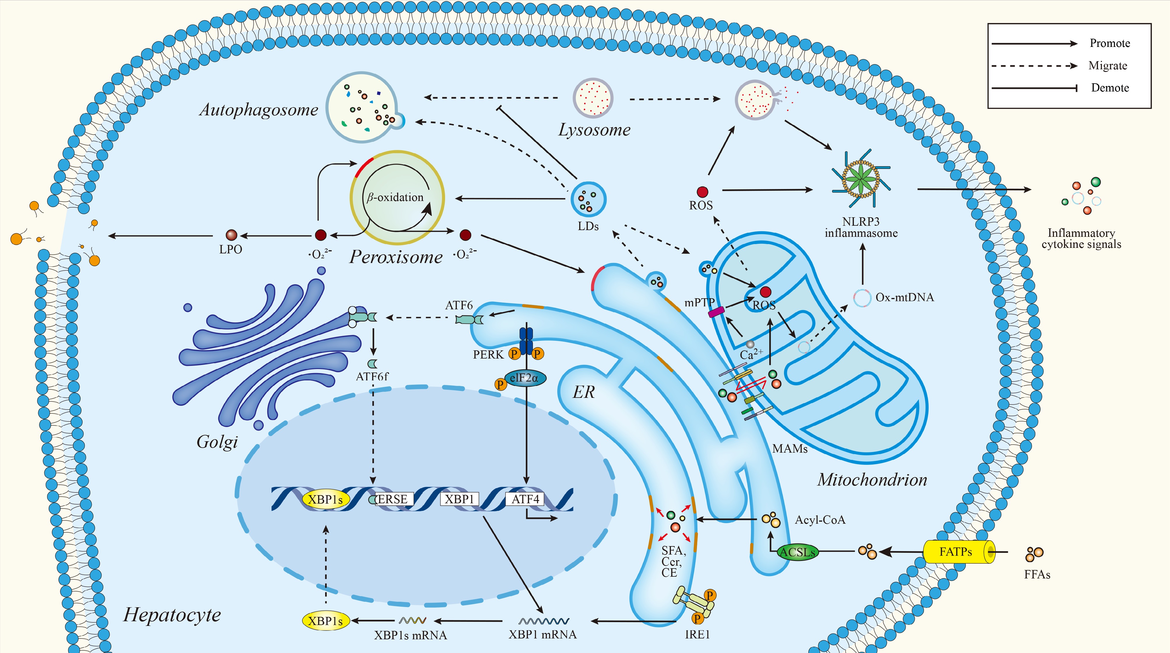
Figure 1.
Lipotoxicity as a spatially and temporally organized lipid signaling network in hepatocytes. A visual representation of hepatocellular lipotoxicity. Lipid metabolism is presented as a spatially established signaling network controlled by inter-organellar communication. Lipid homeostasis is maintained by contact sites between the endoplasmic reticulum (ER), mitochondria, lipid droplets (LDs), lysosomes, and peroxisomes under physiological conditions, coordinating the synthesis, trafficking, storage, and degradation of lipids. Chronic nutrient excess leads to a dysregulation of this coordination, which results in ER stress, mitochondrial dysfunction, lipid droplet overload, impaired lipophagy, and redox imbalance. These interconnected dysfunctions result in oxidative stress, inflammasome activation, inflammatory signaling, and hepatocellular injury, pointing to lipotoxicity as an emerging consequence of dysregulated lipid signaling and organelle crosstalk. ACSLs, acyl-CoA synthetase long-chain family members; Acyl CoA, acetyl-coenzyme A; ATF4/6, activating transcription factors; CE, cholesteryl ester; Cer, ceramide; eIF2α, eukaryotic initiation factor 2α; ERSE, ER stress response element; FATPs, fatty acid transport proteins; FFAs, free fatty acids; IRE1, inositol-requiring enzyme 1; LPO, lipid peroxidation; mPTP, mitochondrial permeability transition pore; Ox mtDNA, oxidized mitochondrial DNA; PERK, protein kinase R-like ER kinase; SFA, saturated fatty acid; XBP1, x-box binding protein 1.
-
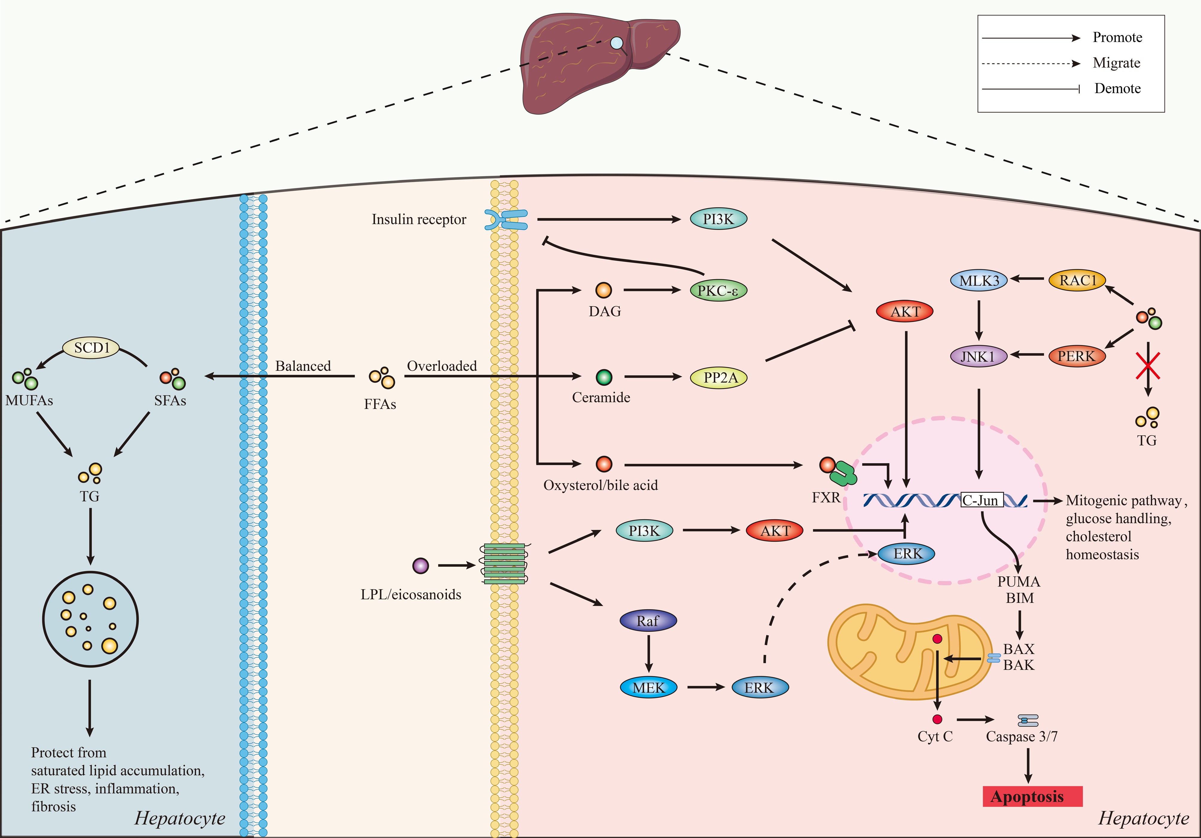
Figure 2.
The intracellular lipotoxic signaling network in hepatocytes. Schematic diagram of lipid class-regulated signaling pathways mediating hepatocellular adaptation or lipotoxicity. Elevated saturated fatty acid (SFA) and SFA metabolite levels (e.g., diacylglycerol [DAG], ceramides, lysophospholipids) activate stress and apoptotic signaling cascades (e.g., c-Jun N-terminal kinase [JNK]-mediated pathways, mitochondrial lipoapoptosis [BAX/BAK activation, cytochrome c release, and caspase-3/7 activation]). DAG accumulation promotes PKCε activation and impairs insulin receptor-PI3K-Akt signaling. Ceramides regulate Akt activity by way of PP2A-dependent signaling. At the same time, oxysterols, bile acids (BAs), and lysophospholipids activate receptor-mediated signaling pathways that regulate metabolic and inflammatory programs. Mono-unsaturated fatty acids (MUFAs), which are partially derived from stearoyl-CoA desaturase 1 (SCD1), facilitate triglyceride synthesis and lipid droplet retention, thus alleviating toxic lipid accumulation. AKT, AKT serine/threonine kinase; ERK, extracellular signal-regulated kinase; LPL, lipoprotein lipase; MEK, mitogen-activated protein kinase kinase; MLK3, mixed lineage kinase 3; PI3K, phosphoinositide 3-kinase; PUMA, p53 upregulated modulator of apoptosis; RAC1, ras-related C3 botulinum toxin substrate 1; Raf, Raf kinase; TG, triglyceride.
-
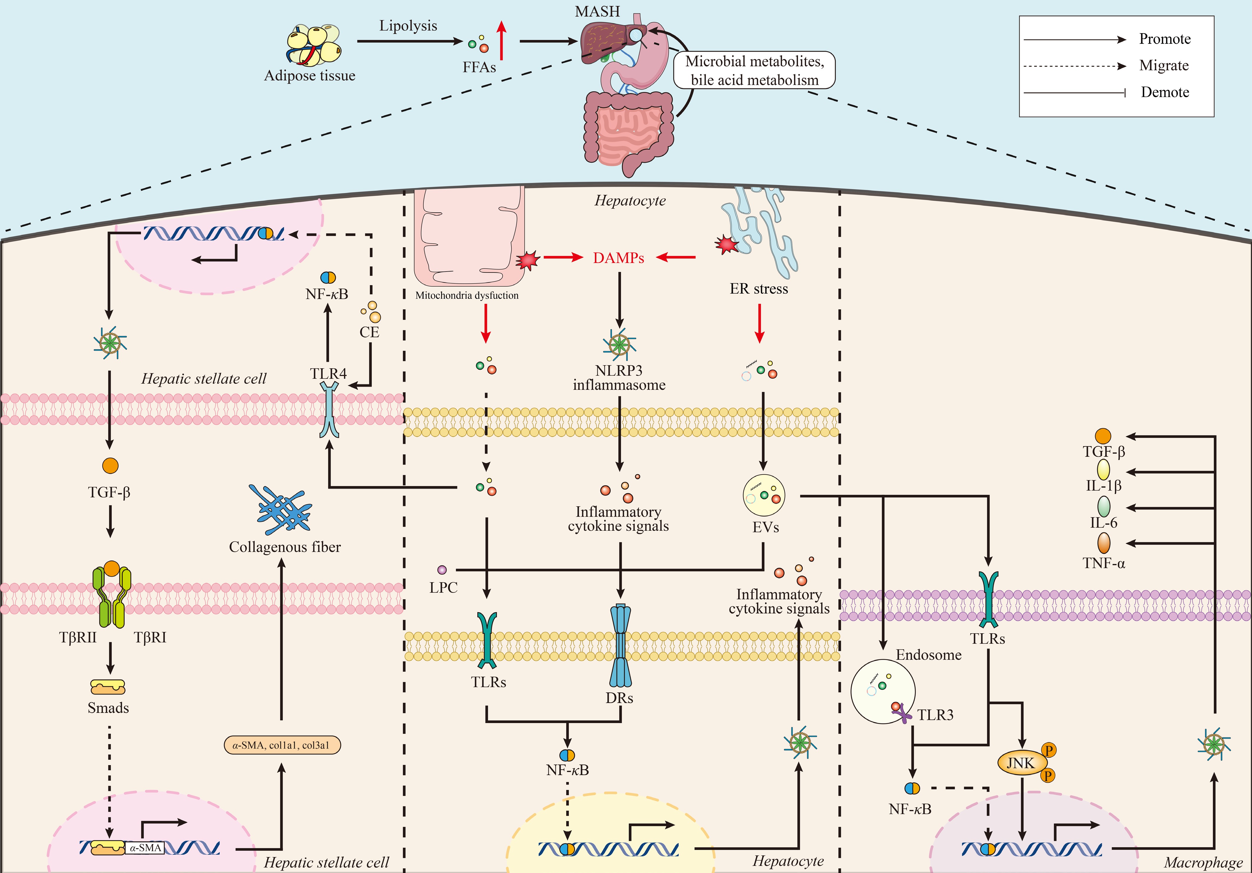
Figure 3.
Lipid-driven intercellular crosstalk promotes hepatic inflammation and fibrosis. Schematic illustration of lipid-mediated intercellular communication in the progression from steatosis to MASH. Lipotoxic stress in hepatocytes leads to the secretion of FFAs, cholesterol crystals, damage-associated molecular patterns (DAMPs), and EVs that serve as paracrine signals in the hepatic microenvironment. These signals activate macrophages by using TLRs, especially TLR4, and NLRP3 inflammasome pathways, leading to activation of NF-κB signaling and production of pro-inflammatory cytokines such as IL-1β, IL-6, and TNF-α. This inflammatory environment aggravates hepatocyte injury and stimulates HSC activation through TGF-β/Smad signaling pathways, which results in collagen deposition and fibrosis. Systemic lipid signals and gut-derived factors further modulate this pathogenic network. α-SMA, alpha-smooth muscle actin; CE, cholesteryl ester; col1a1, collagen type I alpha 1 chain; col3a1, collagen type III alpha 1 chain; DRs, death receptors; JNK-c, Jun N-terminal kinase; LPC, lysophosphatidylcholine; Smads, mothers against decapentaplegic homologs (SMAD family proteins); TLR3, toll-like receptor 3; TβRI, transforming growth factor beta receptor type I; TβRII, transforming growth factor beta receptor type II.
-
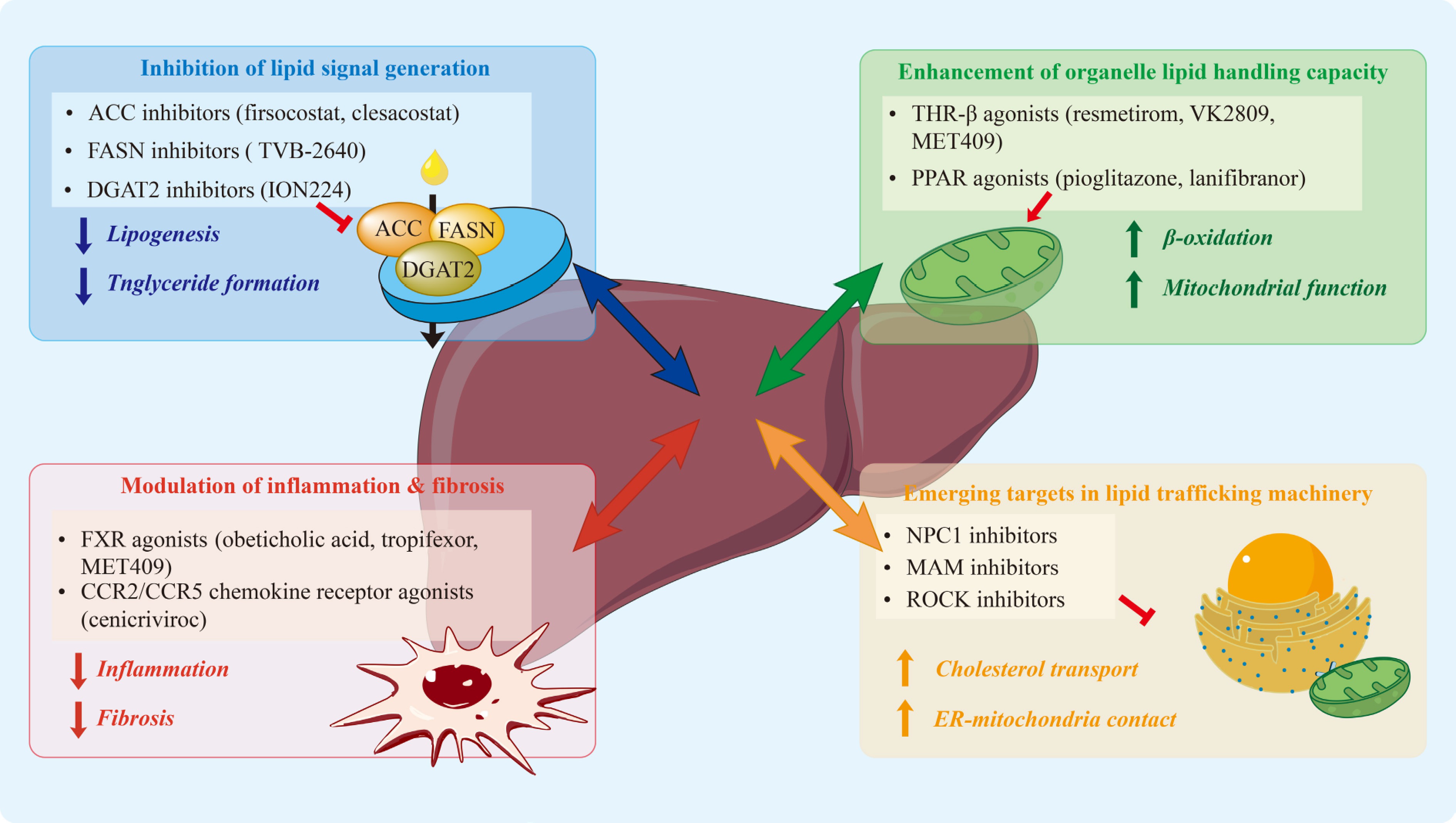
Figure 4.
Therapeutic targeting of lipid signaling and trafficking nodes in MAFLD/MASH. Therapeutic approaches are categorized according to the main hepatocellular processes they modify. Blue (upper left) inhibition of lipid signal generation through acetyl-CoA carboxylase (ACC), fatty acid synthase (FASN), and diacylglycerol acyltransferase 2 (DGAT2) inhibition. Green (upper right), promotion of organelle lipid utilization and mitochondrial function through thyroid hormone receptor β (THR-β) and PPAR pathways. Red (lower left), reduction of inflammatory and fibrogenic signaling through FXR and chemokine pathways. Orange (lower right), modulation of emerging lipid trafficking mechanisms including Niemann-Pick C1 (NPC1), MAMs, and Rho-associated coiled-coil-containing kinase (ROCK) signaling. These complementary interventions reveal targetable nodes that reduce lipotoxic stress, inflammatory amplification, and fibrotic progression. ACC, acetyl-CoA carboxylase; DGAT2, diacylglycerol acyltransferase 2; FASN, fatty acid synthase; PPAR, peroxisome proliferator-activated receptor; THR-β, thyroid hormone receptor beta.
-
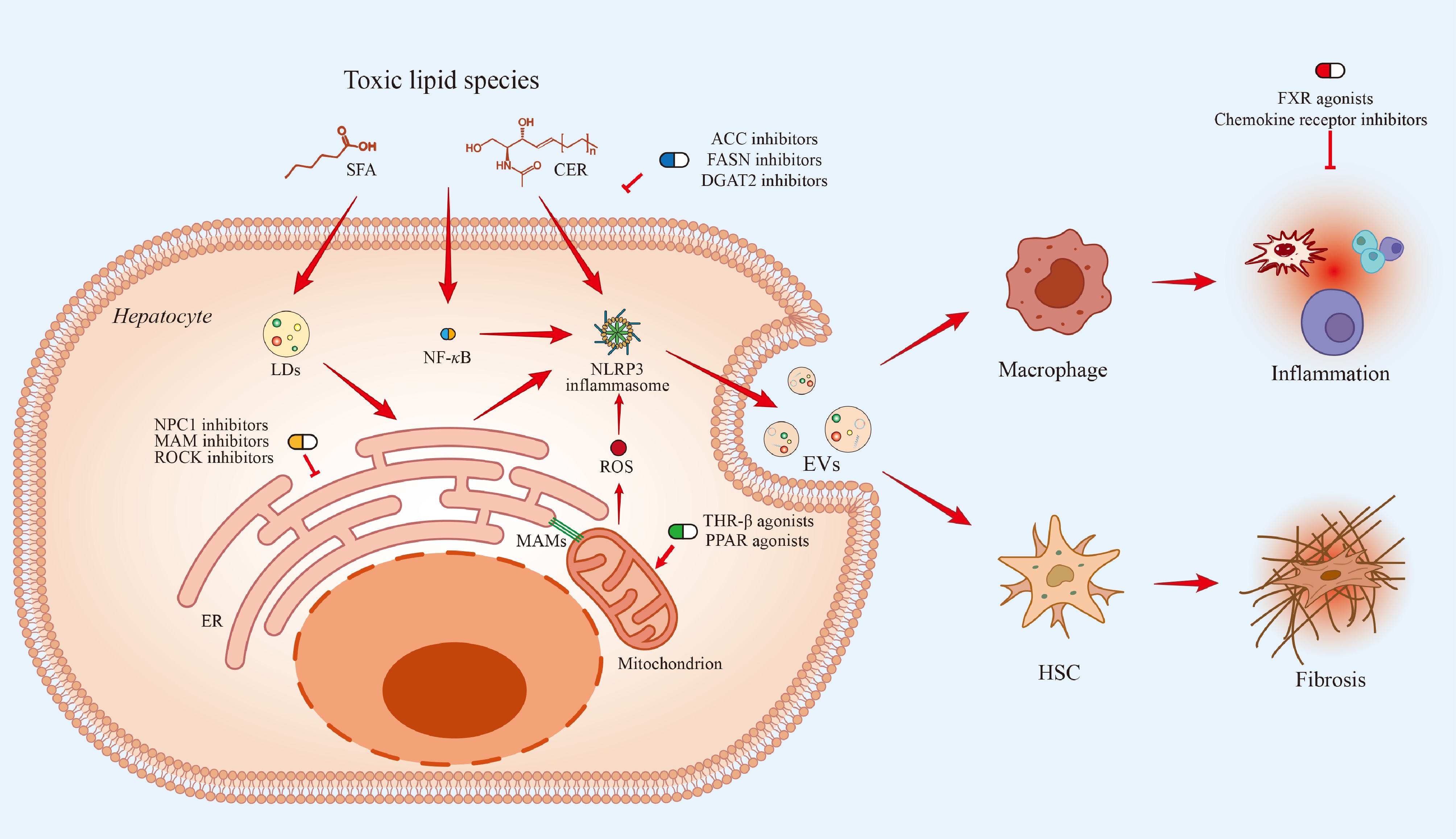
Figure 5.
The accumulation of toxic lipid species, including SFAs and CERs, in hepatocytes drives key pathological processes. Intracellular lipid excess promotes LD expansion, ER stress, mitochondrial dysfunction, and MAM formation, collectively generating ROS and activating the NLRP3 inflammasome and NF-κB signaling pathways. These events trigger inflammatory responses that are spread beyond the hepatocyte through EV-mediated intercellular communication, promoting macrophage activation and HSC-driven fibrosis. Therapeutic strategies are presented at their respective sites of action: ACC, FASN, and DGAT2 inhibitors reduce toxic lipid generation; THR-β and PPAR agonists enhance mitochondrial lipid utilization; NPC1, MAM, and ROCK inhibitors target intracellular lipid trafficking and organelle stress; and FXR agonists and chemokine receptor inhibitors attenuate downstream inflammatory and fibrogenic signaling.
-
Lipid species Classification Primary source(s) Key mechanisms Downstream effects Ref. Palmitate (C16:0) Toxic DNL, dietary intake ER stress (CHOP), JNK activation, mitochondrial dysfunction, ROS Lipoapoptosis, inflammation, insulin resistance [24,25] Stearate (C18:0) Toxic DNL, dietary intake ER stress, mitochondrial uncoupling, ROS Hepatocellular injury, apoptosis [24,35] Oleate (C18:1) Adaptive SCD1 desaturation of SFAs Promotes SFA esterification into triglycerides, lipid droplet sequestration, regulates SFA: MUFA balance Reduces ER stress and apoptosis; protects against lipotoxicity [28,29,31] Ceramides Toxic DNL (serine + palmitate) VDAC2 binding, mitochondrial apoptosis, TGF-β/Smad signaling Insulin resistance, oxidative stress, apoptosis, fibrosis [32,37] Diacylglycerols (DAGs) Toxic DNL, phospholipid remodeling PKCε activation, inhibitory phosphorylation of insulin receptor substrates Hepatic insulin resistance [33,38] Free cholesterol Toxic Dietary intake, de novo synthesis ER stress, mitochondrial dysfunction, inflammasome priming Hepatocyte injury, stellate cell activation, fibrosis [23] Triglycerides Relatively inert (protective) Esterification of FFAs Sequestration of FFAs into lipid droplets Lipid buffering and storage; generally non-toxic in isolation [23] Lysophospholipids (LPC, LPA) Context-dependent (predominantly pro-inflammatory) Phospholipase A2 activity GPCR activation, TLR signaling Inflammation, ER stress, immune cell recruitment [39,40] Oxysterols/bile acids Context-dependent Cholesterol oxidation, bile acid synthesis FXR, LXR, TGR5 activation Metabolic regulation or inflammation depending on context [41] Table 1.
Functional comparison of toxic and adaptive lipid species in MAFLD/MASH.
-
Category Target Agent Mechanism Compartment Hepatic outcomes Status Ref. MAM Mfn2 Photobiomodulation (650 nm) Restores ER-mitochondria contacts; reduces PERK/CHOP signaling MAMs; ER; mitochondria Reduced ER stress; improved mitochondrial function; reduced lipotoxicity Preclinical [226] MAM Mic19-EMC2-SLC25A46 Mic19 overexpression Maintains ER-mitochondria contacts; preserves mitochondrial structure MAMs; mitochondria Reduced steatosis; reversed steatohepatitis and fibrosis Preclinical [227] Cholesterol NPC1 Calycosin Enhances lysosomal cholesterol transport Lysosome; ER Reduced steatosis; reduced inflammation; improved insulin sensitivity Preclinical [235] Cholesterol NPC1-ORP5 Pathway inhibition Reduces lysosome-ER cholesterol transfer;
reduces ER stressLysosome; ER Reduced macrophage activation; reduced inflammation Preclinical [231,232] Cholesterol NPC1L1 Cryptotanshinone Inhibits cholesterol absorption Plasma membrane; ER Reduced hepatic steatosis Preclinical [234] Cholesterol NPC1L1 Pharmacologic inhibition Reduces oxysterol uptake; modulates LXR/ROR signaling Plasma membrane; nucleus Prevented diet-induced steatosis Target validated [233] EV signaling ROCK1 Fasudil Inhibits DR5-ROCK1-dependent EV release Cytosol; plasma membrane Reduced inflammation; reduced fibrosis Preclinical/
repurposing[46] EV signaling ROCK1/2 ROCK inhibitors Inhibits hepatocyte ROCK signaling Cytosol Reduced steatosis; reduced inflammation; reduced fibrosis Preclinical [159] Lipid metabolism SLC25A1 CTPI-2 Inhibits mitochondrial citrate export Mitochondria; ER Reduced steatosis; reduced inflammation; reduced fibrosis Preclinical [236] Lipid metabolism FASN Platensimycin Inhibits de novo
lipogenesisER; lipid droplets Reduced hepatic triglycerides Preclinical [237] Lipid metabolism ACLY Bempedoic acid Reduces cytosolic acetyl-CoA Cytosol; ER Reduced triglycerides; reduced inflammation Preclinical [238] Lipid metabolism THR-β VK2809 Enhances lipid oxidation; promotes lipophagy Nucleus; mitochondria; lysosome Reduced hepatic triglycerides Preclinical [210] Bile acid FXR/GPBAR1 BAR502 + UDCA Modulates bile acid-lipid signaling Liver; intestine;
adiposeReduced steatosis; reduced inflammation; reduced fibrosis Preclinical [239] Metabolic SGLT2 Dapagliflozin/
CanagliflozinModulates metabolic signaling Cytosol; mitochondria Reduced steatosis (preclinical models) Preclinical [240] Energy sensing AMPK Metformin Inhibits lipogenesis; modulates gut-liver axis Mitochondria; ER Reduced steatosis; reduced oxidative stress Preclinical [241] Table 2.
Emerging and preclinical therapeutic targets in lipid trafficking and organelle crosstalk.
-
Category Target Agent Mechanism Compartment Hepatic outcomes Status Ref. Lipogenesis ACC1/2 Firsocostat Inhibits de novo lipogenesis ER; mitochondria Reduced steatosis; increased plasma triglycerides Discontinued (2025) [201,242] Lipogenesis ACC1/2 + DGAT2 Clesacostat + PF-06865571 Dual inhibition of lipogenesis and triglyceride synthesis ER; mitochondria; ER-LD Reduced steatosis; reduced hypertriglyceridemia; MASH resolution without fibrosis worsening in MIRNA trial Phase II (met primary endpoint); in continued development [202,243] Lipogenesis DGAT2 PF-06865571 (monotherapy) Inhibits triglyceride synthesis ER-LD interface Reduced hepatic steatosis; monotherapy did not meet primary endpoint in MIRNA trial Phase II completed (monotherapy did not meet primary endpoint) [202,243] Lipogenesis DGAT2 (ASO) ION224 Inhibits triglyceride synthesis from DAG ER-LD MASH resolution; fibrosis improvement (subset) Phase II completed (met primary endpoint) [208] Lipogenesis FASN Denifanstat (TVB-2640) Inhibits lipogenesis ER; lipid droplets Reduced steatosis; reduced liver injury markers; MASH resolution and fibrosis improvement in Phase IIb (FASCINATE-2) Phase IIb completed (met primary endpoint); Phase III MASH trial withdrawn (2025) [205,207] Lipogenesis KHK PF-06835919 Inhibits fructose-driven lipogenesis Cytosol Reduced liver fat Discontinued (2021) [244,245] Nuclear receptor THR-β Resmetirom Enhances lipid oxidation Nucleus; mitochondria MASH resolution; fibrosis improvement Approved [212] Nuclear receptor PPARγ Pioglitazone Modulates lipid partitioning Nucleus; mitochondria Improved insulin sensitivity; reduced hepatic steatosis and inflammation in MASH trials Approved (T2DM); guideline-recommended off-label use in MASH
[246,247] Nuclear receptor Pan-PPAR Lanifibranor Enhances fatty acid oxidation; reduces inflammation and fibrosis Nucleus; mitochondria MASH resolution; fibrosis improvement Phase III (ongoing) [216,248] Nuclear receptor FXR Cilofexor Inhibits bile acid synthesis Nucleus; ER Reduced steatosis Discontinued (2025) [242,249] Incretin GLP-1R Semaglutide Reduces lipotoxicity and inflammation Hepatocytes MASH resolution; weight loss Approved [250] Incretin GIPR/GLP-1R Tirzepatide Improves metabolic control Hepatocytes MASH resolution; fibrosis improvement Phase III (ongoing) [251,252] Combination FXR + ACC Cilofexor + Firsocostat Dual bile acid and lipid regulation Nucleus; ER Reduced steatosis; reduced inflammation; reduced fibrosis Discontinued (2025) [242,253] Combination GLP-1R + FXR Semaglutide + Cilofexor Combined metabolic and bile acid signaling Nucleus; ER Greater reduction vs monotherapy Discontinued (2025) [242,254] Combination GLP-1R + ACC Semaglutide + Firsocostat Combined lipogenesis inhibition and metabolic modulation Cytosol; lipid droplets Greater reduction in hepatic fat and injury markers Discontinued (2025) [242,254] Combination GLP-1R +
FXR + ACCSemaglutide + Cilofexor + Firsocostat Multi-pathway lipid suppression Nucleus; cytosol; lipid droplets Did not meet primary fibrosis endpoint vs placebo (13.7% vs
8.3%, p = 0.2289)Discontinued (2025) [242,255] Metabolic SGLT2 Empagliflozin Reduces hepatic lipid accumulation Lipid droplets Reduced liver fat Approved (T2DM); Phase IV (non-diabetic MAFLD, ongoing) [256] Table 3.
Current clinical status of therapeutic agents for MAFLD/MASH.
Figures
(5)
Tables
(3)