












-
Non-alcoholic fatty liver disease (NAFLD), recently renamed as metabolic dysfunction-associated fatty liver disease (MAFLD), is the most common chronic liver disease worldwide, with an estimated prevalence of 25%−30% in adults, and is strongly associated with obesity and type 2 diabetes mellitus (T2DM)[1]. Although the multi-society consensus nomenclature now favors metabolic dysfunction-associated steatotic liver disease (MASLD) over MAFLD[2], we retain the terms MAFLD/MASH throughout this review for consistency with the terminology used in the majority of the cited literature, without altering the underlying biological concepts discussed.
Metabolic dysfunction-associated steatohepatitis (MASH), the progressive form of MAFLD, is characterized by hepatocellular injury, inflammation, and fibrosis with an increased risk of cirrhosis, hepatocellular carcinoma (HCC), and liver failure[1,3]. In addition to liver-related complications, MAFLD is highly associated with systemic metabolic dysfunction, and the fibrosis stage is an important predictor of liver-related and all-cause mortality[4,5]. The clinical burden is growing, but therapeutic progress is limited, highlighting the need for better mechanistic frameworks for patient stratification and targeted interventions[6−8].
Lipotoxicity, defined as the accumulation of bioactive lipid molecules that disturb cellular homeostasis, is widely recognized as a major contributor to the progression of MAFLD/MASH[9,10]. Initial studies have shown that certain types of lipid intermediates, including saturated fatty acids (SFAs), ceramides, and diacylglycerols (DAGs), contribute to hepatocyte injury through interconnected stress pathways involving endoplasmic reticulum (ER) and mitochondrial dysfunction, inflammatory signaling, and apoptosis[11,12]. However, these models largely interpret lipotoxicity through individual signaling pathways and provide a relatively static view of disease pathogenesis. They devote limited attention to the spatial organization of lipids and the dynamics within cells.
Recent advances in spatial lipidomics and organelle biology are beginning to shift the view of lipotoxicity as a highly organized and context-dependent condition. These advances demonstrate that the biological effects of lipids are not only determined by their abundance but also by their acyl chain composition, subcellular localization, and transport between organelles[13,14]. The ER, mitochondria, lysosomes, and peroxisomes, as well as lipid droplets (LDs), are interconnected signaling platforms coordinated by membrane contact sites that control lipid flow, calcium signaling, and metabolic adaptation[15,16]. Meanwhile, MAFLD is increasingly recognized as a systemic disease with lipid-mediated communication between hepatocytes, immune cells, adipose tissue, and the gut-liver axis[17]. The spatial and intercellular dimensions of lipid signaling are still not sufficiently integrated into current pathogenic models.
Building on these advances, we propose an alternative conceptual framework in which lipotoxicity in MAFLD/MASH is viewed as a spatially and temporally organized lipid signaling disorder rather than simply a consequence of triglyceride accumulation. In this proposed view, distinct lipid species serve as bioactive mediators in well-defined subcellular microenvironments, such as organelle contact sites, lipid droplet (LD) interfaces, lysosomes, and microdomains of the plasma membrane. These microenvironments function as signaling hubs that coordinate cellular stress responses and metabolic adaptation. These signals can also travel from hepatocytes through extracellular vesicles (EVs) and circulating lipid mediators to modulate inflammation and fibrosis in the liver microenvironment. This model provides a mechanistic explanation for the clinical observation that an excessive accumulation of neutral lipids is often less harmful, while selective enrichment of certain toxic lipids is the driving force behind disease progression.
This review brings together recent advances in defining toxic vs adaptive lipid species, characterizes lipid-activated signaling networks in their spatial context, and discusses how organelle communication and intercellular lipid transport govern the transition from steatosis to steatohepatitis and fibrosis. We integrate these insights with emerging therapeutic approaches targeting lipid metabolic pathways, signaling nodes, and trafficking mechanisms. Here, we aim to complement and extend the current pathogenic understanding and inform the development of more precise, organelle-directed, and rationally combined therapeutic approaches by redefining MAFLD/MASH as a disorder of lipid signaling across cellular space and time.
The review is structured as follows: We first discuss the role of lipid species as active signaling mediators in hepatocytes, followed by a discussion of Intracellular lipid transport and organelle crosstalk. We then examine intercellular and inter-organ lipid communication networks, followed by therapeutic targeting strategies and their translational relevance to the clinic. The review concludes with future perspectives.
What this review adds beyond recent publications
-
Recent reviews have greatly expanded our understanding of MAFLD/MASH pathogenesis, including detailed cataloging of lipotoxic lipid species, analyses of organelle stress pathways, and descriptions of inter-organelle communication networks[9,18,19]. But most of these works consider each process to be standalone and lack an integrated view of how the pathways involved are interconnected.
This review aims to extend recent work in three ways. First, we present a unifying approach that interprets lipotoxicity as a spatially and temporally coordinated lipid signaling disorder. It brings together lipid identity, subcellular localization, intracellular transport, organelle crosstalk, and intercellular communication into a single systems-level model.
Second, we map current and emerging therapeutic approaches systematically onto the same framework. We classify interventions according to the specific lipid signaling nodes and spatial compartments that they target.
Third, we introduce newer tools such as spatial lipidomics and real-time imaging approaches to demonstrate how dynamic lipid fluxes connect to signaling outcomes and to guide future mechanistic investigation.
Together, these advances provide a spatially and temporally integrated framework for understanding lipotoxicity that goes beyond descriptive or pathway-specific models and supports the development of mechanism-based therapeutic approaches.
-
In addition to their role as metabolic substrates and structural components, hepatic lipids are dynamic signaling molecules that modulate hepatocyte fate in MAFLD/MASH[20]. Classical models have focused on the lipid-induced activation of stress and inflammatory pathways, but recent advances suggest that lipid signaling is highly dependent on the identity of lipid species, structural properties, and spatial organization within the cell[21]. Lipidomic and spatial profiling studies have shown that the composition, abundance, and subcellular localization of the acyl chain determine whether hepatocytes adapt to nutrient excess or initiate lipotoxic injury programs[22]. Therefore, disease is better viewed as a dysregulation of lipid signaling networks rather than a simple accumulation of lipids (Fig. 1).
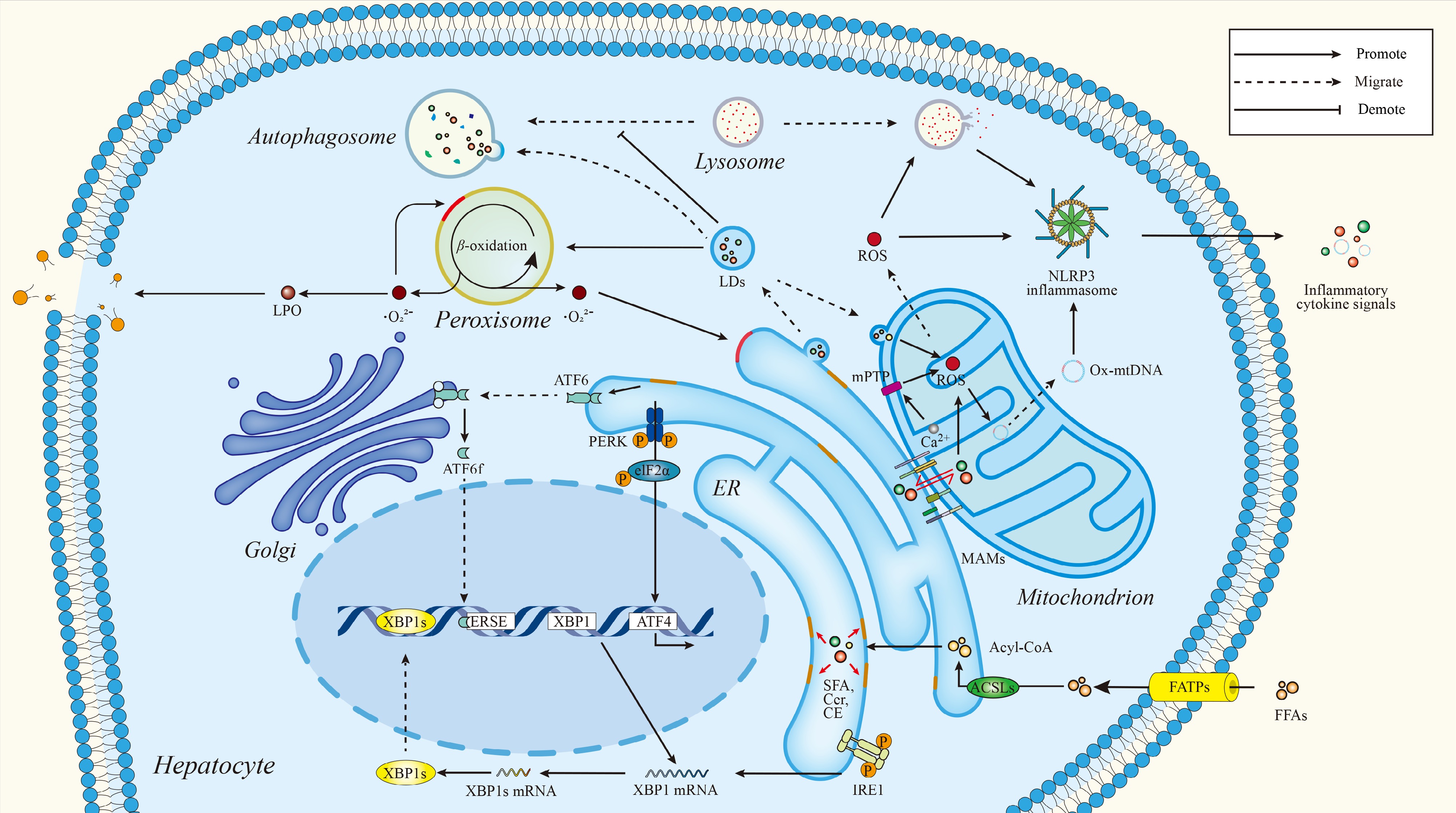
Figure 1.
Lipotoxicity as a spatially and temporally organized lipid signaling network in hepatocytes. A visual representation of hepatocellular lipotoxicity. Lipid metabolism is presented as a spatially established signaling network controlled by inter-organellar communication. Lipid homeostasis is maintained by contact sites between the endoplasmic reticulum (ER), mitochondria, lipid droplets (LDs), lysosomes, and peroxisomes under physiological conditions, coordinating the synthesis, trafficking, storage, and degradation of lipids. Chronic nutrient excess leads to a dysregulation of this coordination, which results in ER stress, mitochondrial dysfunction, lipid droplet overload, impaired lipophagy, and redox imbalance. These interconnected dysfunctions result in oxidative stress, inflammasome activation, inflammatory signaling, and hepatocellular injury, pointing to lipotoxicity as an emerging consequence of dysregulated lipid signaling and organelle crosstalk. ACSLs, acyl-CoA synthetase long-chain family members; Acyl CoA, acetyl-coenzyme A; ATF4/6, activating transcription factors; CE, cholesteryl ester; Cer, ceramide; eIF2α, eukaryotic initiation factor 2α; ERSE, ER stress response element; FATPs, fatty acid transport proteins; FFAs, free fatty acids; IRE1, inositol-requiring enzyme 1; LPO, lipid peroxidation; mPTP, mitochondrial permeability transition pore; Ox mtDNA, oxidized mitochondrial DNA; PERK, protein kinase R-like ER kinase; SFA, saturated fatty acid; XBP1, x-box binding protein 1.
Toxic and adaptive lipid species
-
The development of MAFLD/MASH is fueled by the selective accumulation of bioactive lipid species with distinct functional properties[23]. Classical studies have shown that SFAs, especially palmitate (C16:0) and stearate (C18:0), cause hepatocellular injury through ER stress, as evidenced by activation of C/EBP homologous protein (CHOP) and c-Jun N-terminal kinase (JNK) signaling, mitochondrial dysfunction, reactive oxygen species (ROS) accumulation, and caspase-dependent apoptosis[24,25]. SFAs promote their incorporation into triglycerides (TGs) and phospholipids in experimental models and induce hepatocyte lipotoxicity, ER stress, apoptosis, and inflammatory niche formation. In human MAFLD, ACSL3-related lipid remodeling in hepatocytes is associated with the severity of steatosis and the infiltration of fibrogenesis-related immune cells[26]. Recently, experimental studies demonstrated that hepatocytes secrete small SFA-enriched EVs that activate macrophages via Toll-like receptor 4 (TLR4) and trigger pro-inflammatory responses, aggravating liver injury[27].
On the contrary, monounsaturated fatty acids (MUFAs) like oleate (C18:1) are well tolerated. MUFAs also promote the incorporation of SFAs into triglycerides stored in LDs, decreasing the cytosolic pool of toxic free fatty acids (FFAs) and attenuating stress signaling[28]. MUFA partitioning relieves palmitate-induced ER stress, decreases CHOP expression, and reduces cell death in hepatocytes and in vivo experimental models[29]. Therefore, the intracellular SFA : MUFA ratio, which is partly controlled by stearoyl-CoA desaturase-1 (SCD1), is a major determinant of hepatocyte sensitivity to lipotoxic injury[30]. SCD1 deficiency or MUFA depletion worsens saturated lipid accumulation, ER stress, inflammation, and fibrosis[30], while oleate supplementation is protective[31].
Several lipid classes, in addition to fatty acids, serve as signaling intermediates that connect metabolic overload to specific pathogenic pathways. Ceramides accumulate during MAFLD/MASH progression and are related to oxidative stress, inflammation, apoptosis, fibrosis, and insulin resistance[32]. DAGs serve as second messengers to activate protein kinase C (PKC) isoforms that interfere with insulin receptor signaling. Using a high-fat diet mouse model of MAFLD, decreased hepatic DAG levels following knockdown of cell death-inducing DFF45-like effector B (Cideb) were associated with reduced PKCε activation and lower insulin receptor kinase (IRK T1150) phosphorylation, suggesting that DAG-PKCε signaling impairs insulin receptor function[33]. Free cholesterol induces ER and mitochondrial stress, rendering hepatocytes, Kupffer cells, and hepatic stellate cells (HSCs) more susceptible to inflammatory and apoptotic stimuli[23]. Palmitate stimulates the secretion of lipotoxic hepatocyte-derived extracellular vesicles (sEVs) that induce pro-inflammatory macrophage signaling through TLR4-dependent mechanisms[34]. These lipid species are characteristic of the lipotoxic milieu of progressive MASH, as opposed to the relatively inert accumulation of TGs in simple steatosis.
Notably, lipid bioactivity is not only dictated by class or abundance, but also by structural features and spatial distribution within the cell. The acyl chain length, saturation, and head group composition influence membrane properties, protein interactions, and signaling specificity[21]. The saturated acyl chains of SFAs are responsible for a strong tendency to induce mitochondrial dysfunction, uncoupling, proton leakage, ATP depletion, ROS production, and fragmentation[35]. The functional outcomes of lipid species are determined by their subcellular localization in the ER, mitochondria, LDs, plasma membrane microdomains, or organelle contact sites (Fig. 1)[13]. In particular, mitochondria-associated ER membranes (MERCs) have emerged as major hubs that coordinate lipid synthesis, calcium flux, and stress signaling upon lipotoxic insults[36]. These structural and spatial properties transduce the quantitative excess of lipids into qualitatively different signaling outcomes. The findings here emphasize the functional diversity of lipid species in MAFLD/MASH, as well as their main characteristics what are summarized in Table 1.
Table 1. Functional comparison of toxic and adaptive lipid species in MAFLD/MASH.
Lipid species Classification Primary source(s) Key mechanisms Downstream effects Ref. Palmitate (C16:0) Toxic DNL, dietary intake ER stress (CHOP), JNK activation, mitochondrial dysfunction, ROS Lipoapoptosis, inflammation, insulin resistance [24,25] Stearate (C18:0) Toxic DNL, dietary intake ER stress, mitochondrial uncoupling, ROS Hepatocellular injury, apoptosis [24,35] Oleate (C18:1) Adaptive SCD1 desaturation of SFAs Promotes SFA esterification into triglycerides, lipid droplet sequestration, regulates SFA: MUFA balance Reduces ER stress and apoptosis; protects against lipotoxicity [28,29,31] Ceramides Toxic DNL (serine + palmitate) VDAC2 binding, mitochondrial apoptosis, TGF-β/Smad signaling Insulin resistance, oxidative stress, apoptosis, fibrosis [32,37] Diacylglycerols (DAGs) Toxic DNL, phospholipid remodeling PKCε activation, inhibitory phosphorylation of insulin receptor substrates Hepatic insulin resistance [33,38] Free cholesterol Toxic Dietary intake, de novo synthesis ER stress, mitochondrial dysfunction, inflammasome priming Hepatocyte injury, stellate cell activation, fibrosis [23] Triglycerides Relatively inert (protective) Esterification of FFAs Sequestration of FFAs into lipid droplets Lipid buffering and storage; generally non-toxic in isolation [23] Lysophospholipids (LPC, LPA) Context-dependent (predominantly pro-inflammatory) Phospholipase A2 activity GPCR activation, TLR signaling Inflammation, ER stress, immune cell recruitment [39,40] Oxysterols/bile acids Context-dependent Cholesterol oxidation, bile acid synthesis FXR, LXR, TGR5 activation Metabolic regulation or inflammation depending on context [41] Lipid-activated intracellular signaling networks
-
Lipotoxic species of lipids activate conserved intracellular signaling modules that coordinate hepatocellular stress responses[26].
JNK and stress-activated kinase signaling
-
The activation of ER stress and stress-activated kinase cascades is central to lipotoxic signaling. Palmitate induces stress-responsive kinase cascades, such as c-Jun N-terminal kinase 1 (JNK1/MAPK8), p38 mitogen-activated protein kinase (p38 MAPK), and Janus kinase 2 and signal transducer and activator of transcription (JAK2/STAT) pathways in human hepatocytes in vitro, leading to increased ROS and interleukin-6 (IL-6) production without overt lipid accumulation or metabolic dysfunction[42]. Lysophosphatidylcholine (LPC) induces ER stress and hepatocyte damage, whereas SFAs directly activate Rac1-mixed lineage kinase 3 (MLK3)-c-Jun N-terminal kinase (JNK) signaling, which indicates partially distinct but converging stress pathways[43]. JNK signaling in SFA-treated hepatocytes promotes the induction of the BH3-only proteins p53upregulated modulator of apoptosis (PUMA) and Bcl-2-interacting mediator of cell death (BIM), which activate Bcl-2-associated X protein (Bax) and Bcl-2 antagonist/killer (Bak) to induce mitochondrial outer membrane permeabilization, release of cytochrome c, and caspase-3/7 activation, a process termed lipoapoptosis[43]. Genetic deletion or pharmacologic inhibition of JNK1 prevents PUMA and BIM induction, maintains mitochondrial integrity, decreases caspase activation, and improves hepatocyte survival in preclinical models of lipotoxicity[44].
NF-κB inflammatory signaling
-
In preclinical models, lipotoxic stress and death-receptor signals can induce cell-autonomous activation of nuclear factor kappa B (NF-κB) signaling in hepatocytes, for example, through inhibition of nuclear factor kappa B kinase subunit 2 (IKK2), resulting in chemokine and cytokine transcription that recruits inflammatory myeloid cells and promotes fibrogenesis[45]. Similarly, death receptor 5 (DR5) activated by lipotoxicity triggers the secretion of EVs carrying TNF-related apoptosis-inducing ligand (TRAIL), which binds DR5-receptor-interacting serine/threonine-protein kinase 1 (RIPK1) signaling complexes in macrophages and facilitates NF-κB nuclear translocation. This leads to an increased production of tumor necrosis factor (TNF), IL-6, and interleukin-1 beta (IL-1β), and thus amplifies hepatic inflammatory responses. These mechanisms have been mainly characterized in cellular and pre-clinical models[46].
NLRP3 inflammasome activation
-
NLR family pyrin domain containing three (NLRP3) inflammasomes are primed by lipid-derived danger signals and cytokine-mediated NF-κB activity, inducing NLRP3 and pro-IL-1β expression[47]. Metabolic and oxidative stress then induce inflammasome assembly and caspase-1 activation[48]. The mature IL-1β and interleukin-18 (IL-18) released from immune cells further mediate hepatocyte injury and promote fibrogenic activation of stellate cells, as demonstrated by experimental and human studies[49].
Death receptor-mediated lipoapoptosis
-
Death receptor signaling also plays a direct part in lipoapoptosis. Loading with SFA, especially palmitate, induces ER stress and CHOP-dependent DR5 upregulation and clustering in lipid rafts, leading to the establishment of the death-inducing signaling complex (DISC). This complex recruits Fas-associated death domain (FADD) and activates caspase-8 and/or RIPK1 to engage extrinsic apoptotic signaling and, in some contexts, necroptotic pathways[50,51]. These mechanisms have been mostly characterized in experimental models. Preclinical studies have demonstrated that loss of the anti-apoptotic protein myeloid cell leukemia 1 (Mcl-1) promotes Bax/Bak activation, decreasing the mitochondrial apoptotic threshold and increasing the susceptibility of hepatocytes to lipoapoptosis[52,53].
Lipid second messengers: DAG, ceramide, lysophospholipids, and oxysterols
-
In addition to stress-responsive signaling pathways, lipids act as second messengers to directly link metabolic status to intracellular signaling networks[12]. Specifically, accumulation of DAG in certain membrane compartments activates PKCε in hepatocytes, leading to inhibitory phosphorylation of the insulin receptor and attenuation of downstream phosphoinositide 3-kinase-protein kinase B (PI3K-Akt) signaling[38,54]. However, different PKC isoforms might play context-dependent and even opposing roles. For example, in some contexts, PKCδ has been linked to pro-apoptotic signaling whereas PKCζ might have a protective role in insulin signaling[55,56]. The subcellular localization of DAG at the plasma membrane, ER, or LDs determines which PKC isoforms are activated, adding another layer of spatiotemporal regulation[54]. Such complexities are often underestimated in simplified models of DAG-PKC signaling. Ceramides bind to the voltage-dependent anion channel 2 (VDAC2) and associate metabolic stress with mitochondrial apoptotic pathways by regulating Bax conformational activation and promoting mitochondrial outer membrane permeabilization[37]. Ceramides also enhance transforming growth factor-beta (TGF-β/Smad) signaling in fibrogenic cells, which increases the expression of extracellular matrix genes and promotes progression of fibrosis[57]. G-protein-coupled receptors (GPCRs) are principally activated by extracellular ligands such as lysophospholipids and polyunsaturated fatty acid (PUFA)-derived eicosanoids, which influence MAPK and PI3K-Akt signaling, regulate immune cell recruitment, and impact hepatocyte survival during metabolic stress[39,40]. Oxysterols and bile acid intermediates further broaden lipid signaling by acting as endogenous agonists for nuclear receptors such as the farnesoid X receptor (FXR) and liver X receptor (LXR), thereby linking lipid metabolism to transcriptional control of inflammatory and metabolic programs (Fig. 2)[41].
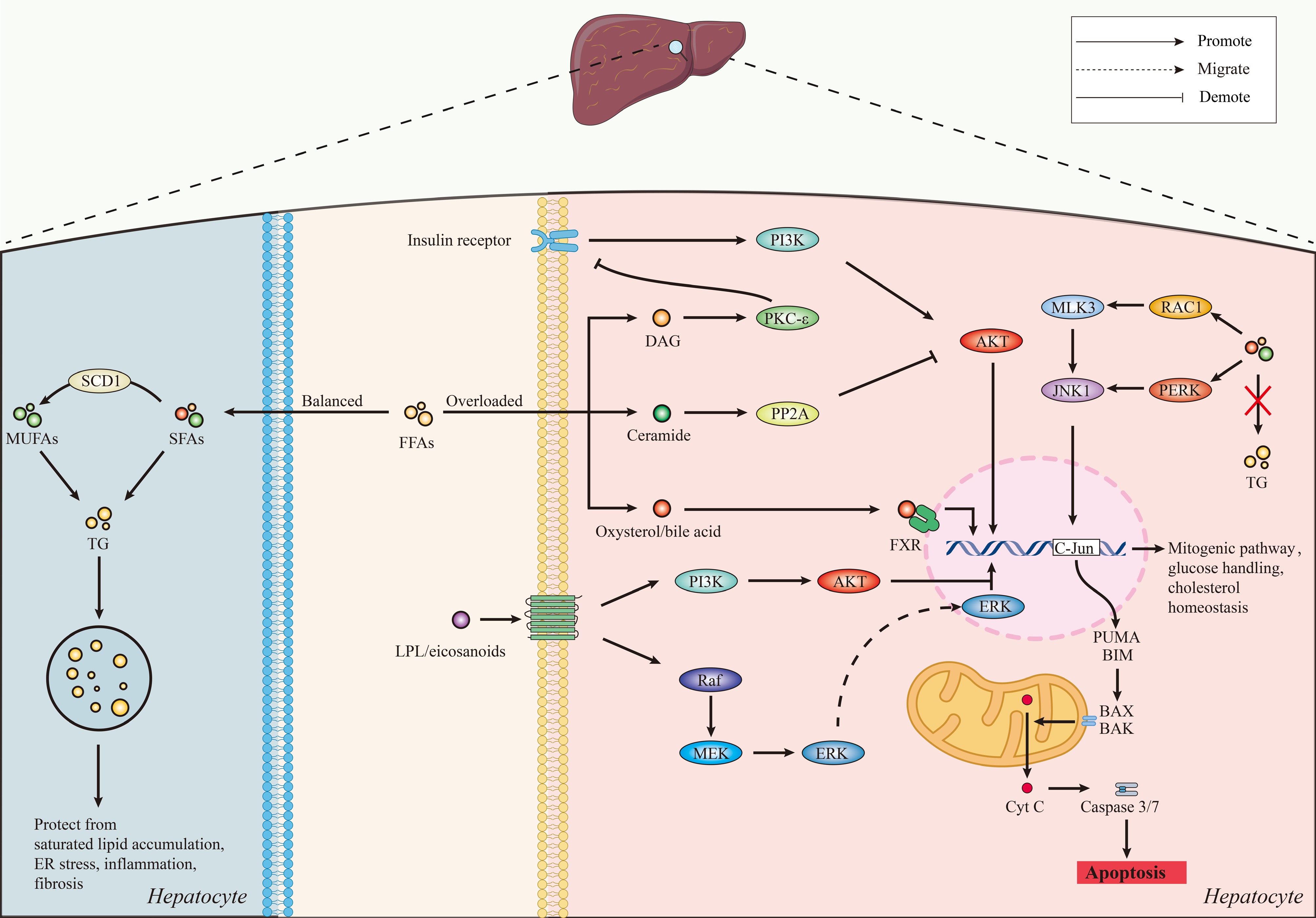
Figure 2.
The intracellular lipotoxic signaling network in hepatocytes. Schematic diagram of lipid class-regulated signaling pathways mediating hepatocellular adaptation or lipotoxicity. Elevated saturated fatty acid (SFA) and SFA metabolite levels (e.g., diacylglycerol [DAG], ceramides, lysophospholipids) activate stress and apoptotic signaling cascades (e.g., c-Jun N-terminal kinase [JNK]-mediated pathways, mitochondrial lipoapoptosis [BAX/BAK activation, cytochrome c release, and caspase-3/7 activation]). DAG accumulation promotes PKCε activation and impairs insulin receptor-PI3K-Akt signaling. Ceramides regulate Akt activity by way of PP2A-dependent signaling. At the same time, oxysterols, bile acids (BAs), and lysophospholipids activate receptor-mediated signaling pathways that regulate metabolic and inflammatory programs. Mono-unsaturated fatty acids (MUFAs), which are partially derived from stearoyl-CoA desaturase 1 (SCD1), facilitate triglyceride synthesis and lipid droplet retention, thus alleviating toxic lipid accumulation. AKT, AKT serine/threonine kinase; ERK, extracellular signal-regulated kinase; LPL, lipoprotein lipase; MEK, mitogen-activated protein kinase kinase; MLK3, mixed lineage kinase 3; PI3K, phosphoinositide 3-kinase; PUMA, p53 upregulated modulator of apoptosis; RAC1, ras-related C3 botulinum toxin substrate 1; Raf, Raf kinase; TG, triglyceride.
Spatial integration of lipid signaling networks
-
Subcellular localization and lipid compartmentalization of lipid signaling pathways determine their activation and functional consequences. Localized DAG pools in membrane microdomains selectively activate PKC isoforms, while ceramide accumulation in mitochondrial membranes promotes apoptotic signaling. Organelle contact sites further coordinate lipid-induced stress responses, where lipid flux intersects with calcium signaling and redox homeostasis.
These observations suggest that lipotoxic signaling pathways function in spatially organized intracellular networks, rather than as isolated linear cascades. These processes define the DAG-PKC, ceramide-mitochondrial, lysophospholipid-GPCR, and sterol-nuclear receptor domains as the central layers of hepatocyte signaling (Fig. 2).
Lipid-receptor interactions and signal amplification
-
Interaction with receptor systems, which integrate metabolic and inflammatory responses at both transcriptional and membrane levels, also amplifies lipid signaling. Lipid-responsive nuclear receptors function as major lipid sensors that regulate gene expression processes that govern lipid metabolism, inflammation, and energy homeostasis[58]. Notably, the peroxisome proliferator-activated receptor alpha (PPARα) is activated by fatty acids and heterodimerizes with the retinoid X receptor (RXR) to regulate transcriptional processes involved in fatty acid uptake, trafficking, β-oxidation and lipoprotein/glucose metabolism, promoting hepatic lipid catabolism, and preventing triglyceride accumulation[59]. Ligand-activated PPARs also inhibit inflammatory gene expression through transrepression of NF-κB signaling[60].
The bile acid-activated receptor FXR decreases hepatic triglyceride levels through complementary hepatic and intestinal mechanisms. In the liver, FXR inhibits lipogenic gene expression via small heterodimer partner (SHP)-sterol regulatory element-binding protein 1c (SREBP-1c) signaling, but in the intestine, it decreases lipid absorption and increases bile acid transport and metabolic regulatory pathways[61]. These effects improve steatosis and inflammation in experimental models and patients with MASH, although context-dependent variability was reported[62]. Oxysterol-activated LXR regulates cholesterol release, transport, and excretion while modulating inflammatory signaling[63]. However, prolonged LXR activation correlates with hypertriglyceridemia and hepatic steatosis[64]. PPARs, FXR, and LXR are central transcriptional nodes that link lipid sensing to metabolic and inflammatory adaptations[60,62].
The functions of PPARs, FXR, and LXR are cell type-specific, a characteristic with profound implications for therapeutic targeting. In hepatocytes, PPARα mainly regulates fatty acid oxidation and ketogenesis[59], while in Kupffer cells, PPARα activation inhibits inflammatory cytokine production through the transrepression of NF-κB signaling[65]. PPARγ is expressed at low levels in healthy hepatocytes, but is upregulated in MASH in both human liver biopsy studies and preclinical dietary models, where its activation may exert protective effects through lipid redistribution away from toxic intermediates, as well as adverse effects through promotion of steatosis[66]; however, PPARγ activation in Kupffer cells and HSCs is generally anti-inflammatory and anti-fibrotic[67]. Hepatocyte FXR activation inhibits SREBP-1c-mediated lipogenesis. FXR signaling in HSCs has direct anti-fibrotic effects by inhibiting profibrogenic gene expression[61,68]. LXR activation in Kupffer cells enhances cholesterol efflux and reduces inflammatory signals, but when activated in hepatocytes, it paradoxically induces lipogenesis, highlighting the context-specificity of its metabolic effects[69,70]. These cell type-specific differences complicate systemic therapeutic targeting, but at the same time provide opportunities for cell-selective interventions exploiting different receptor functions in the hepatic microenvironment.
A further link between lipid excess and activation of innate immunity is provided by pattern-recognition receptors. TLR4 responds to the SFA-induced accumulation of ceramides in hepatocytes, which induces the MyD88-dependent production of pro-inflammatory cytokines, including IL-1β, IL-6, and TNF-α, and promotes liver injury in MASH[71]. In line with this role, mice lacking TLR4 are partially protected from lipid-induced insulin resistance[72]. The endosomal receptor TLR9 induces IL-1β production in Kupffer cells via myeloid differentiation primary response 88 (MyD88) signaling, which subsequently drives hepatocyte triglyceride accumulation, cellular injury, and fibrogenic activation of hepatic stellate cells. Genetic deficiency of TLR9, IL-1β signaling, or MyD88 protects against steatohepatitis, fibrosis, and insulin resistance, thus establishing the TLR9-MyD88-IL-1β axis as a key innate immune pathway linking danger signals to liver damage[73−75].
Growth factor signaling is another important interface between lipid metabolism and signal amplification. Insulin stimulates hepatic de novo lipogenesis (DNL) through insulin receptor substrate (IRS)-PI3K-Akt signaling, leading to activation of SREBP-1c through mechanistic target of rapamycin complex 1 (mTORC1) and repression of insulin-induced gene (Insig)[76,77]. Also, the FAM83A-RAF1-ERK module demonstrated that insulin-induced MAPK signaling increases lipogenic gene expression and causes hepatocellular lipotoxicity in models of diet-induced MASH[78].
Finally, the composition of membrane lipids has a fundamental impact on receptor function. Membrane microdomains rich in cholesterol regulate the localization and activation of insulin receptors; disruption of these domains redistributes the receptor, impairs insulin-induced nuclear Ca2+ signaling, and inhibits proliferative responses[79]. Restoration of cholesterol with raft-forming sterols rescues receptor autophosphorylation, while non-raft-forming sterols do not, indicating that sterol-dependent membrane organization is a key determinant of insulin receptor activation and downstream PI3K-Akt signaling[79,80].
Hepatocyte lipid dynamics influence the progression of MAFLD/MASH through three interconnected processes: (i) the formation of toxic vs adaptive lipid pools, (ii) the induction of intracellular signaling networks, including stress kinases, inflammasomes, death receptors, and second messengers, and (iii) the regulation of receptor-mediated signal amplification, such as nuclear receptors, TLRs, and growth factor receptors. Importantly, these processes are not only regulated by the identity of the lipid but also by their spatial distribution and dynamic trafficking between organelles. Lipotoxicity is thus defined by the spatiotemporal organization of lipid signaling rather than just lipid accumulation, establishing MAFLD/MASH as a lipid signaling disorder that is spatially and temporally organized (Fig. 2).
-
MAFLD/MASH is now considered an inter-organellar communication disorder in which spatial organization of lipid production, storage, transport, and degradation determines whether the previously described signaling pathways will lead to cellular adaptation or stress, inflammation, and apoptosis[19,81]. In addition to quantitative accumulation of lipids, qualitative changes in lipid trafficking between the ER, mitochondria, LDs, lysosomes, and peroxisomes generate signaling microdomains that contribute to the progression from simple steatosis to steatohepatitis[82−84]. These organelles are connected through membrane contact sites, allowing a rapid exchange of lipids, Ca2+, and redox signals. When the regulatory capacity of this network is overwhelmed or dysregulated, hepatocellular injury is more likely to occur[85,86].
Endoplasmic reticulum as a lipid-sensing and signaling hub
-
Triglyceride, phospholipid, and cholesterol synthesis are mostly performed in the ER and are coordinated by the DNL pathway enzymes regulated by SREBP-1c and carbohydrate-responsive element-binding protein (ChREBP)[87]. In MAFLD/MASH, chronic nutrient excess disrupts lipid and protein homeostasis, which manifests as ER stress. An abnormal change in lipid composition and subsequent lipid accumulation is sufficient to trigger unfolded protein response (UPR) signaling cascades, indicating that ER dysfunction can be induced not only by protein misfolding, but also by a disruption of membrane lipid balance[18]. These features make the ER a major lipid sensing organelle. The activation of the UPR is a common finding in liver biopsies from patients with MAFLD/MASH and in experimental dietary and genetic models[88,89]. Elevated levels of SFAs and free cholesterol induce the three canonical UPR pathways: protein kinase R-like ER kinase (PERK)-eukaryotic initiation factor-2α (eIF2α)-activating transcription factor-4 (ATF4), inositol-requiring enzyme-1 (IRE1)-X-box binding protein-1 (XBP1), and activating transcription factor-6 (ATF6), which are characteristic of lipid bilayer stress, Ca2+ homeostasis imbalance, and the accumulation of misfolded proteins[90,91]. For example, cholesterol loading activates the UPR and results in hepatocyte death in vitro[92]. Cholesterol crystallization in hepatocyte LDs has also been linked to necroinflammatory responses in human MASH, including Kupffer cell crown-like structures and inflammasome activation[93].
Under conditions of continuous stimulation, the UPR can change from adaptive recovery of ER homeostasis to the activation of pro-apoptotic programs. For instance, the PERK-ATF4-CHOP signaling cascade induces transcriptional programs promoting apoptotic signaling, including mitochondrial sensitization to apoptotic stimuli[94]. At the same time, IRE1 signaling facilitates inflammatory responses through multiple downstream mechanisms. In addition to stress kinase activation, the IRE1α-XBP1 pathway stimulates ceramide synthesis and enhances the release of ceramide-enriched EVs, linking lipotoxic stress to macrophage recruitment and subsequent inflammatory signaling (Fig. 2)[95].
ER-mitochondria contact sites and lipotoxic signaling
-
As illustrated in Fig. 1, mitochondria-associated membranes (MAMs) physically bind to the ER and mitochondria, allowing for a two-way transfer of lipids and Ca2+ and coupling fatty acid flux to oxidative metabolism[96]. These subdomains house enzymes involved in phospholipid synthesis and the trafficking of cholesterol and ceramide, which are major sites for lipid exchange between organelles[85,97].
Enhanced ER–mitochondria interactions and Ca2+ transfer at the onset of ER stress can promote mitochondrial respiration and ATP production and support adaptive bioenergetic responses[98]. The administration of nicotinamide mononucleotide (NMN) in high-fat diet (HFD) models strengthens ER-mitochondrial contact sites, improves mitochondrial function, increases ATP production, and relieves hepatic steatosis, highlighting the importance of MAMs in maintaining metabolic homeostasis[99]. On the other hand, sustained MAM enrichment induced by chronic overnutrition and hyperinsulinemia leads to excessive mitochondrial Ca2+ uptake, compromised oxidative capacity, and increased ROS production, changes that contribute to cellular stress and insulin resistance[100].
Both hyper-coupling and loss of structural integrity of MAMs are harmful, highlighting the context-dependent role of MAMs in cellular homeostasis[101]. Hypercoupling enhances calcium and lipid flux, resulting in mitochondrial overload, permeability transition, and excess ROS production, which in turn increases ER stress and apoptotic signaling[102]. In contrast, MAM disorganization disrupts the coordinated inter-organelle exchange, impairs lipid trafficking and β-oxidation, hence promoting the accumulation of cytotoxic lipid intermediates[85,103].
Ceramides and free cholesterol accumulating in MAFLD/MASH can be transported via MAMs to mitochondria, where they disrupt mitochondrial membrane stability, leading to cytochrome c release and apoptosis, though direct mechanistic evidence in human liver tissue remains limited[104]. High levels of mitochondrial ROS cause damage to mitochondrial DNA (mtDNA), respiratory chain proteins, and membrane lipids, resulting in feed-forward pathways of oxidative damage[105]. At the same time, ROS act as second messengers activating NF-κB and JNK pathways, thus facilitating inflammatory signaling in hepatocytes[106]. Release of oxidized mtDNA into the cytosol can activate the NLRP3 inflammasome, linking organelle damage directly to innate immune responses[107].
Clinical evidence indicates that mitochondrial oxidative damage and ER-mitochondria contact architecture modifications are associated with disease progression[85]. In experimental models, MAMs serve not only as metabolic regulators but also as sites for insulin signaling and inflammasome control, linking intracellular lipid trafficking to systemic metabolic and inflammatory dysregulation[85,108]. In agreement with these findings, restoration of MAM integrity reduces insulin sensitivity and steatosis in preclinical models, supporting ER-mitochondria contact sites as potential therapeutic targets[109]. However, the optimal degree of MAM coupling is context-dependent and not well-defined, and therapeutic modulation of MAM integrity will require precise validation to avoid pathological hypercoupling or fragmentation.
Lipid droplets as dynamic buffers of lipotoxicity
-
LDs are formed from specialized subdomains of the ER and serve to confine excess FFAs into neutral triglycerides, thereby reducing lipotoxicity[110]. Enzymes such as the diacylglycerol acyltransferase-2 (DGAT2) and the scaffolding protein seipin regulate LD biogenesis, while lipase access to stored lipids is regulated by proteins of the perilipin family[111,112].
In early MAFLD/MASH, hepatocyte LD expansion enriched in neutral triglycerides is generally considered metabolically protective. Simple steatosis, which is defined by an excess of triglycerides and cholesteryl esters, is not usually associated with significant inflammation or fibrosis, but the transition to steatohepatitis is accompanied by rising levels of free cholesterol, DAGs, and ceramides[23,113]. Experimental models show that cholesterol accumulation in hepatocyte LDs contributes to steatohepatitis and early fibrosis, a finding confirmed in human liver samples showing increased LD cholesterol in MASH. By retaining surface fatty acids, LDs prevent their entry into cell membranes and harmful lipid-signaling pathways, thereby suppressing lipotoxic species, ER stress, and apoptosis in experimental systems[114].
This capacity of protection is not unlimited. When the storage capacity of LDs is compromised, or when lipolysis and lipophagy pathways are dysregulated, LDs can be sources of excess FFAs and other toxic lipid species that drive lipotoxicity[110]. The consequence of fatty acid release to mitochondria contributes to oxidative stress and mitochondrial dysfunction. Cholesterol accumulation and crystallization induce Kupffer cell activation and fibrogenic signaling[115].
LDs interact significantly with mitochondria and peroxisomes, influencing fatty acids toward β-oxidation and redox metabolism; however, under conditions of fatty acid overload or mitochondrial dysfunction, this interaction is linked with increased oxidative stress[116,117]. Human liver biopsy studies show that genetic variants related to LD remodeling, including a phospholipase domain containing protein 3 (PNPLA3) I148M variant, are linked with increased hepatic steatosis and a higher risk of progression to MASH and fibrosis[118]. Depending on the balance between the retention of fatty acids in neutral lipids and their physiologically regulated mobilization and degradation, LDs can either decrease or worsen lipotoxicity[117,119].
Lysosomes, lipophagy, and lipid quality control
-
Lipid quality control is sustained through the selective degradation of LDs by the lysosomal system and macroautophagic pathways that allow for the removal of damaged organelles and recycling of stored lipids[120,121]. In mild steatosis, lipophagy provides fatty acids for energy generation while at the same time reducing the accumulation of cell-damaging lipid intermediates[122]. LDs supply membrane lipids needed for autophagosome biogenesis, demonstrating a bidirectional coordination between lipid storage and degradation pathways[123].
Impaired autophagic flux is a common feature of progressive MASH. In human liver biopsy studies and dietary animal models, accumulation of the autophagy adaptor protein p62, a marker of impaired autophagic flux, as well as lysosomal changes such as lysosomal-associated membrane protein 1 (LAMP1) expression alterations and lipolysosome accumulation have been reported[124]. Reduced chaperone-mediated autophagy stabilizes perilipins on the surface of the LD, inhibits the recruitment of lipases, and worsens lipid overload[125].
Recent studies have shown that lipid metabolism and autophagy are bidirectionally linked. Lipophagy is regulated by nutrient-sensing pathways, such as AMP-activated protein kinase (AMPK) and mTORC1, which control autophagy initiation and lysosomal function[120,126]. Transcription factors such as transcription factor EB (TFEB) further integrate these signals by regulating lysosomal biogenesis and the expression of genes involved in autophagy. Under conditions of lipid overload, altered regulation of these pathways leads to reduced lipophagic flux and defective clearance of lipids[127,128]. Contrary to this, restoration of autophagy has shown benefits in lipid handling and cellular metabolic homeostasis in experimental models[129]. However, the protective effect is not universal. Increased or dysfunctional autophagic activity can paradoxically contribute to cell death in some lipotoxic contexts, indicating the context-dependent and dual role of autophagy in the pathogenesis of MAFLD/MASH[130].
Functional interplay between lipophagy and other organelle quality control pathways also influences disease progression. Mitochondrial dysfunction and ROS can hinder autophagic flux, while defective organelle clearance further promotes lipid peroxidation and LD expansion[131]. These interconnected processes indicate the integration of lipophagy in wider organelle stress networks.
Lysosomal membrane damage due to ROS and lipid peroxidation promotes cathepsin release into the cytosol, leading to inflammasome activation and cell death[132]. Defects in autophagy in Kupffer cells and HSCs amplify inflammatory and fibrotic signaling, resulting in feed-forward loops where lipotoxicity inhibits autophagy and defective autophagy further promotes lipotoxic damage[133].
Coordination with peroxisomes
-
Peroxisomes contribute to cellular redox homeostasis and complement mitochondria by β-oxidation of very-long-chain and branched-chain fatty acids[134]. They increase in number and β-oxidation capacity upon nutrient and lipid overload, thereby alleviating metabolic stress[135]. Despite this, peroxisomal β-oxidation generates H2O2 and other ROS, which, if antioxidant defenses are overwhelmed, can result in redox imbalance and oxidative damage that spreads to other organelles[136]. Consistent with this function, peroxisomal dysfunction is associated with increased lipid peroxidation and changes in membrane lipid composition[137].
LDs, mitochondria, and peroxisomes interact at physical contact sites that mediate fatty acid transfer to β-oxidation, supporting metabolic flexibility[138]. At the same time, nuclear receptor signaling, particularly by PPARα, stimulates gene expression associated with fatty acid uptake and oxidation[139]. Genetic defects of peroxisome biogenesis are marked by severe hepatic steatosis and progressive liver damage, indicating the importance of peroxisomes in hepatic lipid homeostasis[140].
The ER, mitochondria, LDs, lysosomes, and peroxisomes together form an interconnected metabolic network within hepatocytes. Cellular homeostasis is usually maintained during initial metabolic stress through lipid buffering, controlled oxidation, and autophagic clearance. Prolonged fatty acid and cholesterol overload, severe ER stress, mitochondrial ROS generation, defective lipophagy, and maladaptive peroxisomal responses progressively transform this adaptive network into an inflammatory, fibrosis-promoting state in chronic MAFLD/MASH. These spatially identified lipotoxicity central points are mechanistically important and represent emerging therapeutic targets in MAFLD/MASH. Together, these findings further support the proposition that lipotoxicity is a spatiotemporally organized disorder of organelle crosstalk.
-
Based on the intracellular organelle crosstalk framework established earlier in the review, the progression from simple steatosis to MASH is driven by both cell-autonomous lipotoxic stress and dynamically regulated intercellular communication between hepatocytes and non-parenchymal liver cells[46,141]. Lipid-stressed hepatocytes release toxic lipid species, bioactive lipid mediators, inflammatory cytokines, and EVs (membrane-enclosed nanoparticles containing lipids, proteins, and nucleic acids) that activate Kupffer cells, recruited monocyte-derived macrophages, and HSCs. These interactions sustain self-renewing fibrogenic and inflammatory loops, as illustrated in experimental models and patient cohorts (Fig. 3)[46,141,142].
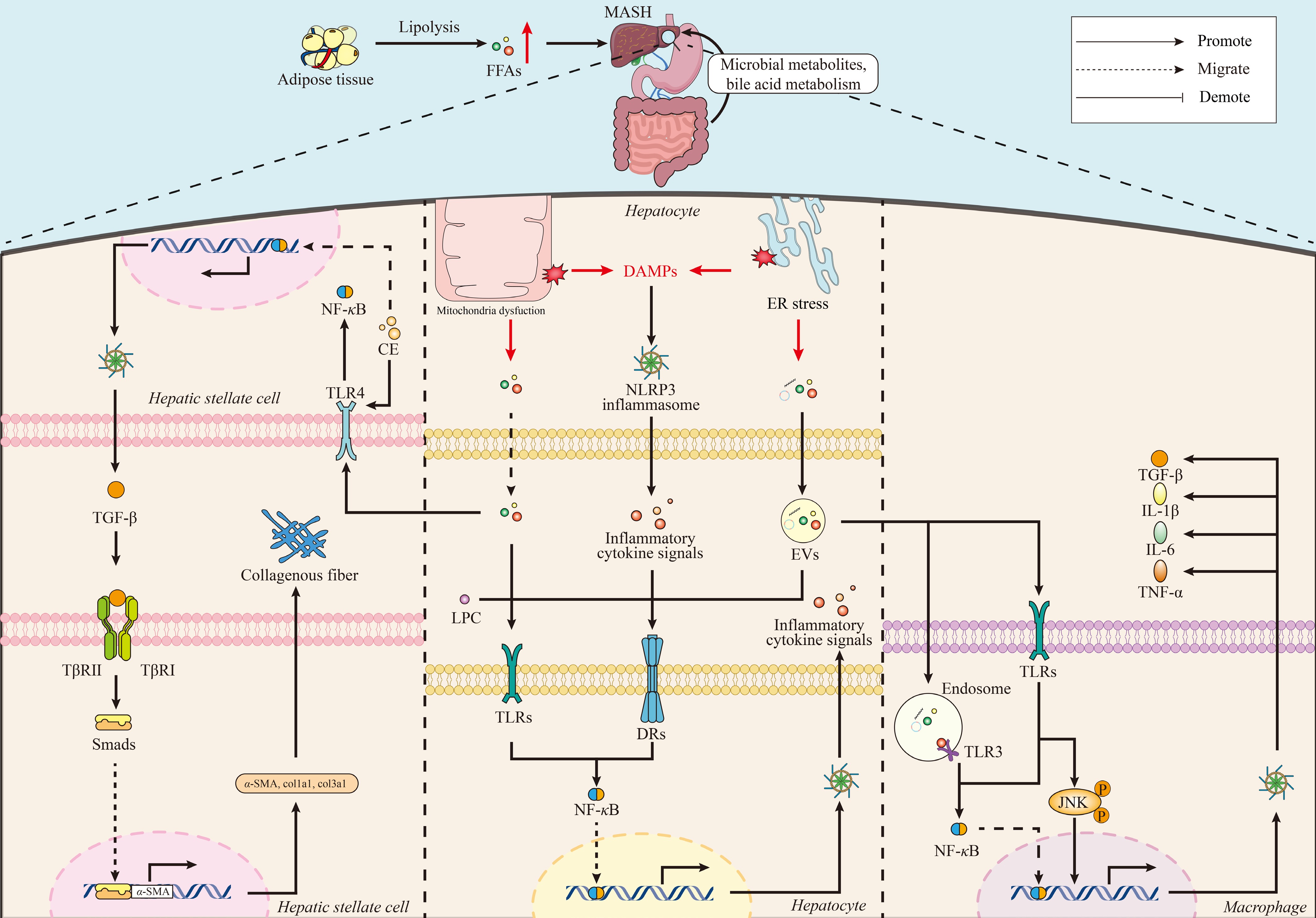
Figure 3.
Lipid-driven intercellular crosstalk promotes hepatic inflammation and fibrosis. Schematic illustration of lipid-mediated intercellular communication in the progression from steatosis to MASH. Lipotoxic stress in hepatocytes leads to the secretion of FFAs, cholesterol crystals, damage-associated molecular patterns (DAMPs), and EVs that serve as paracrine signals in the hepatic microenvironment. These signals activate macrophages by using TLRs, especially TLR4, and NLRP3 inflammasome pathways, leading to activation of NF-κB signaling and production of pro-inflammatory cytokines such as IL-1β, IL-6, and TNF-α. This inflammatory environment aggravates hepatocyte injury and stimulates HSC activation through TGF-β/Smad signaling pathways, which results in collagen deposition and fibrosis. Systemic lipid signals and gut-derived factors further modulate this pathogenic network. α-SMA, alpha-smooth muscle actin; CE, cholesteryl ester; col1a1, collagen type I alpha 1 chain; col3a1, collagen type III alpha 1 chain; DRs, death receptors; JNK-c, Jun N-terminal kinase; LPC, lysophosphatidylcholine; Smads, mothers against decapentaplegic homologs (SMAD family proteins); TLR3, toll-like receptor 3; TβRI, transforming growth factor beta receptor type I; TβRII, transforming growth factor beta receptor type II.
Hepatocyte-derived lipid signals: from intracellular stress to paracrine communication
-
Lipotoxicity-induced hepatocyte injury is a key driver of MASH pathogenesis[143]. Upon sublethal insults, ER stress, mitochondrial dysfunction, and death receptor signaling promote the release of danger-associated molecular patterns, cytokines, and EVs, converting intracellular metabolic stress into paracrine inflammatory signaling[144,145].
The accumulation of saturated FFAs, free cholesterol, and phospholipids containing arachidonic acid induces lipotoxic membrane remodeling and supports the generation and release of bioactive lipid mediators through phospholipase-dependent pathways and EV secretion[27]. Lipid forms such as LPC and oxidized phospholipids activate pattern recognition receptors, death receptors, and inflammasome components in surrounding cells to amplify inflammatory signaling in the hepatic microenvironment[146]. In addition, upregulated expression of phospholipase A2 and lysophosphatidylcholine acyltransferase-2 further promotes arachidonic acid metabolism and eicosanoid production, which are associated with oxidative stress and inflammatory responses[147].
EVs have emerged as important mediators of danger signals from hepatocyte-derived lipids[148]. Lipotoxic stimuli such as palmitate and LPC enhance EV release from hepatocytes, generating vesicles enriched in bioactive lipid species, including ceramides and other sphingolipids, as well as associated signaling molecules[27,46]. Mechanistically, ceramide trafficking by way of lipid transport proteins such as StAR-related lipid transfer domain protein 11 (STARD11) regulates EV biogenesis and cargo composition, linking intracellular lipid metabolism to extracellular signaling outputs[149]. These ceramide-containing EVs are carriers of bioactive lipids, such as sphingosine-1-phosphate, that induce macrophage recruitment and activation, and amplify HSC fibrogenic responses, promoting inflammation and fibrosis[149,150].
Evidence now suggests that membrane microdomain organization impacts EV cargo selection. EVs are similar in composition to lipid raft domains, and the lipid–protein interactions within these ordered membrane regions support selective cargo incorporation[151]. Thus, changes in membrane lipid composition during lipotoxic stress not only promote EV release but also impact the signaling properties of EV cargo.
MicroRNAs (miRNAs) are another class of EV cargo involved in cell-to-cell communication. However, in the context of EVs, miRNAs are less significant than EV lipids, and their effects are context-dependent. MicroRNAs contained in EVs derived from hepatocytes can modulate both fibrogenic and metabolic responses of recipient cells[142,152], while other cell types may release EVs with anti-inflammatory effects[153]. However, bioactive lipid species are transmitted, and this is closely linked to the progression of lipotoxic injury. Hepatocyte-derived EVs are platforms for lipid signaling, translating intracellular lipotoxic stress into a unified inflammatory and fibrotic response at local and systemic levels[154,155].
Lipid signaling to hepatic macrophages: amplifying inflammation
-
Kupffer cells and infiltrating monocyte-derived macrophages are key amplifiers of inflammation in MASH[156]. These cells respond to FFAs, cholesterol, EVs, and lipid-bound pathogen- and danger-associated molecular patterns generated by hepatocytes in the hepatic microenvironment. This activates pattern-recognition receptors, particularly TLR2, TLR4, and nucleotide-binding oligomerization domain-like receptors (NLRs), resulting in NF-κB signaling, NLRP3 inflammasome formation, and secretion of IL-1β, IL-6, and TNF-α. Despite this, the intensity and nature of these responses can vary widely between resident Kupffer cells and infiltrating monocyte-derived macrophages, depending on their activation state and local lipid environment[27,157]. This anti-inflammatory response intensifies hepatocyte injury, initiates immune cell activation, and contributes to steatohepatitis and fibrogenesis (Fig. 3)[158].
Saturated lipid-enriched hepatocyte EVs induce TLR4-mediated macrophage activation, as shown by JNK and NF-κB signaling and increased cytokine expression[27]. Lipotoxic stress in hepatocytes stimulates DR5 signaling and increased EV release, and the uptake of these vesicles by macrophages enhances the production of IL-1β and IL-6, which promote fibrogenic signaling[46]. The inhibition of DR5 or Rho-associated coiled-coil-containing protein kinase 1 (ROCK1) in hepatocytes suppresses lipotoxicity-induced EV release and attenuates EV-mediated macrophage cytokine responses in vitro. Preclinical models have shown that in vivo ROCK1 inhibition decreases the extent of liver damage, inflammation, and fibrosis, but whether these findings are applicable to human MASH still needs to be confirmed in clinical studies[46,159].
EVs also function as lipid transport vehicles. Small EVs enriched in palmitate activate TLR4 in macrophages, leading to JNK phosphorylation, nuclear translocation of NF-κB, and cytokine induction[27]. Myeloid TLR4 inhibition prevents EV-induced inflammation and hepatocyte insulin resistance, demonstrating that vesicle-mediated lipid transfer is sufficient to sustain macrophage-hepatocyte inflammatory loops[27].
MASH macrophages undergo metabolic reprogramming, manifested by cholesterol accumulation, foam cell-like differentiation, and inflammasome activation[160]. Lipidomic and metabolite studies link these manifestations to enrichment of saturated fatty acid, oxysterol, and eicosanoid signaling pathways[161,162]. These findings, based largely on experimental lipidomic and cell-based studies, support a model in which hepatocyte-derived toxic lipids metabolically prime hepatic macrophages and thereby sustain inflammasome activation and chronic liver inflammation; however, the relative contribution of this mechanism to human MASH progression requires further study in translational and clinical settings.
Lipid-mediated hepatic stellate cell activation and fibrogenesis
-
The activation of HSCs is a key transition between chronic inflammation and the deposition of fibrous extracellular matrix[163]. The loss of HSC reactivity and their conversion to extracellular matrix-producing myofibroblasts is mediated by paracrine stimuli released by damaged hepatocytes and macrophages, including unesterified cholesterol, bioactive lipid mediators, EV cargo, and pro-inflammatory cytokines (Fig. 3)[142,164].
Free cholesterol accumulation in HSCs activates TLR4 and increases cell sensitivity to TGF-β signaling, with increased collagen production[165]. Pathological cholesterol loading in hepatocytes, macrophages, and HSCs enhances inflammatory and fibrogenic signaling due to dysregulated cholesterol synthesis, uptake, and esterification in MAFLD/MASH[166].
EV-mediated communication creates a direct mechanistic link between hepatocyte lipotoxicity and HSC activation[142]. Lipotoxic stress stimulates the release of hepatocyte-derived EVs that are absorbed by HSCs, resulting in fibrogenic reprogramming as indicated by enhanced expression of α-smooth muscle actin (α-SMA), collagen-1α1, tissue inhibitor of metalloproteinase-2 (TIMP2), TGF-β, and connective tissue growth factor (CTGF), as well as proliferative and migratory responses[142]. These effects are partially due to surface molecules on EVs, as evidenced by the observation that inhibition of Vanin-1 decreases EV uptake and fibrogenic responses[142]. EVs transport miR-128-3p, which negatively regulates PPARγ and thus promotes HSC activation[142]. Lipotoxic hepatocyte-derived small EVs also transfer profibrotic protein cargo that disrupts intracellular stress and quality-control pathways, thus promoting further stellate cell activation[167].
Not all EV-mediated pathways are pathogenic[168]. Mesenchymal stem cell (MSC)-derived EVs enriched with miR-223-3p attenuated lipid accumulation and fibrogenesis in experimental models of MAFLD through the regulation of E2F transcription factor 1 (E2F1)[169]. Similarly, IL-6 signaling in myeloid cells triggers the secretion of miR-223-rich exosomes that suppress the profibrotic transcriptional coactivator TAZ in hepatocytes[170]. These findings support therapeutic strategies targeting reprogramming of EV cargo for disease resolution[171].
Hepatocyte-derived lipids and EVs are important mediators of the hepatic microenvironment multicellular interaction. These intrahepatic signaling pathways coordinate macrophage activation and HSC reprogramming to drive the inflammatory and fibrotic remodeling that characterizes MASH (Fig. 3). Therapeutic approaches that disrupt, reset, or reprogram lipid-mediated intrahepatic communication thus represent promising avenues for disease modification. These intercellular signaling networks further support the concept of lipotoxicity as a lipid signaling disorder that is spatially and temporally organized in the hepatic microenvironment. In addition to these local networks, systemic lipid signals derived from adipose tissue and the gut-liver axis also regulate hepatic responses.
-
In addition to intrahepatic signaling networks, lipid-mediated communication between the liver and peripheral organs, in particular adipose tissue and the gut, defines the hepatic response to metabolic stress[172]. These inter-organ axes transmit lipid-derived signals that can worsen or suppress lipotoxic injury, inflammation, and fibrosis[15]. To have a full picture of the pathogenesis of MAFLD/MASH, these systemic lipid signaling networks need to be understood.
Adipose tissue-liver axis
-
Visceral adipose tissue-mediated extrahepatic lipid flux has a significant impact on the hepatic microenvironment[173]. In obesity and insulin resistance, defective insulin suppression of lipolysis leads to increased circulating FFA levels and increased hepatic lipid uptake, promoting steatosis and lipotoxic stress[174]. Adipose tissue also emits adipokines and cytokines that influence hepatic metabolism and immune tone[175].
Recent advances have identified adipocyte-derived EVs as key mediators of inter-organ lipid signaling. These EVs transport bioactive lipids, miRNAs, and proteins having direct effects on hepatic insulin sensitivity and inflammatory tone[176]. For instance, the decreased expression of miR-141-3p in adipocyte-derived EVs results in phosphatase and tensin homolog (PTEN) derepression in hepatocytes, thereby blocking hepatic insulin signaling pathways[177]. Adipocyte-derived EVs also promote macrophage polarization toward a pro-inflammatory phenotype and contribute to peripheral insulin resistance, ultimately worsening MAFLD progression[178].
Recent work has begun to unravel the molecular gating of adipocyte lipolysis to protect the liver from FFA overflow. Loss of the purinergic Gi-coupled receptor P2Y13 in white adipose tissue increases cyclic adenosine monophosphate (cAMP) and hormone-sensitive lipase activity, resulting in enhanced lipolysis, higher circulating non-esterified fatty acids (NEFAs), and increased susceptibility to insulin resistance, systemic inflammation, and MAFLD even in the presence of intact hepatic P2Y13[179]. Similarly, loss of activating transcription factor 3 (ATF3) in adipocytes leads to derepression of adipose triglyceride lipase, causing excess FFA release, which is then taken up by hepatocytes via cluster of differentiation 36 (CD36) and converted into triglycerides and pro-inflammatory, pro-fibrogenic lipid species, consequently accelerating MASH development[180]. These findings identify adipocyte intrinsic brakes on lipolysis, such as ATF3 and P2Y13-R, as key determinants of hepatic lipid burden and inflammation in MAFLD/MASH.
In addition to classical adipokines, emerging adipose-derived factors and ceramide species have been identified as important signals in the adipose–liver axis. An example is the adipocyte-specific overexpression of the peptide adrenomedullin-2 (ADM2), which promotes alkaline ceramidase 2 (ACER2)-dependent catabolism of ceramides in adipocytes, resulting in a reduction of the adipose ceramide content and, secondarily, in the amount of hepatic triglycerides in diet-induced MAFLD models, thus identifying the turnover of adipose ceramides as a modifiable driver of liver steatosis[181]. Contrary to this, selective enrichment of long-chain C24:0 ceramide inhibits adipose lipid storage and differentiation, but promotes hepatic steatosis, mitochondrial dysfunction, and ER stress, thus linking an adipose-derived ceramide signal to ectopic liver lipid deposition and glucose imbalance[182]. In addition, newly characterized adipokines, such as brown adipose-derived procollagen C-proteinase enhancer 1 (PCPE-1) and white adipose-derived Isthmin-1 (Ism1), were found to promote liver fibrosis and restrain hepatic steatosis, respectively, emphasizing how the altered adipokine and ceramide environment of dysfunctional adipose tissues can remodel hepatic metabolism and fibrotic tone in MAFLD/MASH[183,184].
Gut–liver axis
-
The gut–liver axis transmits additional lipid-related inflammatory stimuli from the intestinal lumen. Dysbiosis and increased intestinal permeability promote portal delivery of lipopolysaccharide (LPS) and microbial metabolites[185]. LPS activates TLR4 on hepatic immune cells, resulting in the activation of pro-inflammatory signaling pathways that worsen steatohepatitis[186]. At the same time, BAs act as lipid-derived signaling molecules that modulate hepatic metabolism and inflammation through nuclear receptors such as FXR and Takeda G protein-coupled receptor 5 (TGR5) (Fig. 3)[187].
Evidence now shows that gut microbiota-derived lipids and lipid-like metabolites directly influence hepatic lipid handling. SCFAs, produced by microbial fermentation of dietary fiber, modulate hepatic DNL and inflammation through GPCR-dependent and epigenetic mechanisms[188]. On the other hand, dysregulated bile acid metabolism in MAFLD/MASH impacts the intestinal lipid environment, causing further dysbiosis and a feed-forward loop of metabolic dysfunction[189].
Gut and diet-derived EVs have also emerged as structural lipid carriers, conditioning both the intestinal barrier and downstream liver injury. In vivo, EVs derived from milk from bovine and human breast milk withstand gastrointestinal transit to reach the colon and strengthen several components of the gut barrier, including the mucus layer, epithelial junctions, and immune layers[190]. Oral administration of these EVs in diet-induced MASH models prevents endotoxin translocation to the liver and significantly attenuates hepatic inflammation and steatohepatitis, demonstrating that gut-delivered vesicular lipids and cargo can indirectly modulate hepatic inflammatory and metabolic tone by way of stabilizing the intestinal interface[190]. Similarly, ellagic acid-enriched plant-derived exosome-like nanovesicles reduce serum endotoxin, restore dysbiotic microbial profiles, and normalize oxidative stress and fibrosis markers in MAFLD mouse livers, further highlighting EV-mediated lipid signaling as a modifiable component of the gut–liver axis[191].
In addition, microbiome- and bile acid-targeted approaches are starting to mechanistically validate the therapeutic relevance of gut-liver lipid circuits. The small molecule ACT001 improves steatosis and inflammation in both high-fat diet and methionine-choline-deficient MAFLD models, while restoring the intestinal barrier integrity and reshaping the microbiota towards microbial populations that promote secondary bile acid production[192]. ACT001 leads to elevated levels of specific BAs, including 23-nor-deoxycholic acid and lithocholic acid-3-sulfate, leading to reduced activation of the enteral FXR-fibroblast growth factor 15 (FGF15) axis, thus restoring the balance of bile acid-FXR signaling and improving hepatic lipid metabolism[192]. Similarly, ileal FXR knockdown in Western diet-fed rats enhances the reabsorption of secondary and unconjugated BAs into the portal blood, remodels the cecal microbiota to secondary bile acid-producing genera, and attenuates hepatic steatosis and inflammation, indicating that the microbiota-BA-FXR module is a core, druggable axis in gut-liver lipid communication[193].
Integration of inter-organ signals with intrahepatic networks
-
An integrated network is formed from the convergence of adipose-derived FFAs, gut-derived microbial products, and hepatocyte-derived lipid signals that determine disease progression. In simple steatosis, these inter-organ signals may be relatively inactive. However, once lipotoxic stress has developed in the liver, peripheral signals can promote intrahepatic inflammation and fibrosis through positive feedback loops[194].
For example, lipolysis of adipose tissue releases FFAs that not only cause direct injury to hepatocytes but also sensitize Kupffer cells to enhanced inflammatory responses upon subsequent TLR activation[195]. Likewise, LPS and bile acid metabolites from the gut suppress the activation threshold of the NLRP3 inflammasome in hepatic macrophages and act in synergy with hepatocyte-derived EVs to induce progressive fibrosis[196]. Such interactions suggest that the progression from steatosis to MASH is often not caused by a single event, but by several signals from multiple organ systems in succession (Fig. 3).
Therapeutic strategies that disrupt these inter-organ communication pathways, including reducing adipose tissue lipolysis, restoring gut barrier integrity, modulating the gut microbiome, or targeting EV release, represent promising directions for disease modification. Clinical trials of FXR, PPARs, and other lipid-sensing pathways have demonstrated varying effectiveness, emphasizing the need for approaches that address the integrated, multi-organ nature of MAFLD/MASH.
-
Besides the general anti-steatotic approaches that have long been used to manage MAFLD/MASH, there has been a gradual transition toward therapeutic strategies that directly target lipid signaling pathways and intracellular trafficking events to control the condition. This transition aligns with growing evidence that organelle dysfunction and lipotoxic stress, rather than triglyceride accumulation per se, are notable pathophysiological contributors in most patients[6,197,198]. This is why modern strategies aim not only to decrease the hepatic lipid load, but also to restore lipid flux and organelle homeostasis, and to minimize inflammatory and fibrogenic signaling (Fig. 4; Tables 2, 3).
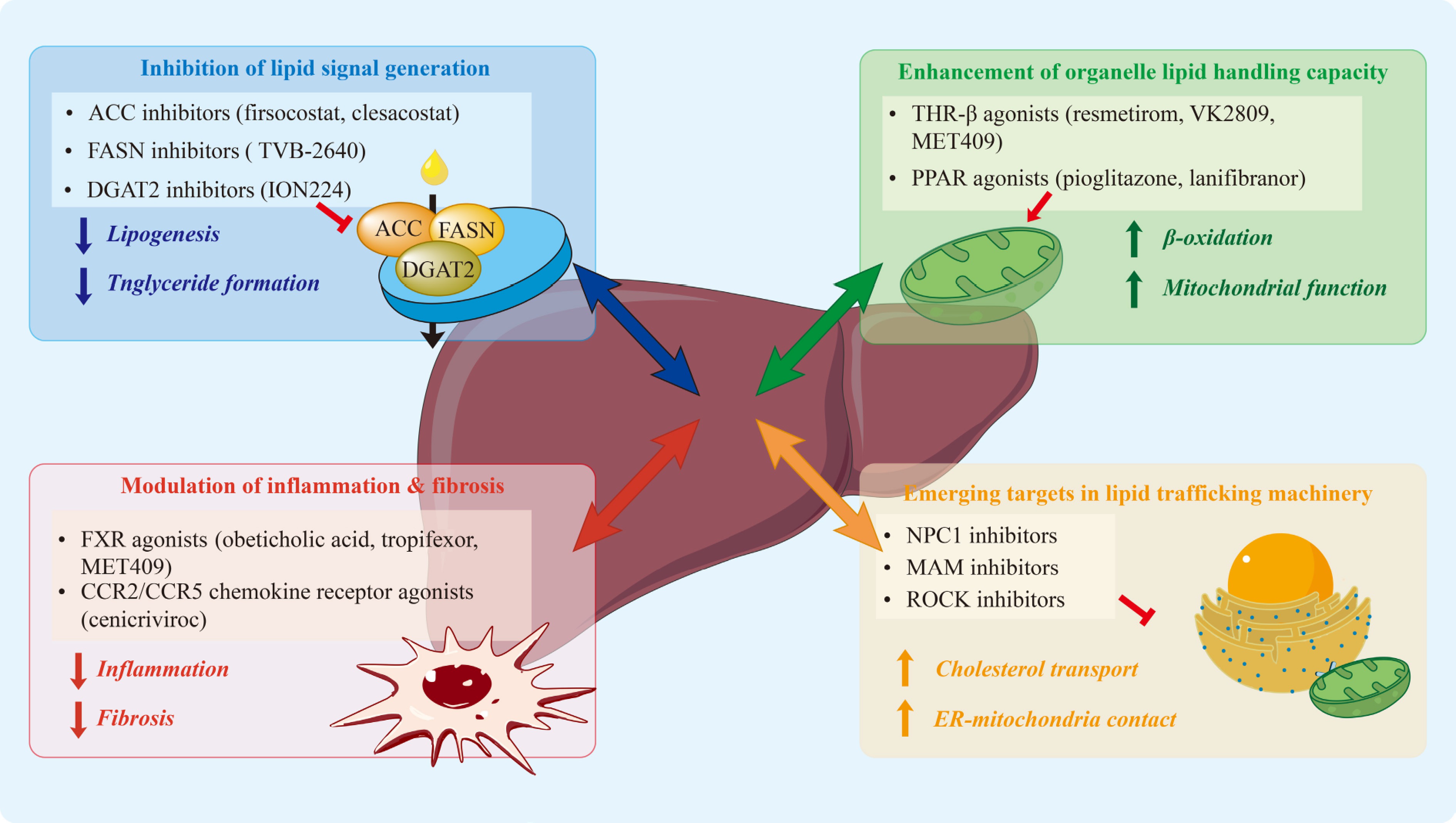
Figure 4.
Therapeutic targeting of lipid signaling and trafficking nodes in MAFLD/MASH. Therapeutic approaches are categorized according to the main hepatocellular processes they modify. Blue (upper left) inhibition of lipid signal generation through acetyl-CoA carboxylase (ACC), fatty acid synthase (FASN), and diacylglycerol acyltransferase 2 (DGAT2) inhibition. Green (upper right), promotion of organelle lipid utilization and mitochondrial function through thyroid hormone receptor β (THR-β) and PPAR pathways. Red (lower left), reduction of inflammatory and fibrogenic signaling through FXR and chemokine pathways. Orange (lower right), modulation of emerging lipid trafficking mechanisms including Niemann-Pick C1 (NPC1), MAMs, and Rho-associated coiled-coil-containing kinase (ROCK) signaling. These complementary interventions reveal targetable nodes that reduce lipotoxic stress, inflammatory amplification, and fibrotic progression. ACC, acetyl-CoA carboxylase; DGAT2, diacylglycerol acyltransferase 2; FASN, fatty acid synthase; PPAR, peroxisome proliferator-activated receptor; THR-β, thyroid hormone receptor beta.
Table 2. Emerging and preclinical therapeutic targets in lipid trafficking and organelle crosstalk.
Category Target Agent Mechanism Compartment Hepatic outcomes Status Ref. MAM Mfn2 Photobiomodulation (650 nm) Restores ER-mitochondria contacts; reduces PERK/CHOP signaling MAMs; ER; mitochondria Reduced ER stress; improved mitochondrial function; reduced lipotoxicity Preclinical [226] MAM Mic19-EMC2-SLC25A46 Mic19 overexpression Maintains ER-mitochondria contacts; preserves mitochondrial structure MAMs; mitochondria Reduced steatosis; reversed steatohepatitis and fibrosis Preclinical [227] Cholesterol NPC1 Calycosin Enhances lysosomal cholesterol transport Lysosome; ER Reduced steatosis; reduced inflammation; improved insulin sensitivity Preclinical [235] Cholesterol NPC1-ORP5 Pathway inhibition Reduces lysosome-ER cholesterol transfer;
reduces ER stressLysosome; ER Reduced macrophage activation; reduced inflammation Preclinical [231,232] Cholesterol NPC1L1 Cryptotanshinone Inhibits cholesterol absorption Plasma membrane; ER Reduced hepatic steatosis Preclinical [234] Cholesterol NPC1L1 Pharmacologic inhibition Reduces oxysterol uptake; modulates LXR/ROR signaling Plasma membrane; nucleus Prevented diet-induced steatosis Target validated [233] EV signaling ROCK1 Fasudil Inhibits DR5-ROCK1-dependent EV release Cytosol; plasma membrane Reduced inflammation; reduced fibrosis Preclinical/
repurposing[46] EV signaling ROCK1/2 ROCK inhibitors Inhibits hepatocyte ROCK signaling Cytosol Reduced steatosis; reduced inflammation; reduced fibrosis Preclinical [159] Lipid metabolism SLC25A1 CTPI-2 Inhibits mitochondrial citrate export Mitochondria; ER Reduced steatosis; reduced inflammation; reduced fibrosis Preclinical [236] Lipid metabolism FASN Platensimycin Inhibits de novo
lipogenesisER; lipid droplets Reduced hepatic triglycerides Preclinical [237] Lipid metabolism ACLY Bempedoic acid Reduces cytosolic acetyl-CoA Cytosol; ER Reduced triglycerides; reduced inflammation Preclinical [238] Lipid metabolism THR-β VK2809 Enhances lipid oxidation; promotes lipophagy Nucleus; mitochondria; lysosome Reduced hepatic triglycerides Preclinical [210] Bile acid FXR/GPBAR1 BAR502 + UDCA Modulates bile acid-lipid signaling Liver; intestine;
adiposeReduced steatosis; reduced inflammation; reduced fibrosis Preclinical [239] Metabolic SGLT2 Dapagliflozin/
CanagliflozinModulates metabolic signaling Cytosol; mitochondria Reduced steatosis (preclinical models) Preclinical [240] Energy sensing AMPK Metformin Inhibits lipogenesis; modulates gut-liver axis Mitochondria; ER Reduced steatosis; reduced oxidative stress Preclinical [241] Table 3. Current clinical status of therapeutic agents for MAFLD/MASH.
Category Target Agent Mechanism Compartment Hepatic outcomes Status Ref. Lipogenesis ACC1/2 Firsocostat Inhibits de novo lipogenesis ER; mitochondria Reduced steatosis; increased plasma triglycerides Discontinued (2025) [201,242] Lipogenesis ACC1/2 + DGAT2 Clesacostat + PF-06865571 Dual inhibition of lipogenesis and triglyceride synthesis ER; mitochondria; ER-LD Reduced steatosis; reduced hypertriglyceridemia; MASH resolution without fibrosis worsening in MIRNA trial Phase II (met primary endpoint); in continued development [202,243] Lipogenesis DGAT2 PF-06865571 (monotherapy) Inhibits triglyceride synthesis ER-LD interface Reduced hepatic steatosis; monotherapy did not meet primary endpoint in MIRNA trial Phase II completed (monotherapy did not meet primary endpoint) [202,243] Lipogenesis DGAT2 (ASO) ION224 Inhibits triglyceride synthesis from DAG ER-LD MASH resolution; fibrosis improvement (subset) Phase II completed (met primary endpoint) [208] Lipogenesis FASN Denifanstat (TVB-2640) Inhibits lipogenesis ER; lipid droplets Reduced steatosis; reduced liver injury markers; MASH resolution and fibrosis improvement in Phase IIb (FASCINATE-2) Phase IIb completed (met primary endpoint); Phase III MASH trial withdrawn (2025) [205,207] Lipogenesis KHK PF-06835919 Inhibits fructose-driven lipogenesis Cytosol Reduced liver fat Discontinued (2021) [244,245] Nuclear receptor THR-β Resmetirom Enhances lipid oxidation Nucleus; mitochondria MASH resolution; fibrosis improvement Approved [212] Nuclear receptor PPARγ Pioglitazone Modulates lipid partitioning Nucleus; mitochondria Improved insulin sensitivity; reduced hepatic steatosis and inflammation in MASH trials Approved (T2DM); guideline-recommended off-label use in MASH
[246,247] Nuclear receptor Pan-PPAR Lanifibranor Enhances fatty acid oxidation; reduces inflammation and fibrosis Nucleus; mitochondria MASH resolution; fibrosis improvement Phase III (ongoing) [216,248] Nuclear receptor FXR Cilofexor Inhibits bile acid synthesis Nucleus; ER Reduced steatosis Discontinued (2025) [242,249] Incretin GLP-1R Semaglutide Reduces lipotoxicity and inflammation Hepatocytes MASH resolution; weight loss Approved [250] Incretin GIPR/GLP-1R Tirzepatide Improves metabolic control Hepatocytes MASH resolution; fibrosis improvement Phase III (ongoing) [251,252] Combination FXR + ACC Cilofexor + Firsocostat Dual bile acid and lipid regulation Nucleus; ER Reduced steatosis; reduced inflammation; reduced fibrosis Discontinued (2025) [242,253] Combination GLP-1R + FXR Semaglutide + Cilofexor Combined metabolic and bile acid signaling Nucleus; ER Greater reduction vs monotherapy Discontinued (2025) [242,254] Combination GLP-1R + ACC Semaglutide + Firsocostat Combined lipogenesis inhibition and metabolic modulation Cytosol; lipid droplets Greater reduction in hepatic fat and injury markers Discontinued (2025) [242,254] Combination GLP-1R +
FXR + ACCSemaglutide + Cilofexor + Firsocostat Multi-pathway lipid suppression Nucleus; cytosol; lipid droplets Did not meet primary fibrosis endpoint vs placebo (13.7% vs
8.3%, p = 0.2289)Discontinued (2025) [242,255] Metabolic SGLT2 Empagliflozin Reduces hepatic lipid accumulation Lipid droplets Reduced liver fat Approved (T2DM); Phase IV (non-diabetic MAFLD, ongoing) [256] Inhibition of lipid signal production
-
DNL is upregulated in MAFLD/MASH, resulting in increased intrahepatic triglyceride, DAG, and ceramide pools, which promote ER stress, mitochondrial dysfunction, and hepatic insulin resistance in experimental models and human disease[199]. Thus, a mechanism-based pharmacological inhibition of the major lipogenic enzymes such as acetyl-CoA carboxylase (ACC) and fatty acid synthase (FASN) provides an approach to limit the production of lipotoxic intermediates at their origin (Fig. 4)[198,200].
ACC inhibitors such as firsocostat (GS-0976) and clesacostat (PF-05221304) decrease hepatic fat and serum alanine aminotransferase levels in phase II studies[201,202]. However, these benefits tend to be followed by increased circulating levels of triglycerides, a marker of disturbed systemic lipid partitioning[201,202]. Combination treatment regimens, including fibrates, can potentially reverse this effect, although the long-term cardiovascular consequences are not fully characterized[203]. From a mechanistic view, inhibition of ACC results in decreased malonyl-CoA levels, suppression of DNL, alleviation of carnitine palmitoyltransferase-1 (CPT-1) inhibition, and therefore increased mitochondrial β-oxidation (Tables 2, 3)[201,202,204].
FASN inhibition decreases palmitate synthesis and reduces triglyceride accumulation in hepatocytes. In a phase IIa trial, a high dose of Denifanstat (TVB-2640) resulted in a reduction of hepatic fat content by approximately 28% as estimated by magnetic resonance imaging proton density fat fraction (MRI-PDFF)[205], without significant compensatory hypertriglyceridemia[206]. A subsequent phase IIb trial (FASCINATE-2) confirmed histological benefit, with denifanstat achieving significant MASH resolution and fibrosis improvement compared to placebo and earning FDA Breakthrough Therapy Designation; however, the planned phase III registrational trials were withdrawn in 2025, and the drug's near-term clinical development has shifted toward a non-hepatic indication[207]. A commonly reported adverse reaction has been reversible alopecia. These outcomes support the upstream inhibition of lipotoxic substrate production as a reasonable, but not risk-free, therapeutic approach.
In addition to these approaches, inhibition of DGAT2, which facilitates the final step in triglyceride synthesis at the interface between the ER and lipid droplets, offers another approach. The phase II study of ION224, a hepatocyte-targeted antisense oligonucleotide, showed improvements in steatosis, inflammation, and fibrosis, and a reduction in lipid levels, without changes in body weight, highlighting the clinical relevance of selective regulation of triglyceride formation[208].
Enhancement of organelle lipid handling capacity
-
Given that lipotoxicity results from an imbalance between lipid influx and oxidative disposal, cellular tolerance to lipid excess can be improved by interventions that upregulate mitochondrial and peroxisomal fatty acid oxidation. Thyroid hormone receptor-β (THR-β) agonists such as resmetirom (MGL-3196) and VK2809 activate selectively hepatic thyroid hormone signaling, which results in increased β-oxidation, improved LDL clearance, and reduced toxic lipid pools[209,210]. In 2024, Resmetirom became the first approved therapy for MASH after showing statistically significant reductions in steatohepatitis and fibrosis compared to placebo in phase III studies[211,212]. These outcomes provide clinical evidence that enhancing organelle lipid-handling capacity can alter disease progression in selected patient populations.
Peroxisome proliferator-activated receptor (PPAR) agonists have greater metabolic effects on hepatocytes, adipose tissue, and immune cells. The PPARγ agonist pioglitazone improves insulin sensitivity and steatohepatitis, and some of its metabolic effects appear to be linked to the redistribution of lipid storage into adipose depots rather than ectopic sites[213,214]. Dual and pan-PPAR agonists, such as lanifibranor, demonstrate more consistent anti-inflammatory and anti-fibrotic effects by simultaneously influencing fatty acid oxidation pathways, adipose remodeling, and immune modulation[215]. In line with these mechanisms, lanifibranor achieved resolution of MASH without progression of fibrosis in a phase II trial and is currently being evaluated in a phase III trial[216,217]. THR-β and PPAR agonists improve metabolic flexibility and organelle resilience, complementing strategies to restrict lipid synthesis (Fig. 4; Tables 2, 3).
Modulation of lipid-driven inflammation and fibrosis
-
In chronic steatohepatitis, the inflammatory and fibrogenic cascades can become partially self-renewing and thus require direct therapeutic intervention[62]. FXR agonists are bile acid-activated nuclear receptors that regulate lipid and glucose metabolism; they reduce DNL, enhance intestinal barrier integrity, and exert anti-inflammatory and anti-fibrotic activity in hepatocytes and non-parenchymal cells. Obeticholic acid showed fibrosis improvement in advanced clinical trials but was associated with dose-dependent pruritus and elevations in LDL cholesterol[218]. Next-generation FXR agonists are in clinical development. Notably, Tropifexor (phase II) and MET409 have shown significant hepatic fat reduction in preclinical studies (Fig. 4)[219,220].
Immune cell recruitment has also been targeted therapeutically. Cenicriviroc, a dual C-C chemokine receptor type 2/type 5 (CCR2/CCR5) antagonist, suppresses monocyte recruitment and macrophage-stellate cell exchange. However, later-phase trials did not show sufficient histological benefit despite evidence of target engagement[221,222]. These outcomes indicate that chemokine-based approaches may not work as monotherapy, but instead may be beneficial in combination with agents that reduce upstream lipotoxic stress.
Emerging targets in lipid-trafficking machinery
-
More recent therapeutic approaches are increasingly focusing on intracellular lipid routing and organelle contact sites as regulatory nodes of lipotoxic signaling. Selective inhibition of DGAT2 decreases triglyceride formation at the ER-lipid droplet interfaces and alleviates hepatic steatosis; however, excessive inhibition of DGAT2 may increase cytotoxic DAG and ceramide pools, emphasizing the importance of precise dose optimization and patient stratification (Tables 2, 3)[223].
MAM modulators: restoring ER-mitochondria communication
-
MAMs are emerging as key centers integrating lipid metabolism, calcium signaling, and cellular stress responses. These contact sites regulate the exchange of lipids between ER and mitochondria, and house enzymes involved in triglyceride and cholesterol metabolism, thereby directly impacting mitochondrial function and redox balance[224,225]. Disruption of MAM stability leads to impaired formation of lipid droplets and induces cellular stress, highlighting the significance of the contact site architecture in buffering lipid overload[103]. MAMs are under the control of several structural regulators and represent potential therapeutic targets. Mitofusin 2 (Mfn2) is a major mediator of ER-mitochondria bonding and maintenance of contact site stability. Restoration of Mfn2 expression improves ER-mitochondria communication and attenuates lipotoxic stress signaling[226]. Also, the Mic19-EMC2–SLC25A46 axis is important for mitochondrial architecture and lipid homeostasis. Disruption of this axis reduces ER-mitochondria contacts, changes the balance of mitochondrial lipids, and promotes steatohepatitis and fibrosis, while restoration of this axis reverses these pathological changes[227].
Additional MAM regulators, including phosphofurin acidic cluster sorting protein 2 (PACS-2), caveolin-1, and sigma-1 receptor, further modulate lipid trafficking, calcium transfer, and redox signaling at these contact sites. For example, PACS-2 organizes ER-mitochondria lipid-synthesizing domains, directly connecting the contact site structure to local lipid metabolic capacity[228]. These findings identify MAMs as targetable platforms integrating lipid flux with stress signaling, although clinical application of MAM-targeted therapies remains in the early stages.
NPC1 and NPC1L1: targeting cholesterol trafficking pathways
-
A further important axis in the pathogenesis of MAFLD/MASH is cholesterol trafficking. The lysosomal cholesterol transporter Niemann-Pick disease type C1 protein (NPC1) regulates intracellular cholesterol distribution, and its dysfunction contributes to free cholesterol accumulation, ER stress, inflammasome activation, and fibrogenesis[229]. NPC1 is also relevant for immune metabolism regulation beyond hepatocyte autonomous effects. Myeloid NPC1 deficiency was shown to affect macrophage efferocytosis and promote the release of inflammatory mediators, such as high mobility group box 1 (HMGB1) and mitochondrial DNA, leading to activation of stimulator of interferon genes (STING)/NF-κB signaling and worsening of liver injury and fibrosis[230]. NPC1-dependent cholesterol transport between lysosomes and the ER, partially through the NPC1-oxysterol-binding protein-related protein 5 (ORP5) axis, connects cholesterol trafficking with ER redox stress and pro-inflammatory macrophage polarization[231,232].
Niemann-Pick C1-like 1 (NPC1L1) is a complementary regulator at the level of cholesterol uptake. Hepatic NPC1L1-directed uptake of biliary oxysterols promotes steatosis through the activation of nuclear receptor pathways, and circulating oxysterol levels are linked with hepatic lipid accumulation[233]. Pharmacological inhibition of NPC1L1 has been shown to decrease hepatic cholesterol burden and ameliorate steatosis in experimental models, suggesting therapeutic potential[234,235]. At the same time, modulation of NPC1 expression has shown beneficial metabolic and anti-inflammatory effects in preclinical studies, further supporting the concept that targeting cholesterol trafficking pathways can decrease both lipotoxic stress and inflammatory signaling[235]. These findings identify NPC1/NPC1L1-controlled pathways as key nodes linking lipid metabolism to immune activation and fibrosis.
ROCK1 inhibition: disrupting lipotoxic EV signaling and disease amplification
-
ROCK1 has emerged as an important mediator of lipotoxic signal transmission. As was explained above, ROCK1 inhibition suppresses the release of saturated fatty acid-enriched hepatocyte-derived EVs, leading to reduced macrophage activation and fibrogenesis in experimental MASH models[159]. Mechanistically, lipotoxic stimuli such as palmitate and lysophosphatidylcholine activate DR5, which triggers ROCK1-dependent EV biogenesis. These EVs are loaded with pro-inflammatory cargo such as TRAIL and bioactive lipids such as ceramides that activate macrophage signaling pathways and increase hepatic inflammation[46,149].
Recent studies further indicate that ROCK1 is significantly required for disease progression. Diet-induced MASH is associated with increased hepatic ROCK1 expression and activity, and hepatocyte-specific ROCK1 deletion reduces steatosis, inflammation, hepatocyte death, and fibrosis. Pharmacological inhibition of ROCK1/2 similarly improves metabolic parameters and reduces liver injury in preclinical models[159]. The translational potential of this approach is supported by the availability of clinically approved ROCK inhibitors such as Fasudil, although liver-targeted delivery strategies may be required to minimize systemic effects[46].
These emerging approaches draw attention to intracellular lipid trafficking, organelle contact sites, and intercellular signal transmission as interconnected governing layers of MAFLD/MASH pathogenesis. By targeting MAM integrity, cholesterol pathways via NPC1/NPC1L1, and ROCK1-dependent EV signaling, we are moving toward modulating spatially organized lipid signaling networks. The present evidence suggests that long-lasting clinical benefit will likely require rational combination therapies that are tailored to specific manifestations of lipid dysregulation, organelle stress responses, and disease stage (Fig. 4; Tables 2, 3). Therefore, effective therapies will need to target multiple nodes within this spatially and temporally organized lipotoxic signaling network.
-
Despite the strong mechanistic foundation and the rapid progress in the understanding of lipid-mediated pathogenesis, translation of lipid-targeted therapies to the clinic has been challenging in MAFLD/MASH. The failure of multiple late-stage programs to achieve primary histologic endpoints highlights a continued gap between mechanistic understanding and therapeutic efficacy across patient populations.
Despite promising early data, the apoptosis signal-regulating kinase 1 (ASK1) inhibitor selonsertib did not demonstrate benefit over placebo for fibrosis improvement in two phase III trials (STELLAR-3 and STELLAR-4). Post-hoc analysis suggested that target participation may have been insufficient, or that inhibition of a single stress pathway is insufficient to reverse established fibrosis[257]. Similar to this, the pan-caspase inhibitor emricasan failed to improve liver histology in phase IIb trials, possibly reflecting the limitation of targeting apoptosis without addressing upstream metabolic and lipotoxic factors[258,259]. The dual CCR2/CCR5 antagonist cenicriviroc did not demonstrate significant histologic benefit in the phase III AURORA trial despite evidence of pharmacologic activity[221,222].
Beyond these particular examples, the broader therapeutic landscape in MASH illustrates the challenges of clinical translation. Obeticholic acid, the first FXR agonist to show phase III histological improvement, was not approved due to limited antifibrotic efficacy and safety concerns, including pruritus and dyslipidemia. In addition to elafibranor and selonsertib, other agents with promising preclinical and early-phase data have failed in late-stage trials, even with evidence of target engagement[222,257]. These outcomes likely reflect the redundancy and adaptive nature of lipid and inflammatory signaling networks, together with limitations of current clinical trial design and endpoint selection.
Histological assessment is considered the gold standard, but is often prone to inter- and intra-observer variability and may weaken measurable treatment effects, especially in slowly progressive disease. High placebo response rates, prolonged disease kinetics, and differences in patient populations between phase II and phase III trials may complicate interpretation and contribute to inconsistent outcomes[222,260,261].
Unresolved biomarker and stratification complications further limit progress. Most non-invasive tests were developed for advanced fibrosis, not for fibrotic MASH, and have only moderate diagnostic accuracy, with AUROCs generally ranging from 0.7−0.8 and significant errors in classification at clinically relevant thresholds[262,263]. Newer biomarker panels (e.g., NIS2+, MACK-3, and FAST) improve performance, but are still affected by demographic and metabolic factors and will require further validation before alternative endpoints can be used[264]. The marked heterogeneity in genetic risk, metabolic traits, and extrahepatic disease burden suggests that a single biomarker is unlikely to capture treatment-relevant differences.
Another challenge is translating mechanisms at the organelle level into clinically actionable interventions. MAMs, mitochondrial quality control, and lipophagy pathways have revealed promising therapeutic targets, but most of the evidence is derived from simple experimental systems. Interventions directed at these processes, including MAM-associated kinases, mitochondrial modulators, and ferroptosis regulators, are hindered by off-target effects, pathway redundancy, and the lack of validated clinical readouts of organelle function[84,265].
Collectively, these limitations establish that mechanistic precision alone is not enough to ensure successful clinical translation. Future trial design will need to include biomarker-driven patient stratification, rational combination therapies targeting multiple pathogenic nodes, and treatment durations that are consistent with the slow kinetics of fibrosis regression.
Current therapeutic strategies and clinical outcomes
-
Lipid-targeted approaches that have been translated to clinical evaluation in MAFLD/MASH include lifestyle modification, metabolic and endocrine therapies, nuclear receptor modulators, and emerging biologic approaches. Despite these advances, durable histological improvement with acceptable long-term safety remains a major clinical challenge across diverse patient populations[6,7,200]. Hepatic steatosis is often reversible, while fibrosis regression is slower, less predictable, and requires a coordinated modulation of multiple pathogenic processes[266].
The complexity of pathway-directed approaches is shown by clinical experience. The FXR agonist obeticholic acid appeared to improve fibrosis in a subset of patients but was associated with dose-dependent pruritus and increased LDL cholesterol levels, highlighting the systemic effects of bile acid pathway modulation[218,267]. Similarly, ACC inhibitors potently suppress DNL, but may lead to hypertriglyceridemia and more general dyslipidemia, which may be a result of systemic abnormalities of lipid flux[268].
However, the THR-β agonist resmetirom has shown significant reductions in hepatic steatosis, resolution of MASH, and improvement of fibrosis, leading to regulatory approval and confirming mitochondrial fatty acid oxidation and cholesterol metabolism as therapeutic targets (Fig. 4; Table 3)[211]. PPAR agonists (pioglitazone, dual or pan-PPAR agents such as lanifibranor and saroglitazar) improve steatosis and hepatic inflammation, but with variable antifibrotic effects. These agents also have adverse effects, including weight gain and fluid retention, requiring careful patient selection and monitoring[216,269].
Therapeutic agents targeted to systemic and enterohepatic pathways, such as analogues of fibroblast growth factor 19 (FGF19) and fibroblast growth factor 21 (FGF21), glucagon-like peptide-1 (GLP-1) receptor agonists, and sodium-glucose cotransporter 2 (SGLT2) inhibitors, all reduce hepatic fat content, but the histologic results have been inconsistent across trials[256,270,271]. These results highlight the importance of matching therapeutic mechanisms with the major disease drivers in particular patient subgroups.
Collectively, these findings suggest that monotherapy directed at a single signaling pathway will be insufficient to restore the complex, spatially and temporally coordinated lipid signaling networks underlying MAFLD/MASH. There is considerable inter-individual variability in genetic background, metabolic phenotype, gut microbiota composition, and immune response, which results in differences in disease progression and treatment response[272−275].
Recent clinical evidence increasingly supports a shift toward rational combination strategies. Significant decreases in hepatic steatosis and improvements in disease activity have been associated with combinations of metabolic, lipid-regulatory, and anti-inflammatory mechanisms rather than with monotherapy[254]. These approaches agree with a stratified treatment paradigm where systemic metabolic drivers are dealt with first, followed by liver-directed therapies to restore hepatocellular lipid homeostasis, with selective use of antifibrotic or immunomodulatory agents in patients with confirmed fibrosis. At the same time, the design of multi-target agents capable of modulating different lipid signaling nodes within a single molecule may reduce the need for polypharmacy while improving efficacy[216,217].
Lifestyle intervention is still a cornerstone of therapy. Maintaining weight loss through dietary changes and physical activity is the intervention most consistently associated with histological improvement and, in some cases, MASH resolution[276]. Long-term adherence, however, remains challenging, and complementary pharmaceutical approaches are needed to strengthen adaptive metabolic pathways, such as AMPK activation[277,278]. These complementary approaches have been schematically illustrated in Fig. 4, with specific therapeutic agents, targets, and clinical status specified in Tables 2 and 3.
-
Reconceptualizing MAFLD/MASH as a spatially and temporally organized disorder of lipid signaling, rather than a passive accumulation of triglycerides, offers a unifying model that integrates metabolic stress, inflammation, and fibrogenesis (Figs 1−3, 5). In this model, the effects of lipid species depend on the context, including their molecular identity, subcellular localization, and trafficking through interconnected organelle networks. The ER, mitochondria, LDs, lysosomes, and peroxisomes act as an interconnected metabolic system coordinated by dynamic membrane contact sites that regulate lipid flux, calcium exchange, and redox balance. Disruption of this spatial organization activates stress kinases, death receptor pathways, and inflammatory cascades. Signals originating from hepatocytes, such as EVs, lipid mediators, and cytokines, propagate injury in the hepatic microenvironment and reinforce fibrogenic chain reactions. These processes are also shaped by systemic lipid flux and gut-derived signals, supporting the classification of MAFLD/MASH as a multisystem metabolic disorder.
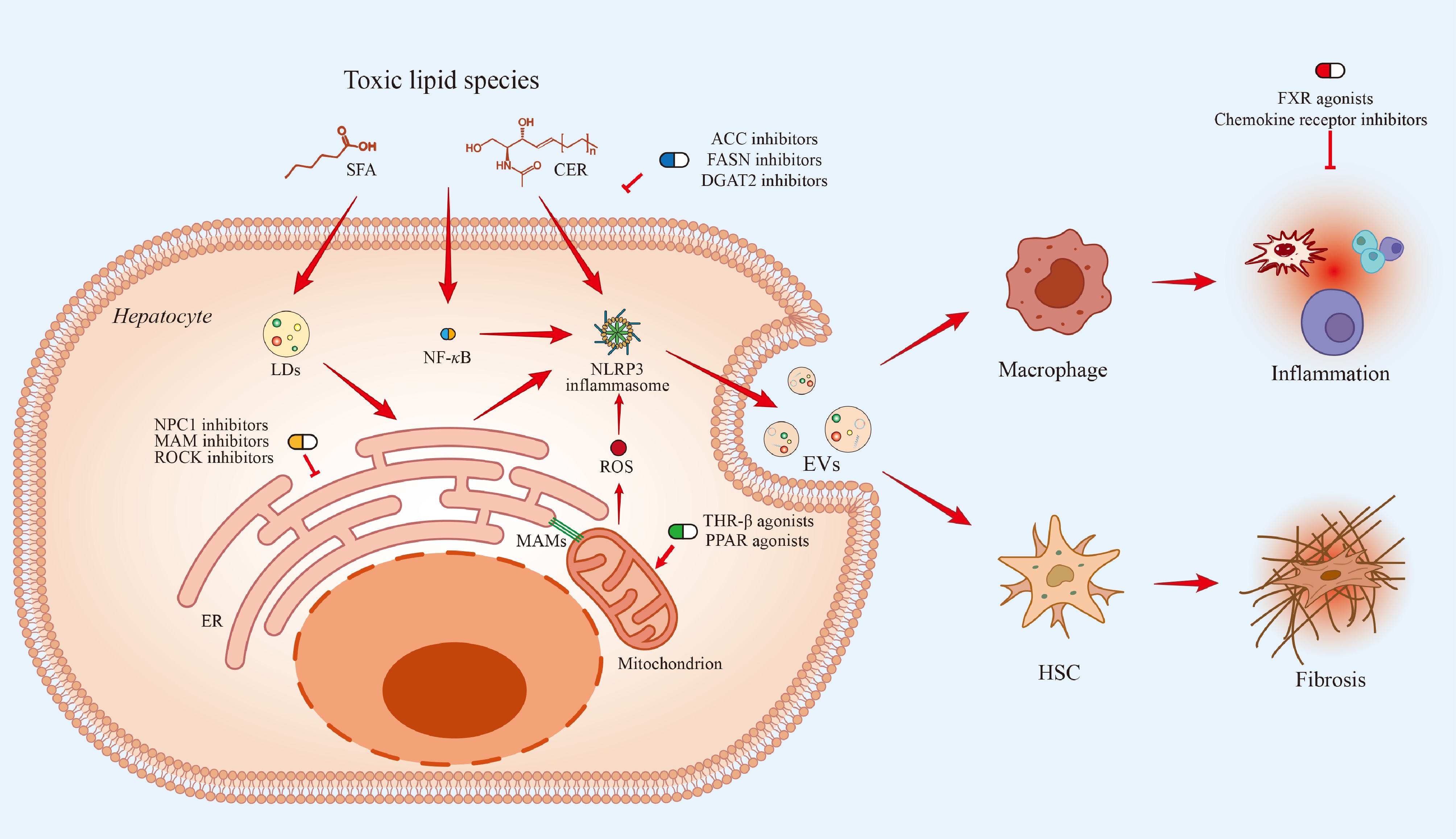
Figure 5.
The accumulation of toxic lipid species, including SFAs and CERs, in hepatocytes drives key pathological processes. Intracellular lipid excess promotes LD expansion, ER stress, mitochondrial dysfunction, and MAM formation, collectively generating ROS and activating the NLRP3 inflammasome and NF-κB signaling pathways. These events trigger inflammatory responses that are spread beyond the hepatocyte through EV-mediated intercellular communication, promoting macrophage activation and HSC-driven fibrosis. Therapeutic strategies are presented at their respective sites of action: ACC, FASN, and DGAT2 inhibitors reduce toxic lipid generation; THR-β and PPAR agonists enhance mitochondrial lipid utilization; NPC1, MAM, and ROCK inhibitors target intracellular lipid trafficking and organelle stress; and FXR agonists and chemokine receptor inhibitors attenuate downstream inflammatory and fibrogenic signaling.
This framework highlights three key priorities for future research and clinical translation:
First, real-time spatial lipid imaging. A significant limitation is the inability to directly visualize lipid dynamics in living systems with adequate spatial and temporal resolution. Emerging technologies now provide complementary approaches across four major areas: (1) spatial lipidomics (including MALDI-MSI, DESI-MSI, and NanoSIMS imaging) to map lipid distributions at subcellular resolution in tissues; (2) organelle-resolved and proximity lipidomics (including TurboID- and APEX-based proximity labelling coupled with LC-MS/MS) to profile lipids within defined protein microenvironments such as ER-mitochondria contact sites; (3) live-cell lipid imaging (including Laurdan GP imaging, BODIPY-based fluorescent probes, and Flipper-TR reporters) to visualize lipid movement and membrane dynamics in real time in living cells; and (4) functional lipid signaling readouts (including genetically encoded biosensors for DAG, PIP2, and ceramide, along with Raman-based imaging platforms such as SRS and CARS) to directly link lipid flux to downstream signaling outcomes (Supplementary Fig. S1)[14,279]. The combined use of these complementary platforms will be vital for defining the transition between adaptive and lipotoxic states with the necessary spatial and temporal resolution to advance mechanistic understanding and therapeutic development.
Second, rational combination therapies. Lipid signaling networks are interconnected, suggesting that monotherapies directed at single pathways are unlikely to provide sustained benefit. There is growing clinical evidence to support combination approaches that concurrently target lipid synthesis, organelle lipid handling, and inflammatory signaling. For example, multi-agent treatment regimens targeting complementary pathways have shown greater reductions in hepatic steatosis and disease activity than single agents alone (Fig. 4; Tables 2, 3). Future efforts should focus on systematically identifying synergistic combinations, optimizing treatment sequencing, and balancing efficacy with safety, in order to achieve sustained disease treatment improvement.
Third, patient stratification biomarkers. MAFLD/MASH shows relevant inter-individual heterogeneity, reflecting diverse genetic, transcriptomic, and metabolic drivers. Mechanistically, different subsets of the disease have been defined by variants in genes such as PNPLA3, TM6SF2, and MBOAT7, as well as different lipidomic profiles, which may require tailored therapeutic approaches[280,281]. Combining molecular profiling with non-invasive imaging methods such as MRI-based steatosis assessment and elastography is expected to categorize patients based on prevailing pathogenic pathways. Prospective validation of biomarker-guided treatment strategies will be vital to the progress of precision medicine in MAFLD/MASH.
These priorities suggest a shift towards mechanism-based, spatially informed, and patient-tailored therapeutics. Future interventions will likely move beyond monotherapy to integrated approaches combining rational drug combinations with biomarker-driven patient selection. The key challenge now is to translate the growing mechanistic advances into safe, durable, and broadly effective treatments for a biologically diversified patient population.
-
Not applicable.
-
The authors confirm their contributions to the paper as follows: conceptualization: Xiong J; investigation and writing—original draft: Tawai J; visualization: Zhou K; writing—review and editing: Xiong J; resources, funding acquisition, and supervision: Xiong J. All authors reviewed the results and approved the final version of the manuscript.
-
Data sharing is not applicable to this review, as no datasets were generated or analyzed.
-
The authors have no conflict of interest to declare.
-
accompanies this paper online at: https://doi.org/10.48130/targetome-0026-0047.
-
#Authors contributed equally: Joseph Tawai, Kairan Zhou
- Supplementary Fig. S1 Emerging technologies for spatiotemporal mapping of lipid signaling in MAFLD/MASH.
- Copyright: © 2026 by the author(s). Published by Maximum Academic Press on behalf of China Pharmaceutical University. This article is an open access article distributed under Creative Commons Attribution License (CC BY 4.0), visit https://creativecommons.org/licenses/by/4.0/.
-
About this article
Cite this article
Tawai J, Zhou K, Xiong J. 2026. Lipotoxicity in MAFLD/MASH: cellular mechanisms and targetable nodes. Targetome 2(5): e049 doi: 10.48130/targetome-0026-0047 

Lipotoxicity in MAFLD/MASH: cellular mechanisms and targetable nodes
- Received: 25 February 2026
- Revised: 25 July 2026
- Accepted: 13 August 2026
- Published online: 29 September 2026
Abstract: Metabolic dysfunction-associated fatty liver disease (MAFLD) and its more advanced form, metabolic dysfunction-associated steatohepatitis (MASH), are now considered to be systemic metabolic disorders driven by dysregulated lipid signaling, not simply by neutral lipid accumulation, which has traditionally been regarded as the principal mechanism underlying MAFLD/MASH. Recent research evidence indicates that bioactive lipid species function within an organized system of space and time in which their subcellular origin, trafficking, and intercellular transfer determine cellular fate. Despite this, the present models of lipotoxicity that have shaped the understanding of MAFLD/MASH are still largely pathway-centric and do not fully reflect the importance of the intracellular organization of lipids and the dynamic communication between organelles. Here we review emerging insights into the spatial and time-dependent architecture of lipid signaling, with an emphasis on how organelle crosstalk and membrane contact sites coordinate lipid metabolism and cellular stress responses. Then we discuss the role of lipid-mediated intercellular communication in inflammation and fibrotic remodeling through extracellular vesicles and systemic metabolic interactions. Finally, we address therapeutic approaches targeting lipid signaling pathways and organelle-specific processes. By reframing MAFLD/MASH as a disorder of disrupted intra- and intercellular lipid communication, this review provides a conceptual framework for understanding disease progression and developing more precise, mechanism-based therapies.