












-
Agaricomycetes are macroscopic fungi commonly known as mushrooms and bracket fungi[1] and are also noted for their extensive enzymatic production[2]. They are responsible for the degradation of the major complex chemical structures in wood, namely cellulose, hemicellulose, and lignin, one of the most abundant and recalcitrant polymers in nature[3]. The enzymatic production of Agaricomycetes, including oxidases, peroxidases, and laccases, holds significant industrial potential due to their efficiency in degrading organic and phenolic compounds such as anthraquinone dyes[4,5]. Many species of Agaricomycetes are known as white-rot fungi due to their production of ligninolytic enzymes capable of degrading lignin and cellulose, which are effective against diverse phenolic and complex structures considered pollutants when present in the environment[6].
Biotechnological studies have also explored the use of Agaricomycetes to remove pollutants from the textile and pharmaceutical industries[7−9]. In a context where a wide range of recalcitrant compounds are released into the environment in large quantities every day, the search for strategies to mitigate their harmful effects is crucial. Among these compounds, synthetic dyes stand out for widespread use in industry and persistence in natural ecosystems. The disposal of synthetic dyes into the environment is an example of a substance that, when discarded, alters the photosynthetic activity of aquatic organisms and the gas-solubility regime, which affects the photosynthesis of aquatic plants and the activity of aerobic organisms[10,11]. However, studies on Brazilian Agaricomycetes that evaluate the degradation of dyes and other xenobiotic substances are scarce[12−15], as many Brazilian species remain unexplored for their lignocellulolytic potential, given the vast fungal diversity in Brazil[2].
Recent studies have investigated strategies to increase enzyme production to improve the efficiency of degrading recalcitrant pollutants. Some recent studies show that carbon dots (C-dots) can enhance enzyme activity, thereby increasing the degradation of recalcitrant pollutants[16]. C-dots are carbon-based nanomaterials, measuring less than 10 nm, characterized by high biocompatibility, water solubility, and low toxicity. In general, C-dots are studied as inhibitors of fungi and bacteria, but surprisingly, recent studies have shown that they can also increase resistance to thermal and chemical stress (from heavy metals) in plants and enhance the enzymatic antioxidant defense system[16]. These properties enable their use in bioimaging, photocatalysis, biosensing, and molecular delivery[17], such as the use of manganese-containing nanocompounds due to their extensive catalytic properties[18]. Studies also indicate that C-dots can increase photosynthesis, nutrient uptake, and microbial interactions, demonstrating potential to influence fungal growth and metabolism, as well as the enzymatic production of fungal species with high bioremediation potential[19,20].
Some of the few reports involving C-dots and fungal growth employed C-dots doped with iron (Fe), copper (Cu), and silver (Ag) on arbuscular mycorrhizal fungi (AMF, phylum Glomeromycota) to investigate their influence, inhibition, and stress reduction in plants through fungal assimilation[21,22]. Both studies observed increases in AMF biomass and ecological functions. The influence of Cu- and Ag-doped C-dots on the growth of wood-degrading fungi and pathogenic species was also studied, showing that these nanoparticles can generally stimulate growth and mycorrhizal colonisation. Another study indicated that foliarly applied pure (metal-free) C-dots can promote corn exudates (e.g., succinic acid, pyruvate, and betaine) and microbial community abundance (e.g., Pseudomonas, Sphingomonas, Nitrospira, and Conocybe)[23]. Photosynthetic rate and carbohydrate content increased significantly, suggesting that C-dots can further regulate root exudates and microorganisms, thereby improving photosynthesis. Finally, positive results were observed in their ability to suppress mycotoxin synthesis in filamentous fungi, and, despite variation in antifungal activity among isolates, the results demonstrated the usefulness of nanoparticles as an alternative method against some pathogenic fungi[24].
Although still scarce, the literature demonstrates the potential of C-dots for use in fungi. This potential appears broad and includes the possibility of increasing fungal enzyme production in a relatively straightforward manner (since the synthesis of C-dots is simple, inexpensive, and does not require specialised equipment) and in an environmentally friendly manner, since nanoparticles are typically composed of carbon and metals that play roles in the enzymatic process.
To our knowledge, no studies have investigated the effect of C-dots on enzyme production in Brazilian Agaricomycetes. Therefore, this study aimed to expand the available data by conducting an initial investigation into the effects of C-dots containing key metals (Cu, Fe, and Mn) on enzyme production, to identify the compositions with the greatest potential to enhance enzyme yields.
-
Frozen fragments of basidiomata stored at 4 °C at the Laboratório de Basidiomycota, Departamento de Micologia, UFPE, and morphologically identified as Psilocybe sp., Schizophyllum commune Fr., Trametes flavida (Lév.) Zmitr. and Trametes sanguinea (L.) Lloyd were isolated in Potato Dextrose Agar (PDA) medium plate and incubated at room temperature (28 °C), pH 7, and 50% humidity for up to two weeks or until mycelial growth[25]. They were then deposited in the culture collection Micoteca URM, UFPE (URM8772, URM8773, URM8774, and URM8775). All subsequent assays were conducted in triplicate to observe variations in growth and enzymatic production halos among the strains. The experiments were conducted in 2022 at the Laboratory of Basidiomycota (LabB) of the Department of Mycology at the Federal University of Pernambuco (UFPE).
Molecular identification
-
DNA extraction was performed for the four strains according to Goés-Neto et al.[26], and the ITS barcode marker was amplified for taxonomic identification[27,28]. The phylogenetic tree of the ITS dataset was constructed using Maximum Likelihood (ML) and Bayesian Inference (BI) methods in MEGA 11[29], with 1,000 bootstrap resamples for ML and 1 × 10⁶ generations for BI[30]. Nemania serpens (Pers.) Gray and Penicillium expansum Link were used as an outgroup. Sequences of each genus were recovered from GenBank using the BLAST platform and selected among those materials from the type locality or from reference material. The generated sequences were deposited in GenBank (
www.ncbi.nlm.nih.gov/genbank ).Nanoparticles description
-
C-dots modified with the respective metal ions (Cu2+, Fe3+, and Mn2+) were synthesized using the hydrothermal method[31,32]. Each C-dot composition contained only one type of metal ion (Fe3+, Cu2+, or Mn2+). For that, the nanoparticles were synthesized using 0.02 mol of each metal from the selected precursor [CuSO4, Fe(NO3)3, or MnSO4], dissolved in 100 mL of distilled water under constant stirring for 2 min to ensure complete solubilization. Subsequently, 6 g of citric acid and 6 mL of ethylenediamine were added to the solution, and the mixture was stirred for an additional 2 min to ensure complete dissolution[33,34]. The mixture was then subjected to ultrasonication for 15 min. The resulting solution was transferred to a hydrothermal reactor and heated to 180 °C for 3.5 h, then filtered to remove insoluble particulate matter. The filtrate was used for characterisation and subsequent testing.
Nanoparticles characterisation
-
Nanoparticles (C-dots, C-dots-Fe, C-dots-Mn, and C-dots-Cu) absorption spectra were acquired using a UV-Vis Spectrometer (Shimadzu, Kyoto, Japan - 1650PC) in a range of 200 to 700 nm, using deionised water as reference (blank). Each sample was diluted 5,000 times with deionized water. For each sample, 2 mL of the diluted sample was taken in a quartz cuvette. The photoluminescent spectra were recorded for water solutions of the nanoparticles diluted 2,000-fold from the initial concentration (C-dots, C-dots-Fe, C-dots-Mn, and C-dots-Cu). The measurements were carried out using a spectrofluorometer (model Fluorolog-3 ISA; Horiba Jobin Yvon, Kyoto, Japan). The apparatus is equipped with a monochromator double excitation and emission, model FL-1039/40. Continuous xenon lamps with a power of 450 W and pulsed xenon lamps of 150 W, photomultiplier R928P. Data were collected at a 90° angle in relation to the emission.
The Attenuated Total Reflection/Fourier Transform Infrared (ATR/FTIR) spectra of nanoparticles (C-dots, C-dots-Fe, C-dots-Mn, and C-dots-Cu) were acquired at room temperature using a Shimadzu IR Spirit, Japan FTIR spectrometer equipped with a Specac Golden Gate (Diamond) ATR and the Opus 6.5 software. All spectra were collected in a 4,000–1,000 cm−1 range through averaging 45 scans using a resolution of 4 cm−1.
Metal content was determined by ICP-MS (Agilent 7500 Series), operated under the following conditions: RF power of 1,500 W; nebulization gas flow rate of 0.80 L/min; ion lens potentials of 1.2 and −118.0 V; and detector potentials of 1,730 (analog) and 1,070 (pulse). The analyses were performed using the isotopes Fe 57, Cu 63, and Mn 55, using Rh 103 as an internal standard.
Physiological and biochemical assays
Bioassimilation of nanoparticles
-
The isolates were inoculated into six media: the negative control (pure agar), the positive control (PDA), and four different nanoparticles as the sole sources of carbon and nitrogen. Medium was poured into petri dishes until a 5 mm layer formed (15 to 25 mL, depending on the dish size). The pure nanoparticle medium (C-dots) and nanoparticle combined with Fe3+ (C-dots-Fe), Cu2+ (C-dots-Cu), and Mn2+ (C-dots-Mn) were separately used at a concentration of 2 mg/mL. This concentration was chosen to provide adequate nanoparticle availability and visible pigmentation for depigmentation/assimilation assessment while remaining compatible with mycelial growth and halo measurements in our assay conditions. Subsequently, the culture inocula were standardized to 6 mm diam for comparative analysis of isolate growth. The growth halos were measured with a caliper, and the isolates were categorized based on their depigmentation capacity after 7 and 28 d, which suggested nanoparticle assimilation by the isolates.
Biostimulation
-
Carboxymethylcellulose (CMC) and starch agar media, both at 1%, were prepared to detect cellulose and amylase biostimulation, respectively. Fifteen to 25 mL of the pure nanoparticle medium (C-dots) and nanoparticle combined with Fe3+ (C-dots-Fe), Cu2+ (C-dots-Cu), and Mn2+ (C-dots-Mn) were added to these media at a concentration of 2 mg/mL, except for the controls. Six-millimeter discs of the isolates were inoculated onto both media, and culture growth was measured with a caliper after 120 h. Once the aerial mycelium was measured, iodine solution was added to the Petri dishes to reveal enzymatic production halos, which were then measured afterward[35,36].
Phenoloxidase qualitative tests
-
The Bavendamm method was used to test polyphenol oxidase activity, which is usually associated with enzymes such as laccases and peroxidases that oxidise phenolic compounds[37]. To verify the biostimulation of polyphenoloxidase activity, tannic acid (0.5%) was added as the sole source of nutrients to the PDA medium in 90 mm petri dishes, in whose center 6 mm discs of the cultivated inocula were transferred. Carbon dots were not added to the medium because nanoparticle pigmentation would hinder the positive reaction of phenoloxidase.
-
The ITS rDNA region dataset included 35 sequences, of which four were generated here. The best evolutionary model estimated for the alignment was T92+G. The ML and BI analyses produced similar topologies, and the ML tree was chosen to represent the phylogenetic placement of the specimens (Fig. 1). The four specimens were placed in three clades representing two orders, and the isolates' original identification was confirmed. The strains represent Psilocybe sp. (URM8775) and Schizophyllum commune Fr. (URM8772) collected in the Brazilian Amazon, and Trametes flavida (Lév.) Zmitr. (URM8773) and Trametes sanguinea (L.) Lloyd (URM8774) collected in the Brazilian Northeastern Atlantic Rain Forest.
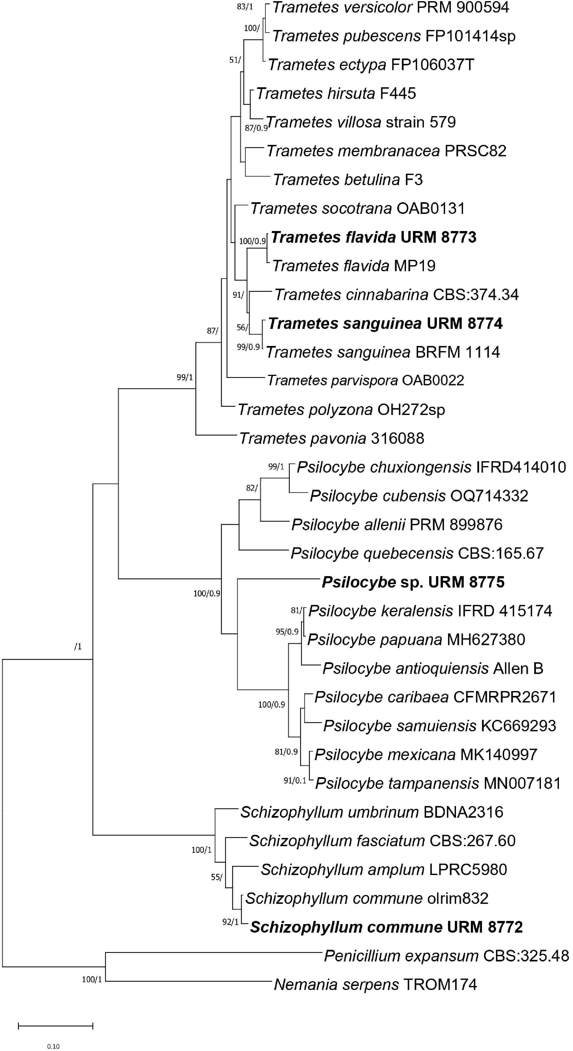
Figure 1.
Maximum likelihood (ML) tree from ITS data. Bootstrap values equal to or above 50% and BI posterior probability equal to or above 0.9 are shown. ITS accession number in GenBank: S. commune URM8772 = PP873655, T. flavida URM8773 = PP873649, T. sanguinea URM8774 = PP873650, and Psilocybe sp. URM8775 = PP873652. Nemania serpens and Penicillium expansum were used as an outgroup.
Nanoparticle characterisation
-
The metal quantification analysis showed concentrations of 33.67 mg/L (Fe3+), 37.84 mg/L (Cu2+), and 19.54 mg/L (Mn2+). No metal was found in the C-dot (pure) solution. The nanoparticles exhibited absorption bands at 272, 335, and 400 nm for pure C-dots; 243 and 350 nm for C-dots-Fe3+; 247 and 350 nm for C-dots-Mn2+; and 238 and 350 nm for C-dots-Cu2+ (Fig. 2c), and emission maxima at 530 nm for pure C-dots and at 455, 455, and 445 nm for C-dots doped with Fe3+, Mn2+, and Cu2+, respectively (Fig. 2a). These findings indicate that the selective doping of carbon dots with metal ions, particularly transition metals, enables precise modulation of their emission spectra, thereby broadening and strengthening their potential for biotechnological applications in diverse contexts. FTIR spectroscopy was employed to analysed the surface functional groups of the synthesized pure (C-dots) and metal-doped nanoparticles (C-dots-Fe, C-dots-Mn, and C-dots-Cu). The resulting spectra reveal characteristic absorption bands corresponding to different vibrational modes characteristic of C-dots (Fig. 2b).
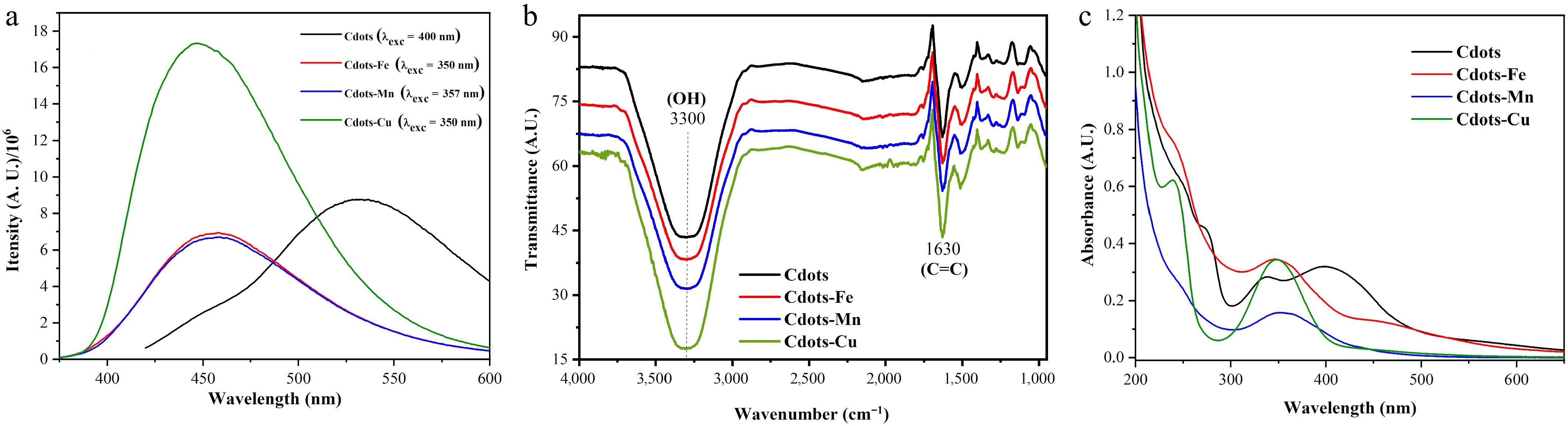
Figure 2.
Characterisation of nanoparticle solution. (a) Photoluminescence (emission) spectra. (b) FTIR vibrational spectra. (c) UV-vis spectra.
Bioassimilation
-
The isolates were grown in agar-agar (negative control) and nanoparticles medium for 168 h. The colonies were all white on the front and reverse of the plate, cottony with regular margins.
Every isolate had C-dots-Fe as the most effective source for growth induction, followed by C-dots-Cu for T. flavida URM8773 (61.0 ± 2.0 mm) and S. commune URM8772 (55.5 ± 2.2 mm) (Fig. 3). C-dots-Fe was also the only medium to stimulate the growth of Psilocybe sp. URM8775 (16.6 ± 7.2 mm), besides agar-agar control, suggesting that this might be the only metal that significantly stimulates the growth of this isolate. Each isolate showed reduced halo growth in the C-dots-Mn medium when compared to the control, suggesting Mn2+ induced growth inhibition. The presence of sole carbon dots for the C-dots medium stimulated growth only for S. commune URM8772 (50.0 ± 6.1 mm) and T. sanguinea URM8774, which showed the highest growth (78.0 ± 2.0 mm). Visual depigmentation was observed in Psilocybe sp. URM8775 after 7 d and in T. sanguinea URM8774 after 28 d of growth, because of C-dots enzymatic breakdown and assimilation for cellular use of the specimens.
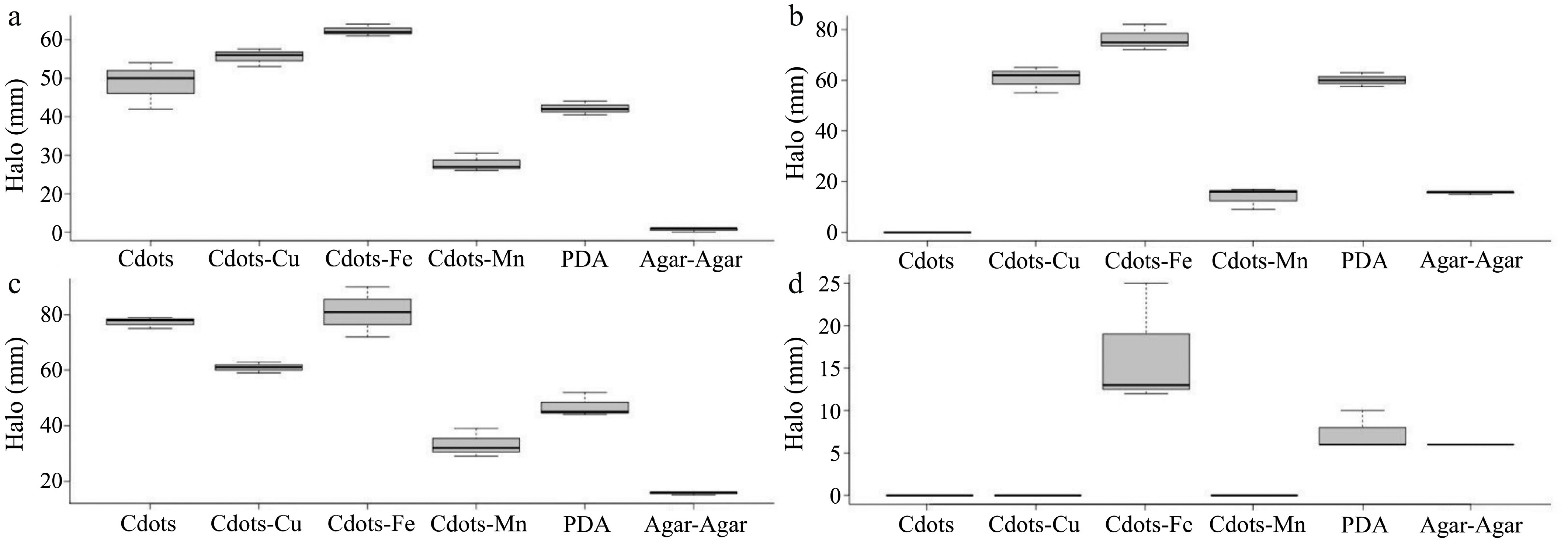
Figure 3.
Bioassimilation of C-dots with or without different metals at a 2 mg/mL concentration. PDA medium was used as a positive control, and agar-agar as a negative control. (a) S. commune URM8772. (b) T. flavida URM8773. (c) T. sanguinea URM8774. (d) Psilocybe sp. URM8775.
Biostimulation
-
Within 7 d, it was possible to observe the isolates that exhibited the highest growth and most positive reactions for enzymatic production in different media (starch and CMC) combined with nanoparticles.
In the CMC medium (Fig. 4), most isolates displayed mycelial growth, except for S. commune URM8772 and T. sanguinea URM8774 in C-dots, and Psilocybe sp. URM8775 in C-dos-Fe and in CMC without C-dots. Schizophyllum commune URM8772 and Trametes flavida URM8773 exhibited the highest growth and substrate degradation halo in the C-dots-Fe medium (29.0 ± 1.0 mm and 48.6 ± 2.5 mm, respectively), indicating that Fe3+ might be an important cofactor and an easier metal to assimilate compared to Cu2+ and Mn2+.
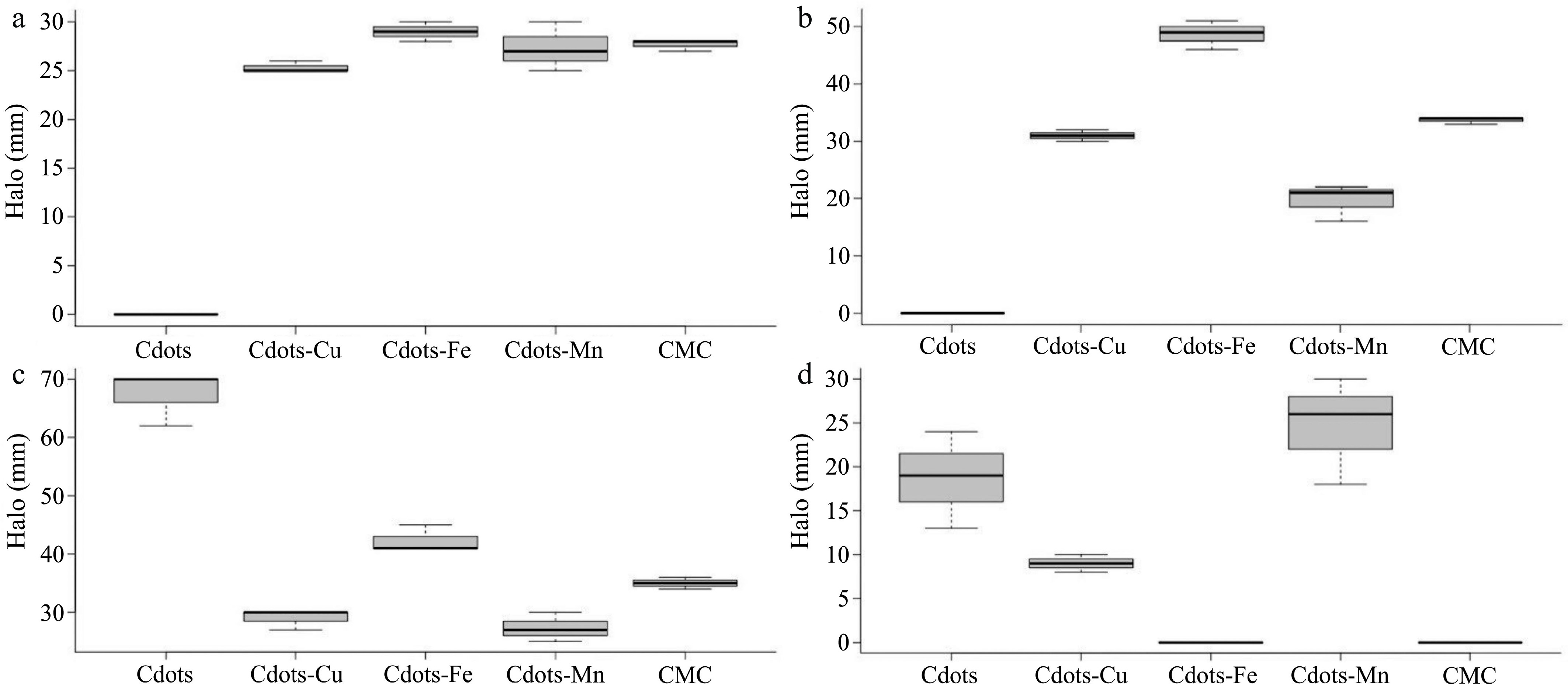
Figure 4.
Biostimulation assay of enzymatic production in CMC (1%) with C-dots with or without different metals in a 2 mg/mL concentration. CMC medium without C-dots was used as a positive control. (a) S. commune URM8772. (b) T. flavida URM8773. (c) T. sanguinea URM8774. (d) Psilocybe sp. URM8775.
Regarding the starch medium (Fig. 5), C-dots media showed the largest depigmentation halo by T. sanguinea URM8774 (64.6 ± 1.5 mm), followed by S. commune URM8772 (46.6 ± 5.8 mm), while the best results among C-dots-metals were achieved by T. flavida URM8773 and Psilocybe sp. URM8775 (20.3 ± 2.3 mm and 13.0 ± 6.0 mm, respectively) in C-dots-Cu media, being the only medium to stimulate the growth of the latter. It is possible that, in those media, the mycelial growth investment was directed towards internal penetration and enzyme production, given the degradation halos in CMC media for C-dots-Mn and C-dots, which were larger than the superficial layer growth halos.
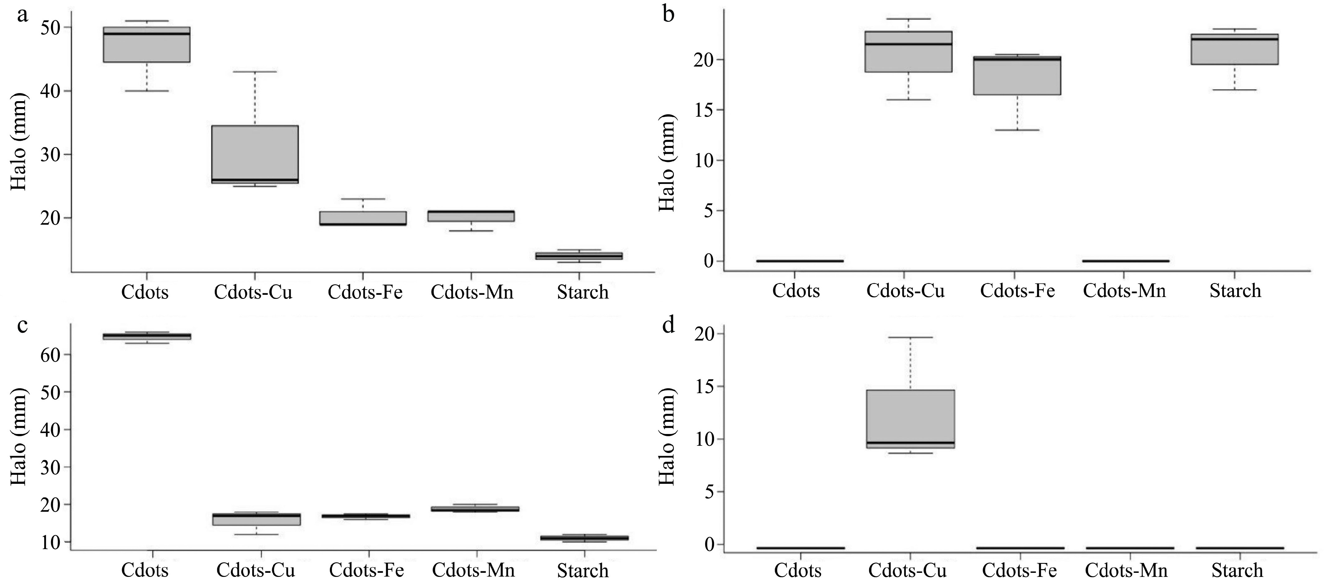
Figure 5.
Biostimulation assay of enzymatic production in starch (1%) with C-dots with or without different metals in 2 mg/mL concentration. Starch medium without C-dots was used as a positive control. (a) S. commune URM8772. (b) T. flavida URM8773. (c) T. sanguinea URM8774. (d) Psilocybe sp URM8775.
Phenoloxidase induction
-
All isolates displayed dark brown halos on the reverse of the plate (Fig. 6). Schizophyllum commune URM8772 showed robust growth but a smaller tannic acid reaction halo, while the other isolates presented smaller mycelium growth halos but larger phenoloxidase production halos. Trametes flavida URM8773 phenoloxidase halo was proportional to the growth halo, while T. sanguinea URM8774 exhibited a larger phenoloxidase production halo when compared to its thinner aerial mycelium growth (Fig. 6), similar to Psilocybe sp. URM8775, which did not show apparent growth on the surface of the media, but demonstrated strong phenoloxidase production, indicated by the darker halos compared to other isolates.
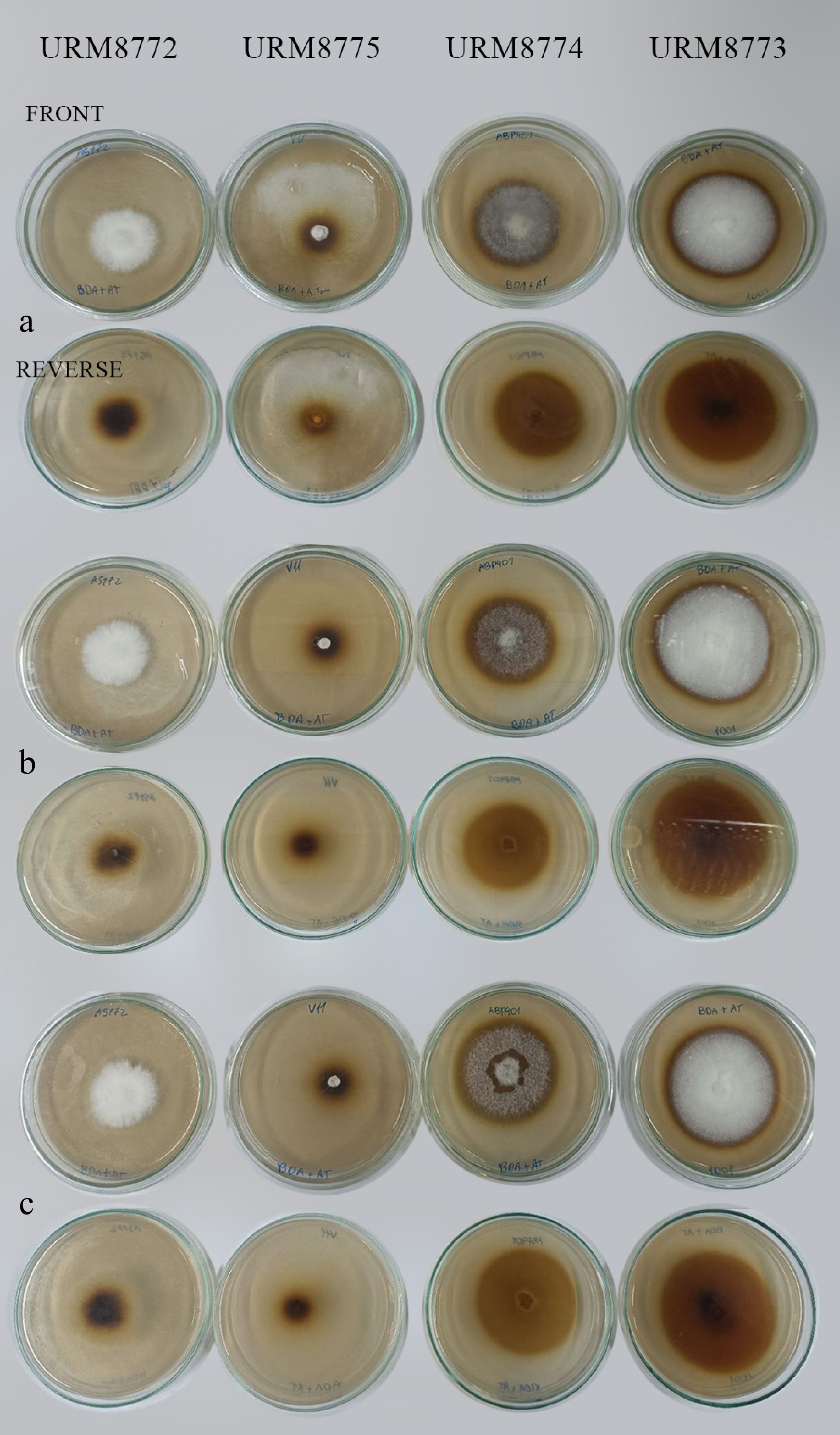
Figure 6.
Isolates growth and brownish halo formations in PDA with tannic acid (0.5%) concentration medium. (a) Triplicate 1. (b) Triplicate 2. (c) Triplicate 3.
Among the isolates, T. flavida URM8773 presented the largest phenoloxidase production halo, higher than 60 mm, followed by T. sanguinea URM8774, S. commune URM8772, and lastly Psilocybe sp. URM8775 with a little above 20 mm (Fig. 7).
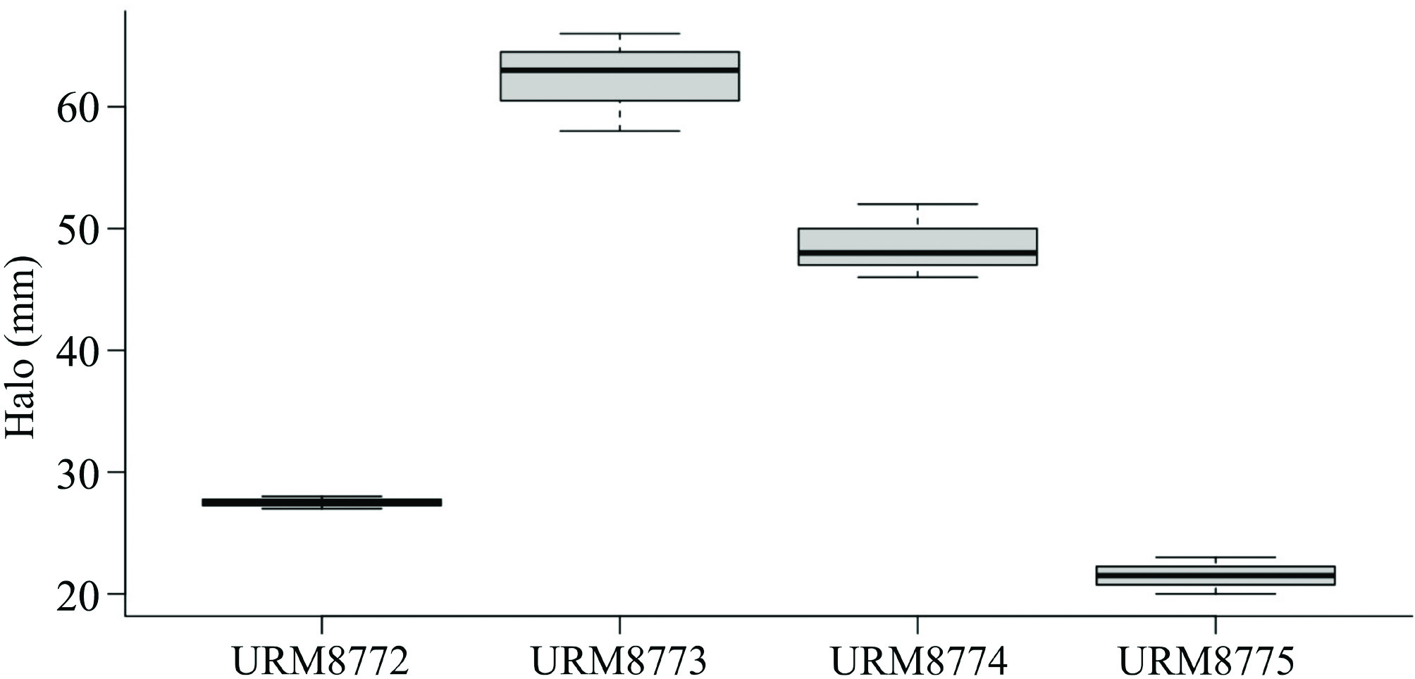
Figure 7.
Phenoloxidase enzymatic production in PDA with tannic acid (0.5%) medium. S. commune URM8772. T. flavida URM8773. T. sanguinea URM8774. (d) Psilocybe sp.
-
According to our results, both pure and doped-C-dots exhibit strong photoluminescent emission in the blue region, with a characteristic peak between 450 and 500 nm[38,39], confirming the formation of C-dots. The incorporation of metal ions modified the photoluminescent properties of C-dots, promoting a shift in emission towards shorter wavelengths (blueshift). Notably, C-dots-Cu2+ exhibited the greatest shift compared to pure C-dots, a behavior also reported in previous studies[38]. The FTIR data show a broad absorption band at 3,300 cm−1 in all samples, which is attributed to the stretching vibrations of hydroxyl (-OH) groups[38,40]. Additionally, a prominent peak approximately at 1,630 cm−1 is present in all spectra, which corresponds to the C=C stretching vibrations of aromatic sp2-hybridized carbon structures. This signal confirms the presence of conjugated carbon frameworks, characteristic of graphitic or partially oxidized carbon-based materials. In UV-vis spectra (Fig. 2a), a discrete peak observed in the 240–270 nm range is attributed to the π–π* electronic transition of sp2 carbons present on the C-dots surface, associated with C=C and C=N bonds[38,41].
Additionally, a peak around 345 nm is due to the n–π* transition, characteristic of C=O groups, as well as to contributions from other surface functional groups[39]. Another peak in the 360–400 nm range is associated with the n–π* orbital transition involving sp3 carbons, indicative of interactions between carbon atoms and functional groups containing nitrogen and oxygen[39]. For the metal-doped samples, displacements of the main peak were observed, shifting from 400 nm in pure C-dots to 350 nm in doped C-dots. This behavior, widely reported in the literature[38,40,41], indicates effective doping of the nanomaterials.
Considering the decolorisation tests, Psilocybe sp. URM8775 might have been able to break the nanoparticle chemical bonds faster, but it did not properly consume the resulting by-products as T. sanguinea URM8774 did. Trametes species are known to produce higher levels of ligninolytic enzymes, such as laccase. In contrast, apparently only Machado et al.[12] investigated the enzymatic production and decolorization of Psilocybe spp. collected in Brazil, and reported laccase for the first time in P. castanella Peck, currently considered P. apelliculosa P.D. Orton (
www.mycobank.org/details/708/53053 ) or Deconica castanella (Peck) Noordel. (www.speciesfungorum.org/Names/SynSpecies.asp?RecordID=515382 ). The current study may be the first to detect the enzymatic production of Psilocybe sp. and its reaction with the C-dots medium.In the study by Machado et al.[42], both T. villosa and T. sanguinea exhibited growth that reached the edges of the plates by the seventh day, similar to the current study. It was visible that none of the isolates displayed growth in the presence of C-dots greater than the positive controls on PDA medium, except for Psilocybe sp. URM8775. It is possible that the ligninolytic enzymes produced by this isolate presented Cu2+ as a cofactor, thus increasing its growth capacity. Baldrian et al.[43] investigated Pleurotus ostreatus by introducing several carbon and nitrogen sources, along with metal ions and aromatic compounds. They confirmed the induction of laccase production in the presence of Cu2+ under limited hydrogen conditions, as previously documented by Palmieri et al.[44] Furthermore, Baldrian et al.[45] reported, for the first time, the induction of laccase production in the presence of cadmium (Cd), specifically when added during the final growth stages. It was also observed that heavy metals exert an influence not only on laccase activity but also on manganese peroxidase (EC 1.11.1.13), displaying a pronounced sensitivity to heavy metal-enriched media like Cu2+ and Cd, as demonstrated in tests involving Stereum hirsutum and Phanerochaete chrysosporium[43].
The isolates' susceptibility to certain metals, such as Mn2+, was also evident in the current study, in which the species demonstrated enhanced development in the presence of C-dots. Regardless of the inhibitory effect on white-rot fungi species when Cu2+ was used[46], S. commune URM8772 has shown higher growth in the presence of Cu2+ and Fe3+, while T. flavida URM8773 has shown similar growth in C-dots-Cu2+ when compared to the PDA control. All isolates showed the greatest growth induction in the presence of C-dots-Fe3+.
The sensitivity of white-rot fungi to certain heavy metal ions, according to Palmieri et al.[44], is due to physiological stress induced by the generation of chemical stressors, which, in turn, trigger the production of reactive oxygen species, leading to oxidative cellular damage. Therefore, further investigations may unravel the intricate metal interactions, such as those involving Mn, in the enzymatic production of the white-rot fungi, especially those from tropical areas[46,47].
As for the biostimulation assays, in CMC media (Fig. 4), T. sanguinea URM8774 exhibited the highest growth and enzymatic production halo, particularly in C-dots media. Enzymes of the cellulase complex include endo 1,4-β-glucanase (EC 3.2.1.4), exo cellobiohydrolase (EC 3.2.1.91), and β-glucosidase (EC 3.2.1.21). These complexes and the resulted by-products from the cellulose degradation, as the registered activity from endo 1,4-β glucanase, have a variety of biotechnological applications in the textile, pulp and paper industries[48], food industry as wine stabilization, denim finishing and cotton bleaching, bioremediation of pollutants in soil and water bodies, beer filtration and oil extraction[49−52], as well as the cellulosic hydrolyzation of materials to sugars in the biofuel production and many others[53,54].
As for the current study, except for Psilocybe sp. URM8775, all isolates showed larger cellulase enzymatic halos in the presence of C-dots-Fe, suggesting that this metal is used in the biosynthesis of cell components. This higher uptake of iron was also registered in yeasts, Aspergillus spp, ectomycorrhizal basidiomycetes, and Coprinopsis cinerea, for which production of siderophores, known as iron-chelating compounds, was detected[55−57].
In previous studies, S. commune has been shown to preferentially degrade carbohydrates, with higher levels of cellulolytic, xylanolytic, and pectinolytic activities than the other three Agaricomycetes[58]. Genes to produce laccase and other oxidase enzymes were detected in S. commune; however, according to Tovar-Herrera et al.[50] and the CAZy database, the species lacks lignin peroxidases (LiP), manganese peroxidases (MnP), and versatile peroxidases (VP) genes. This observation highlights the variability and range of phenotypic plasticity within species, as seen in S. commune, and emphasizes the importance of comparative essays, which provide a better understanding of the range of enzyme production across different isolates.
The polyphenoloxidase complex is also widely used in industry. As listed by Banerjee & Mahapatra[59], the biotechnological application of the tannase complex includes the production of gallic acid, an important pharmaceutical compound. Furthermore, it includes its activity in animal feed production, the reduction of turbidity in beer production, the detoxification of tannery effluents, and others, indicating the success in tannic acid assimilation and therefore production of phenoloxidases, a diverse enzymatic group belonging to the oxidoreductases responsible for the oxidation of phenolic compounds that have Cu2+ ions in their active site[46,59].
The enzyme production ability of Brazilian isolates of Agaricomycetes in the presence of carbon-based nanoparticles containing some metals that play important roles in catalytic sites of enzymatic cofactors was analyzed. Among the tested nanomaterials, C-dots-Fe showed the most pronounced biostimulatory effect, promoting mycelial growth and increasing amylase and cellulase production in T. sanguinea URM8774 and S. commune URM8772. Interestingly, Psilocybe sp. URM8775 showed atypical behavior: although its growth was modestly stimulated by C-dots-Fe, its phenoloxidase production was significant in medium containing tannic acid, a relevant finding given the lack of studies on the enzymatic pathways of species in this genus.
-
These results indicate that C-dots-Fe can be used as effective inducers of enzymatic activity in Agaricomycete isolates, with potential applications in biotechnological processes such as the biodegradation of lignocellulosic residues and pollutant remediation. This work also sheds light on studies of species with unknown enzymatic pathways, such as Psilocybe species and T. flavida, and proposes that new studies be conducted to better identify and enrich the basis for future biotechnological studies, including evaluating different metal concentrations to identify the optimal concentration. The concentrations evaluated in this work were relatively high and may have been above the optimal point for stimulating enzyme production. Future research should explore optimized nanoparticle concentrations, broader metal combinations, and nanoparticle-fungus interactions to fully harness the enzymatic capabilities of unexplored and novel Agaricomycetes species.
-
The authors confirm contribution to the paper as follows: study conception and design: Wanderley SS, de Sousa Filho IA, Mendes-Alvarenga RL; material preparation, data collection and analysis: Wanderley SS, de Sousa Filho IA, Pires ICB, de Oliveira VRT, Weber IT, Junior SA, Gibertoni TB, Mendes-Alvarenga RL; draft manuscript preparation: Wanderley SS; manuscript review and editing: de Sousa Filho IA, Pires ICB, de Oliveira VRT, Weber IT, Junior SA, Gibertoni TB, Mendes-Alvarenga RL; funding acquisition: Gibertoni TB, Weber IT, Junior SA. All authors reviewed the results and approved the final version of the manuscript.
-
The datasets generated during and/or analysed during the current study are available from the corresponding author upon reasonable request.
-
The authors have no relevant financial interests to disclose.
- Copyright: © 2026 by the author(s). Published by Maximum Academic Press, Fayetteville, GA. This article is an open access article distributed under Creative Commons Attribution License (CC BY 4.0), visit https://creativecommons.org/licenses/by/4.0/.
-
About this article
Cite this article
Wanderley SS, de Sousa Filho IA, Pires ICB, de Oliveira VRT, Weber IT, et al. 2026. Analysis of Agaricomycetes' growth and enzyme production under the influence of C-dots doped with metals. Studies in Fungi 11: e029 doi: 10.48130/sif-0026-0027 

Analysis of Agaricomycetes' growth and enzyme production under the influence of C-dots doped with metals
- Received: 19 September 2025
- Revised: 24 April 2026
- Accepted: 14 May 2026
- Published online: 08 October 2026
Abstract: Agaricomycetes, a class of wood-decay fungi, are known for their enzymatic activities and have been investigated for various biotechnological applications, including the degradation of recalcitrant and xenobiotic compounds via laccases and phenoloxidases. Despite this, the biotechnological potential of Agaricomycetes, particularly Brazilian species, remains underexplored. This study analysed and compared the enzymatic production potential of Brazilian Agaricomycetes isolates in the presence of carbon-based nanoparticles (C-dots), either enriched with Fe3+, Cu2+, and Mn2+ or unenriched. Isolate identification was confirmed using ITS rDNA sequencing, as well as Maximum Likelihood and Bayesian Inference analyses. Assimilation and biostimulation tests were performed on solid media supplemented with 2 mg/mL of C-dots. Phenoloxidase, cellulase, and amylase production was observed in isolates of Psilocybe sp., Schizophyllum commune, Trametes flavida, and Trametes sanguinea. Furthermore, Fe3+ and Cu2+ were identified as promising, cost-effective medium enrichers, whereas Mn2+ exhibited inhibitory effects.
-
Key words:
- Bioprospecting /
- Biotechnology /
- Nanoparticle /
- Psilocybe /
- Schizophyllum /
- Trametes